KEY POINTS
Define thecomplement system.
Describe the three pathways of complement
activation.
Understand the function of complement system.
Recognise the regulatory mechanisms.
recognise the disorder in the complement
system.
Define the complement system.
Describe the three pathways of complement
activation.
Understand the function of complement system.
Recognise the regulatory mechanisms.
recognise the disorder in the complement
system.
3.
HISTORICAL BACKGROUND OF
COMPLEMENT
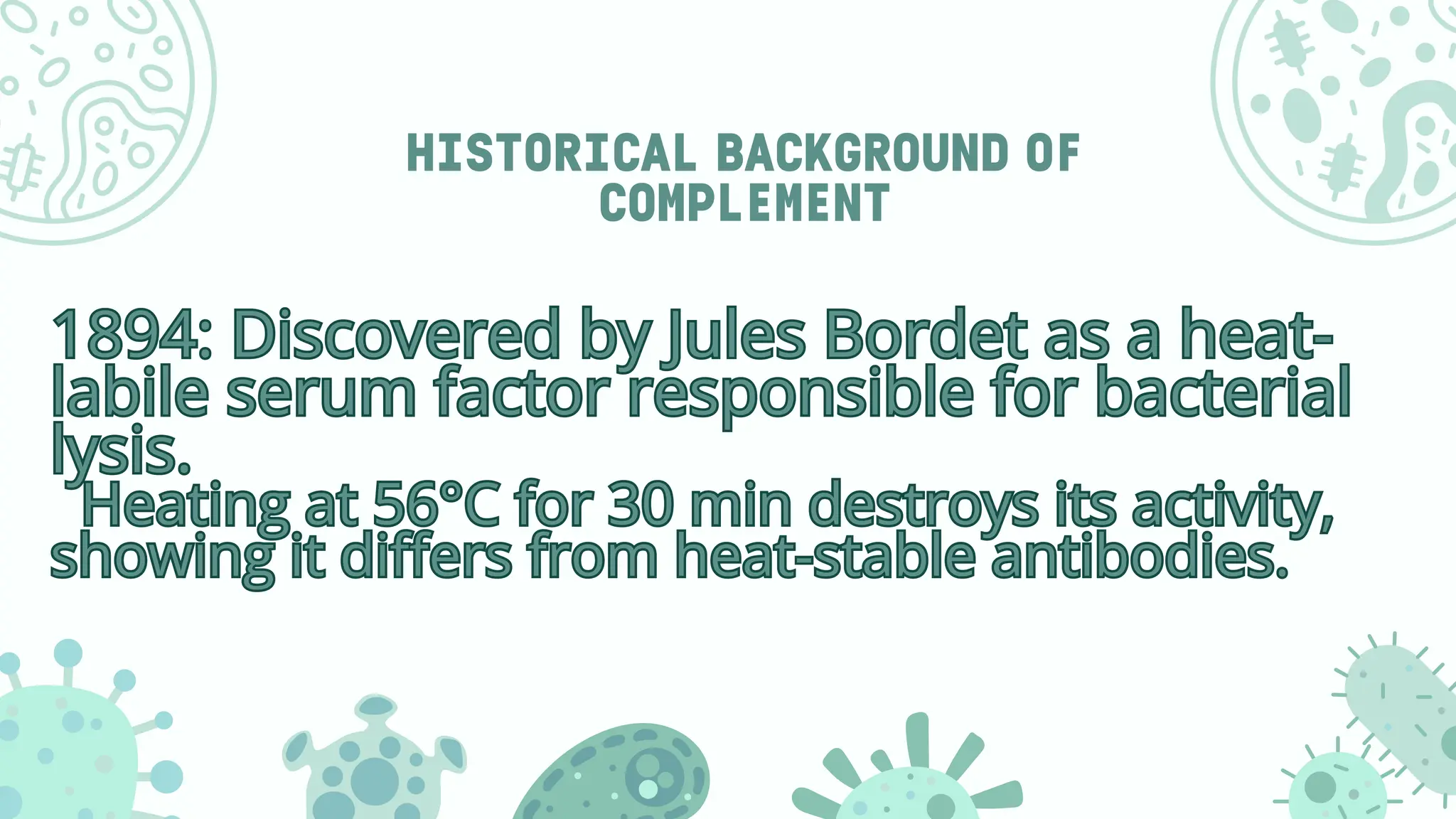
1894:Discovered by Jules Bordet as a heat-
labile serum factor responsible for bacterial
lysis.
Heating at 56°C for 30 min destroys its activity,
showing it differs from heat-stable antibodies.
1894: Discovered by Jules Bordet as a heat-
labile serum factor responsible for bacterial
lysis.
Heating at 56°C for 30 min destroys its activity,
showing it differs from heat-stable antibodies.
4.
INTRODUCTION
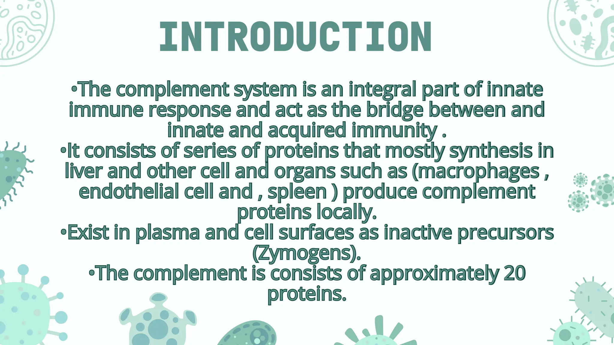
•The complement systemis an integral part of innate
immune response and act as the bridge between and
innate and acquired immunity .
•It consists of series of proteins that mostly synthesis in
liver and other cell and organs such as (macrophages ,
endothelial cell and , spleen ) produce complement
proteins locally.
•Exist in plasma and cell surfaces as inactive precursors
(Zymogens).
•The complement is consists of approximately 20
proteins.
•The complement system is an integral part of innate
immune response and act as the bridge between and
innate and acquired immunity .
•It consists of series of proteins that mostly synthesis in
liver and other cell and organs such as (macrophages ,
endothelial cell and , spleen ) produce complement
proteins locally.
•Exist in plasma and cell surfaces as inactive precursors
(Zymogens).
•The complement is consists of approximately 20
proteins.
5.
CONTINUE
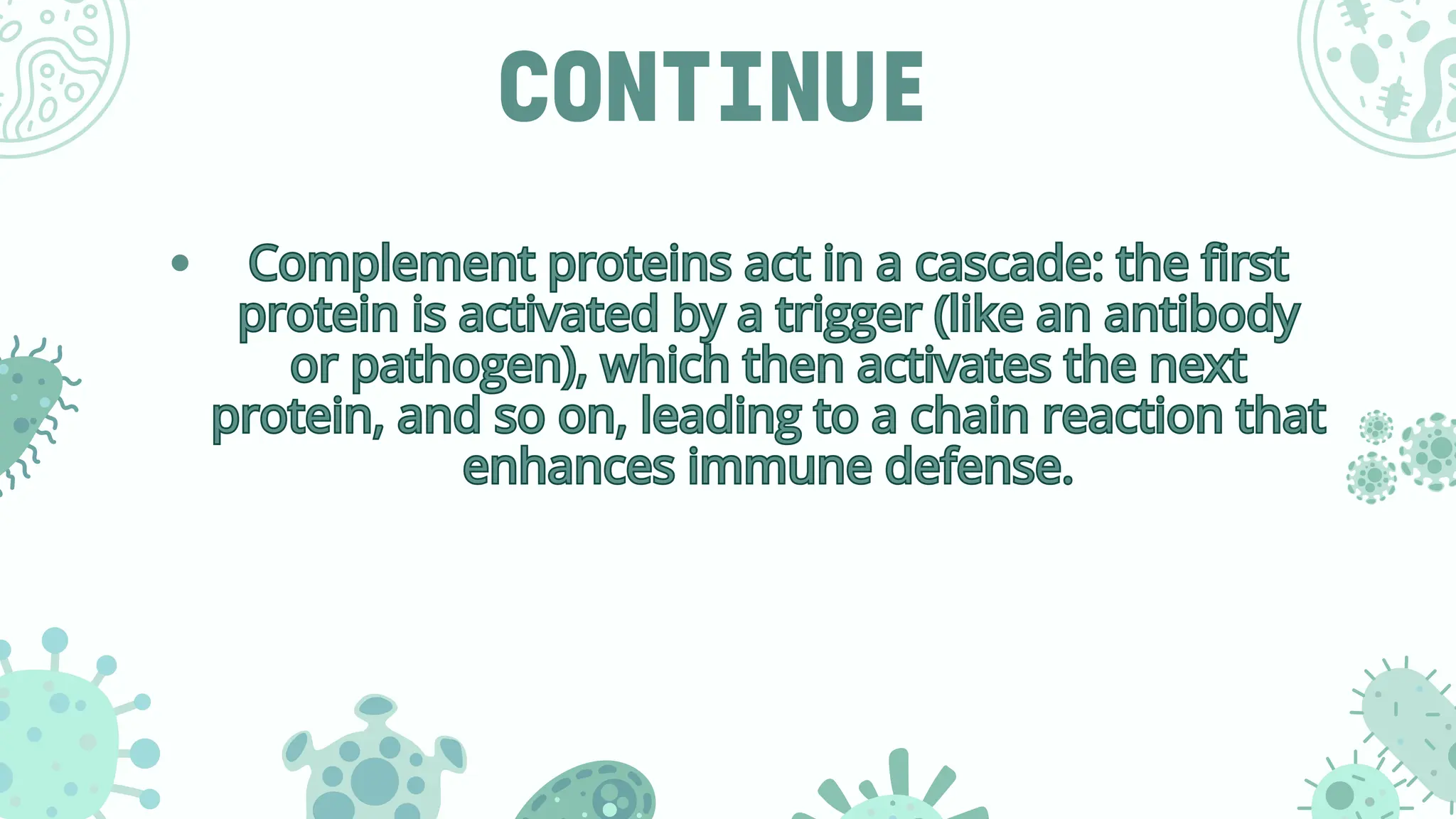
Complement proteins actin a cascade: the first
protein is activated by a trigger (like an antibody
or pathogen), which then activates the next
protein, and so on, leading to a chain reaction that
enhances immune defense.
Complement proteins act in a cascade: the first
protein is activated by a trigger (like an antibody
or pathogen), which then activates the next
protein, and so on, leading to a chain reaction that
enhances immune defense.
6.
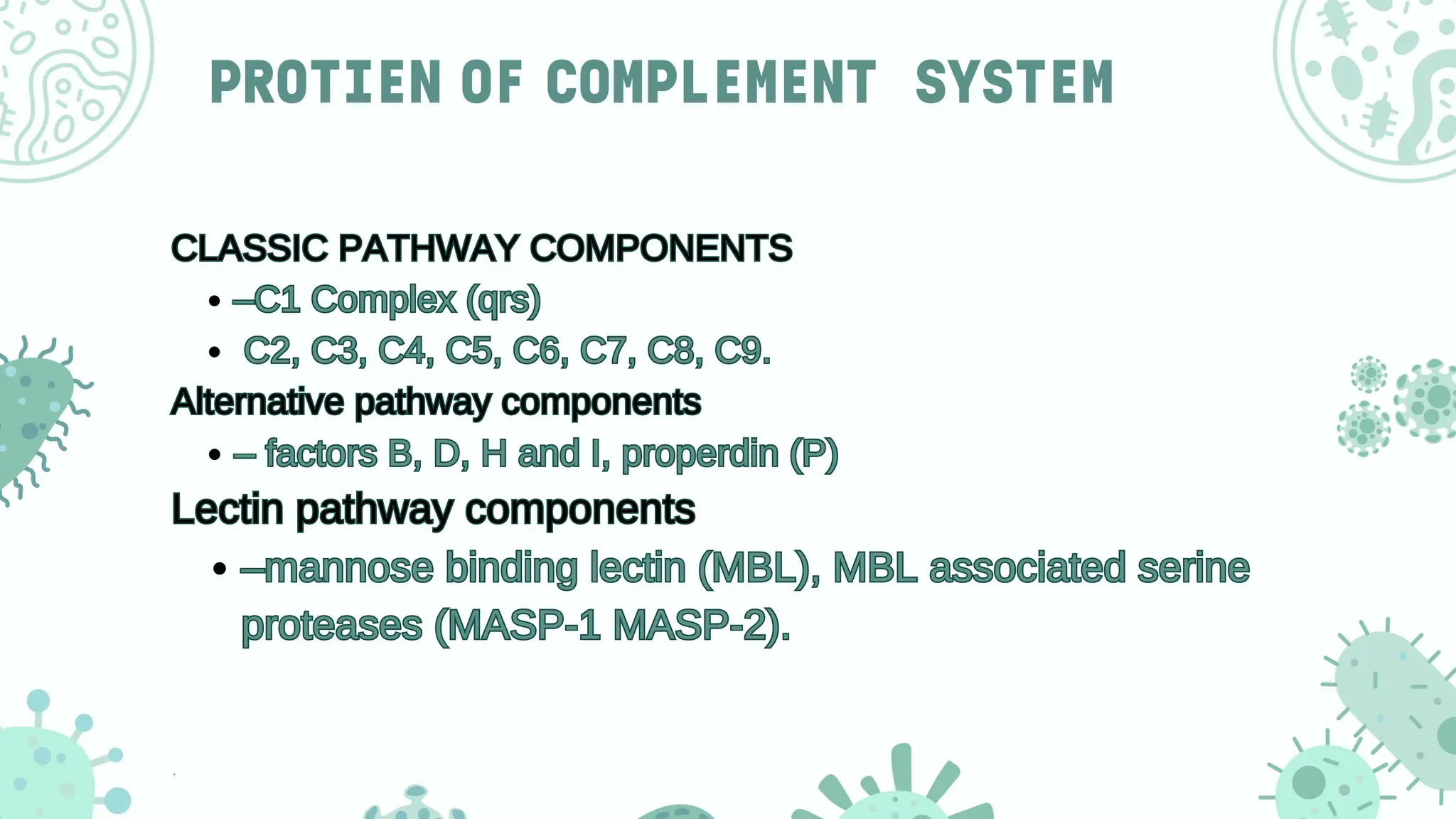
PROTIEN OF COMPLEMENTSYSTEM
CLASSIC PATHWAY COMPONENTS
–C1 Complex (qrs)
C2, C3, C4, C5, C6, C7, C8, C9.
Alternative pathway components
– factors B, D, H and I, properdin (P)
Lectin pathway components
–mannose binding lectin (MBL), MBL associated serine
proteases (MASP-1 MASP-2).
CLASSIC PATHWAY COMPONENTS
–C1 Complex (qrs)
C2, C3, C4, C5, C6, C7, C8, C9.
Alternative pathway components
– factors B, D, H and I, properdin (P)
Lectin pathway components
–mannose binding lectin (MBL), MBL associated serine
proteases (MASP-1 MASP-2).
7.
ACTIVATINO OF
COMPLEM SYSTEM
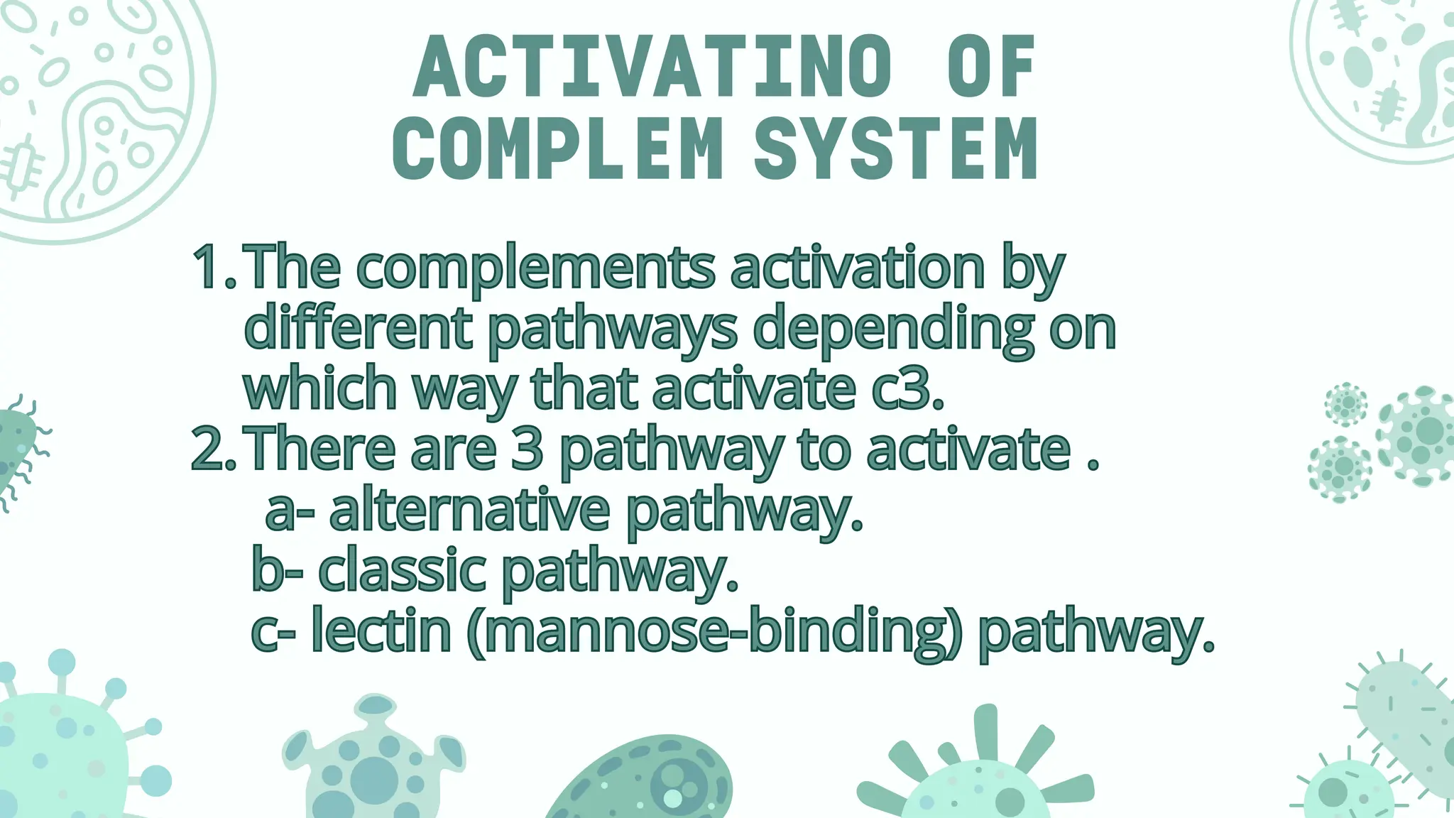
1.Thecomplements activation by
different pathways depending on
which way that activate c3.
2.There are 3 pathway to activate .
a- alternative pathway.
b- classic pathway.
c- lectin (mannose-binding) pathway.
1.The complements activation by
different pathways depending on
which way that activate c3.
2.There are 3 pathway to activate .
a- alternative pathway.
b- classic pathway.
c- lectin (mannose-binding) pathway.
8.
CONTINUED
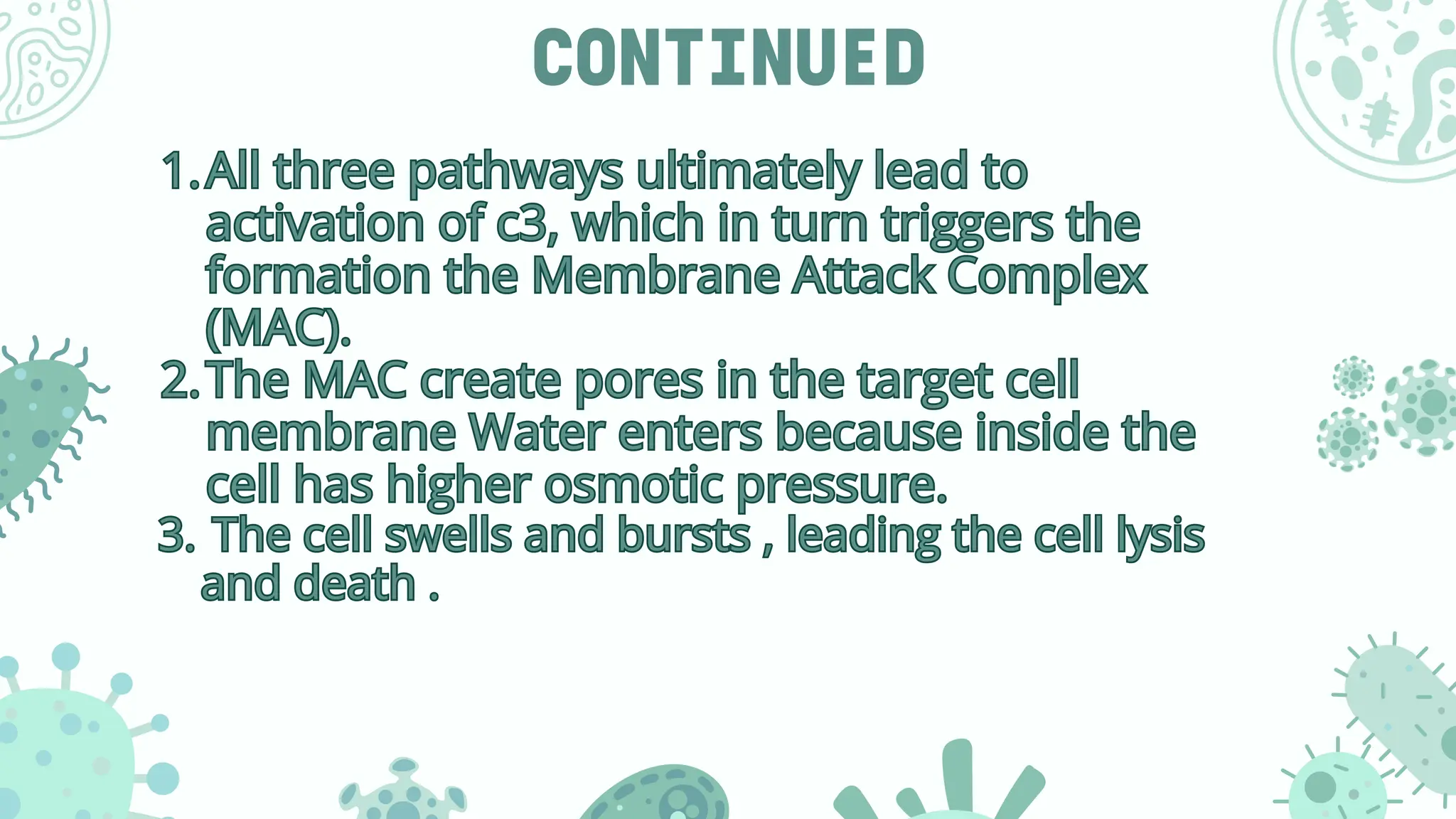
1.All three pathwaysultimately lead to
activation of c3, which in turn triggers the
formation the Membrane Attack Complex
(MAC).
2.The MAC create pores in the target cell
membrane Water enters because inside the
cell has higher osmotic pressure.
3. The cell swells and bursts , leading the cell lysis
and death .
1.All three pathways ultimately lead to
activation of c3, which in turn triggers the
formation the Membrane Attack Complex
(MAC).
2.The MAC create pores in the target cell
membrane Water enters because inside the
cell has higher osmotic pressure.
3. The cell swells and bursts , leading the cell lysis
and death .
9.
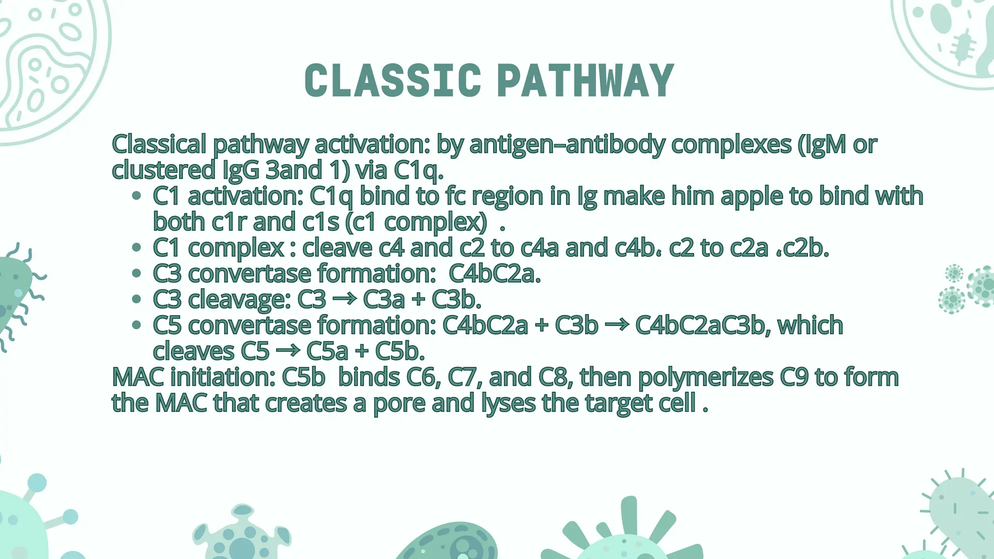
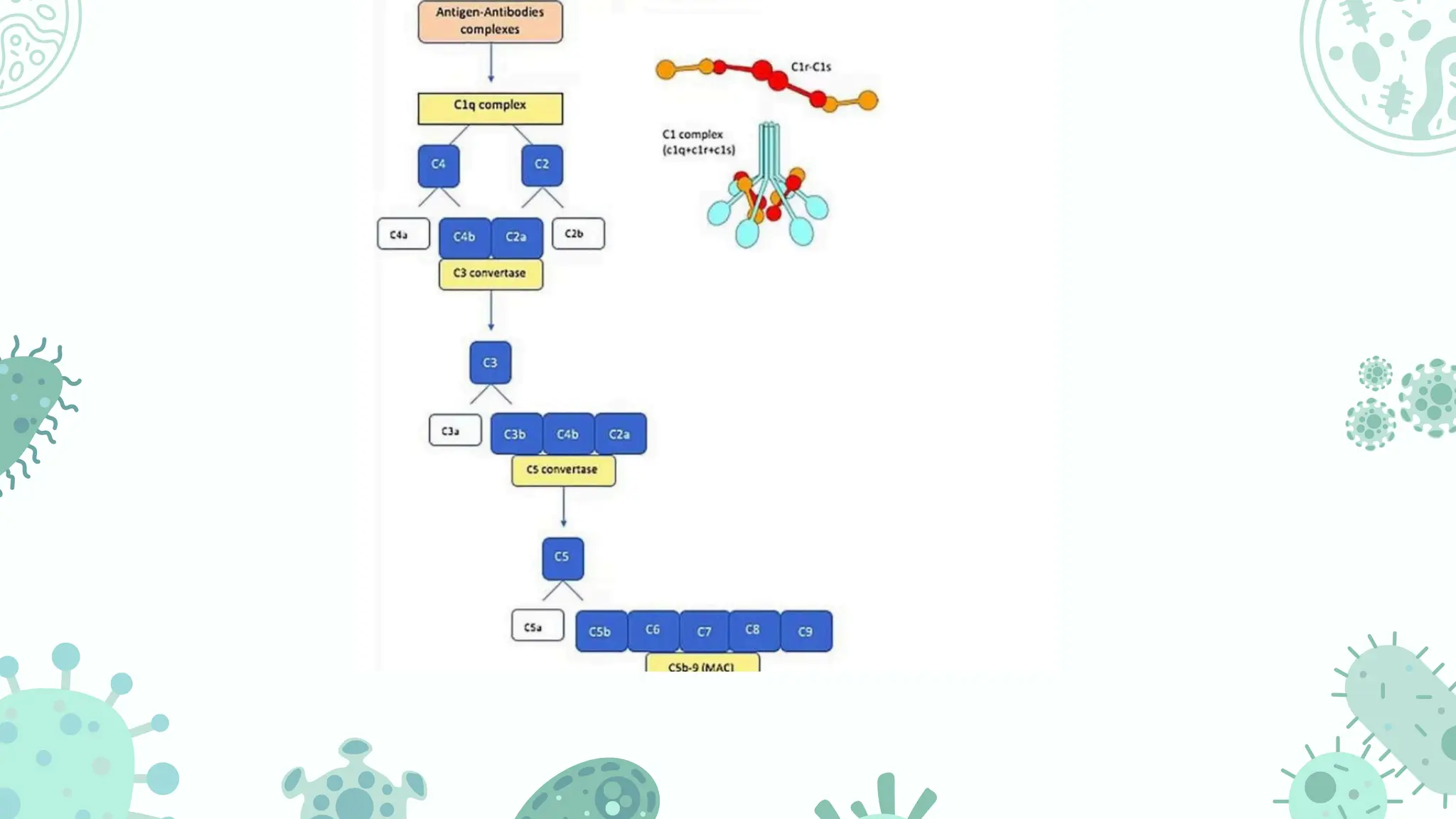
CLASSIC PATHWAY
Classical pathwayactivation: by antigen–antibody complexes (IgM or
clustered IgG 3and 1) via C1q.
C1 activation: C1q bind to fc region in Ig make him apple to bind with
both c1r and c1s (c1 complex) .
C1 complex : cleave c4 and c2 to c4a and c4b، c2 to c2a ،c2b.
C3 convertase formation: C4bC2a.
C3 cleavage: C3 →C3a + C3b.
C5 convertase formation: C4bC2a + C3b →C4bC2aC3b, which
cleaves C5 →C5a + C5b.
MAC initiation: C5b binds C6, C7, and C8, then polymerizes C9 to form
the MAC that creates a pore and lyses the target cell .
Classical pathway activation: by antigen–antibody complexes (IgM or
clustered IgG 3and 1) via C1q.
C1 activation: C1q bind to fc region in Ig make him apple to bind with
both c1r and c1s (c1 complex) .
C1 complex : cleave c4 and c2 to c4a and c4b، c2 to c2a ،c2b.
C3 convertase formation: C4bC2a.
C3 cleavage: C3 →C3a + C3b.
C5 convertase formation: C4bC2a + C3b →C4bC2aC3b, which
cleaves C5 →C5a + C5b.
MAC initiation: C5b binds C6, C7, and C8, then polymerizes C9 to form
the MAC that creates a pore and lyses the target cell .
10.
C0NTINUE
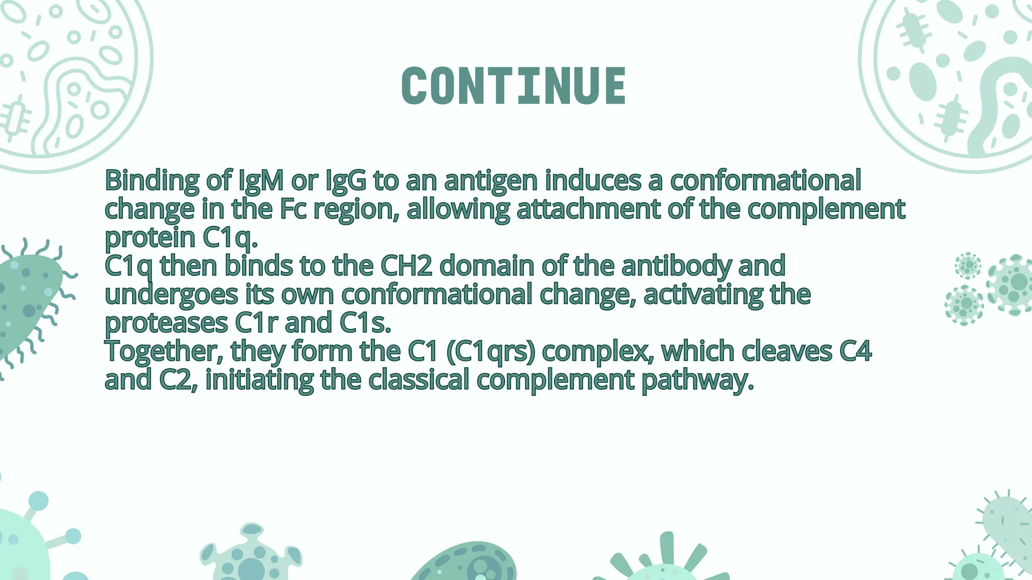
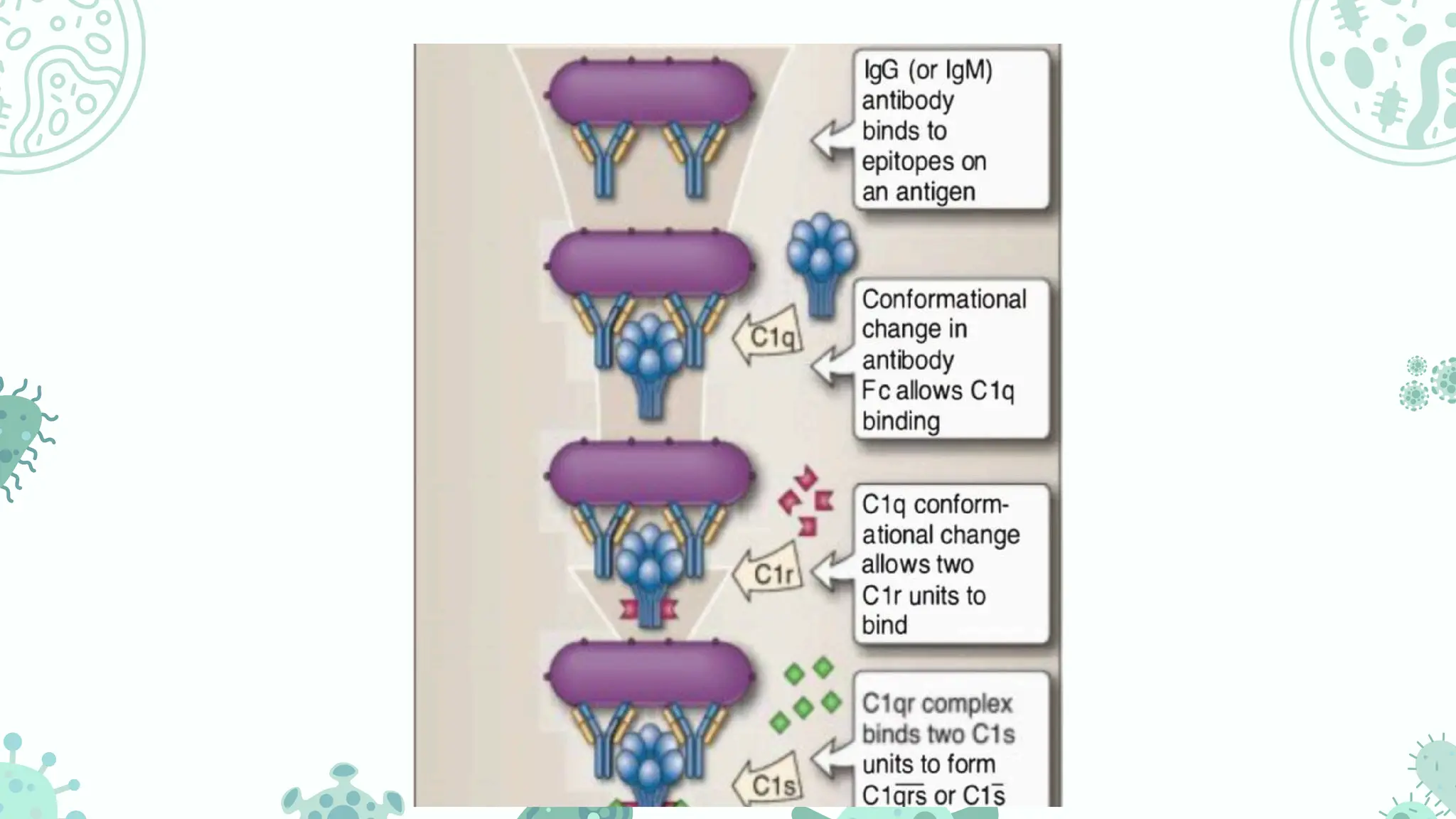
Binding of IgMor IgG to an antigen induces a conformational
change in the Fc region, allowing attachment of the complement
protein C1q.
C1q then binds to the CH2 domain of the antibody and
undergoes its own conformational change, activating the
proteases C1r and C1s.
Together, they form the C1 (C1qrs) complex, which cleaves C4
and C2, initiating the classical complement pathway.
Binding of IgM or IgG to an antigen induces a conformational
change in the Fc region, allowing attachment of the complement
protein C1q.
C1q then binds to the CH2 domain of the antibody and
undergoes its own conformational change, activating the
proteases C1r and C1s.
Together, they form the C1 (C1qrs) complex, which cleaves C4
and C2, initiating the classical complement pathway.
13.
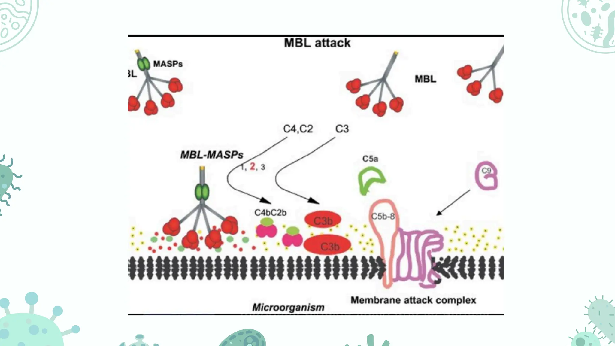
MANNOSE-BINDING LECTIN PATHWAY
Activation:The lectin pathway begins when MBL protein (mannose-binding
lectin) bind to specific carbohydrate called mannose on microbial surfaces.
• MASP activation: Binding recruits MASP-1, MASP-2 (MBL-Associated
Serine Protease) which become activated .
• C4 & C2 cleavage: Activated MASPs cleave C4 and C2 to form the C3
convertase (C4bC2a) — same enzyme as in the classical pathway.
• C3 activation: C3 convertase cleaves C3 →C3a + C3b.
• C5 convertase formation: C4bC2aC3b is formed, which cleaves C5 →C5a
+ C5b.
• MAC formation: C5b initiates assembly of the MAC leading to target cell
lysis.
Activation: The lectin pathway begins when MBL protein (mannose-binding
lectin) bind to specific carbohydrate called mannose on microbial surfaces.
• MASP activation: Binding recruits MASP-1, MASP-2 (MBL-Associated
Serine Protease) which become activated .
• C4 & C2 cleavage: Activated MASPs cleave C4 and C2 to form the C3
convertase (C4bC2a) — same enzyme as in the classical pathway.
• C3 activation: C3 convertase cleaves C3 →C3a + C3b.
• C5 convertase formation: C4bC2aC3b is formed, which cleaves C5 →C5a
+ C5b.
• MAC formation: C5b initiates assembly of the MAC leading to target cell
lysis.
15.
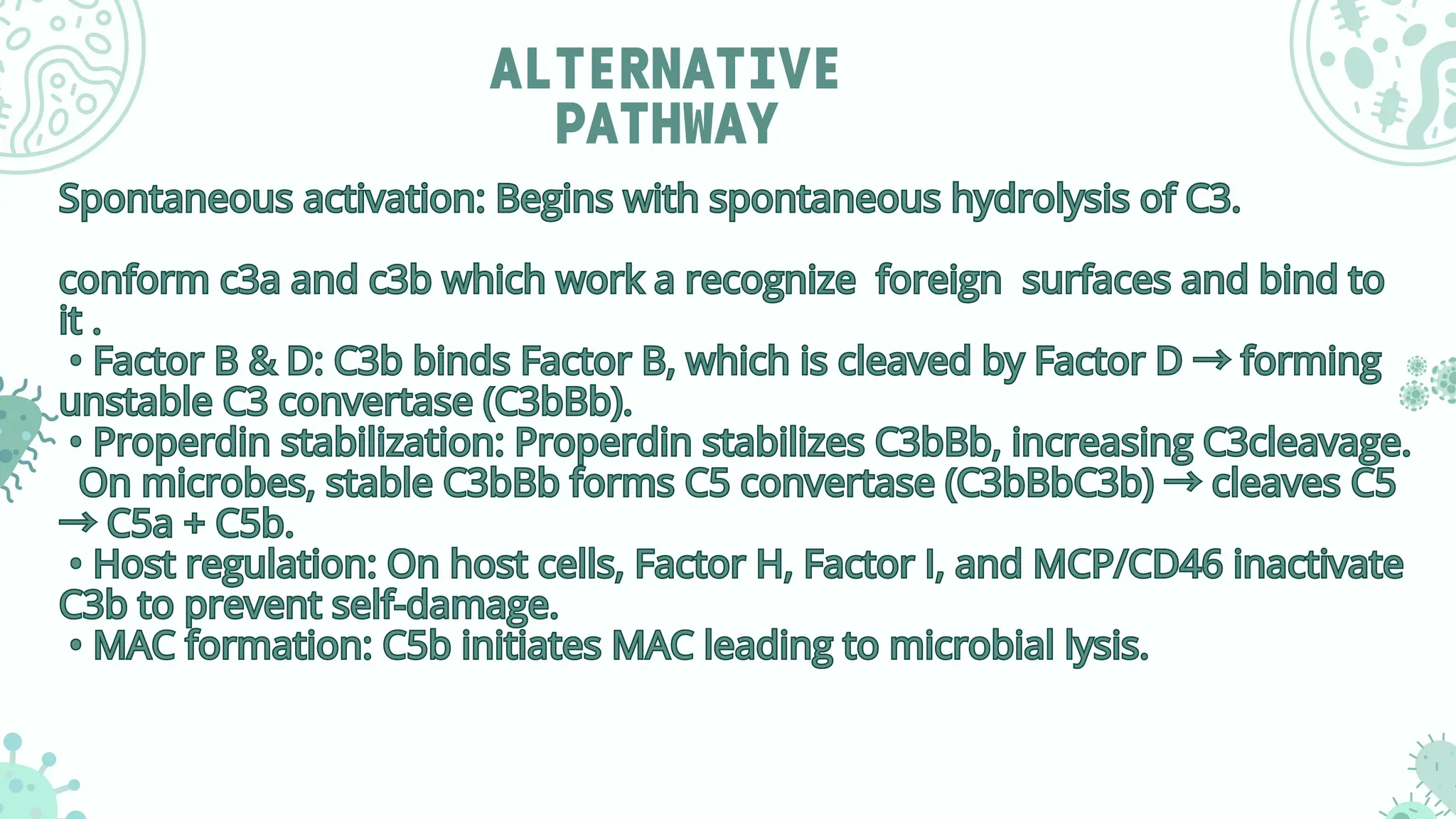
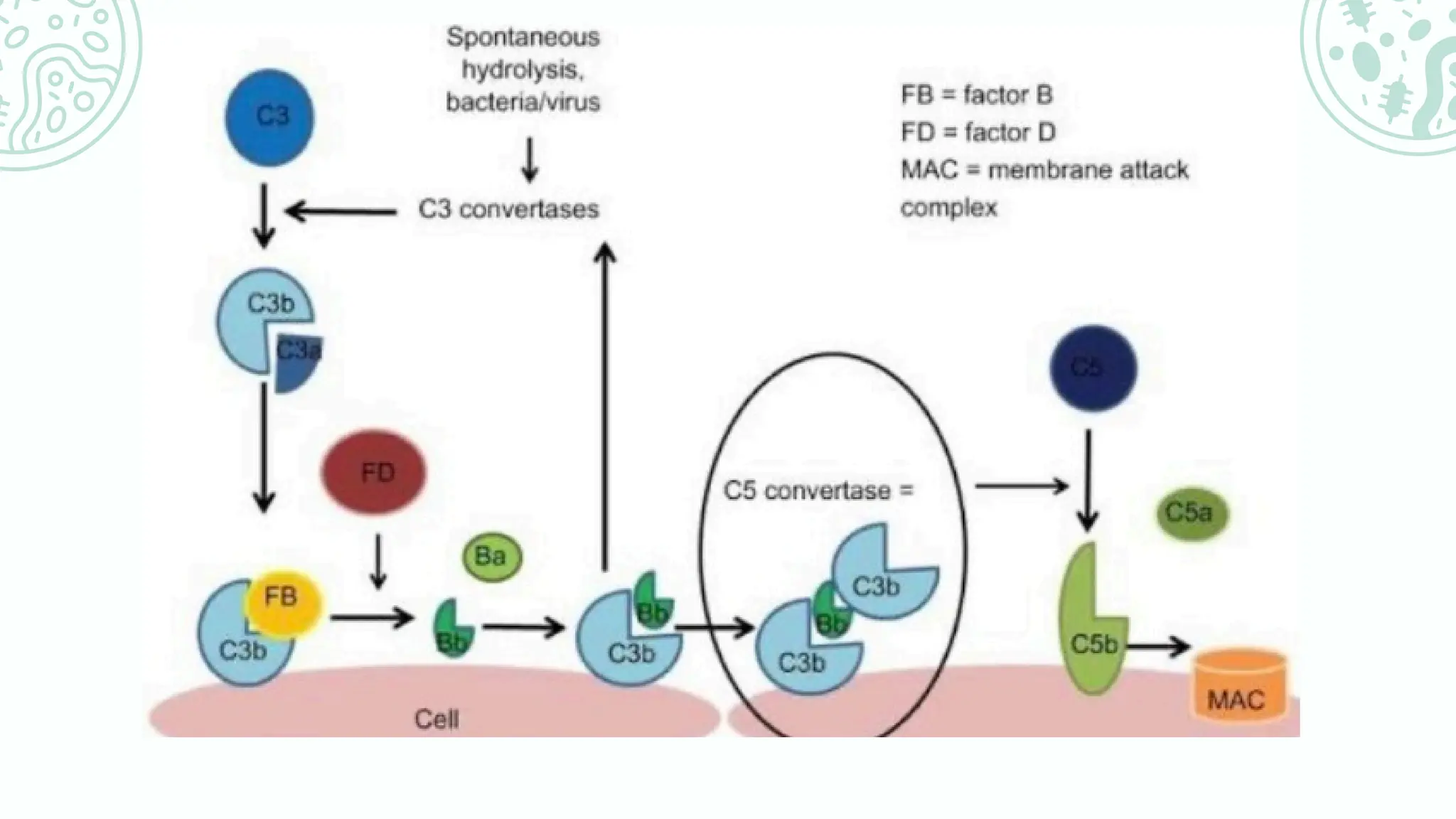
ALTERNATIVE
PATHWAY
Spontaneous activation: Beginswith spontaneous hydrolysis of C3.
conform c3a and c3b which work a recognize foreign surfaces and bind to
it .
• Factor B & D: C3b binds Factor B, which is cleaved by Factor D →forming
unstable C3 convertase (C3bBb).
• Properdin stabilization: Properdin stabilizes C3bBb, increasing C3cleavage.
On microbes, stable C3bBb forms C5 convertase (C3bBbC3b) →cleaves C5
→C5a + C5b.
• Host regulation: On host cells, Factor H, Factor I, and MCP/CD46 inactivate
C3b to prevent self-damage.
• MAC formation: C5b initiates MAC leading to microbial lysis.
Spontaneous activation: Begins with spontaneous hydrolysis of C3.
conform c3a and c3b which work a recognize foreign surfaces and bind to
it .
• Factor B & D: C3b binds Factor B, which is cleaved by Factor D →forming
unstable C3 convertase (C3bBb).
• Properdin stabilization: Properdin stabilizes C3bBb, increasing C3cleavage.
On microbes, stable C3bBb forms C5 convertase (C3bBbC3b) →cleaves C5
→C5a + C5b.
• Host regulation: On host cells, Factor H, Factor I, and MCP/CD46 inactivate
C3b to prevent self-damage.
• MAC formation: C5b initiates MAC leading to microbial lysis.
16.
continue
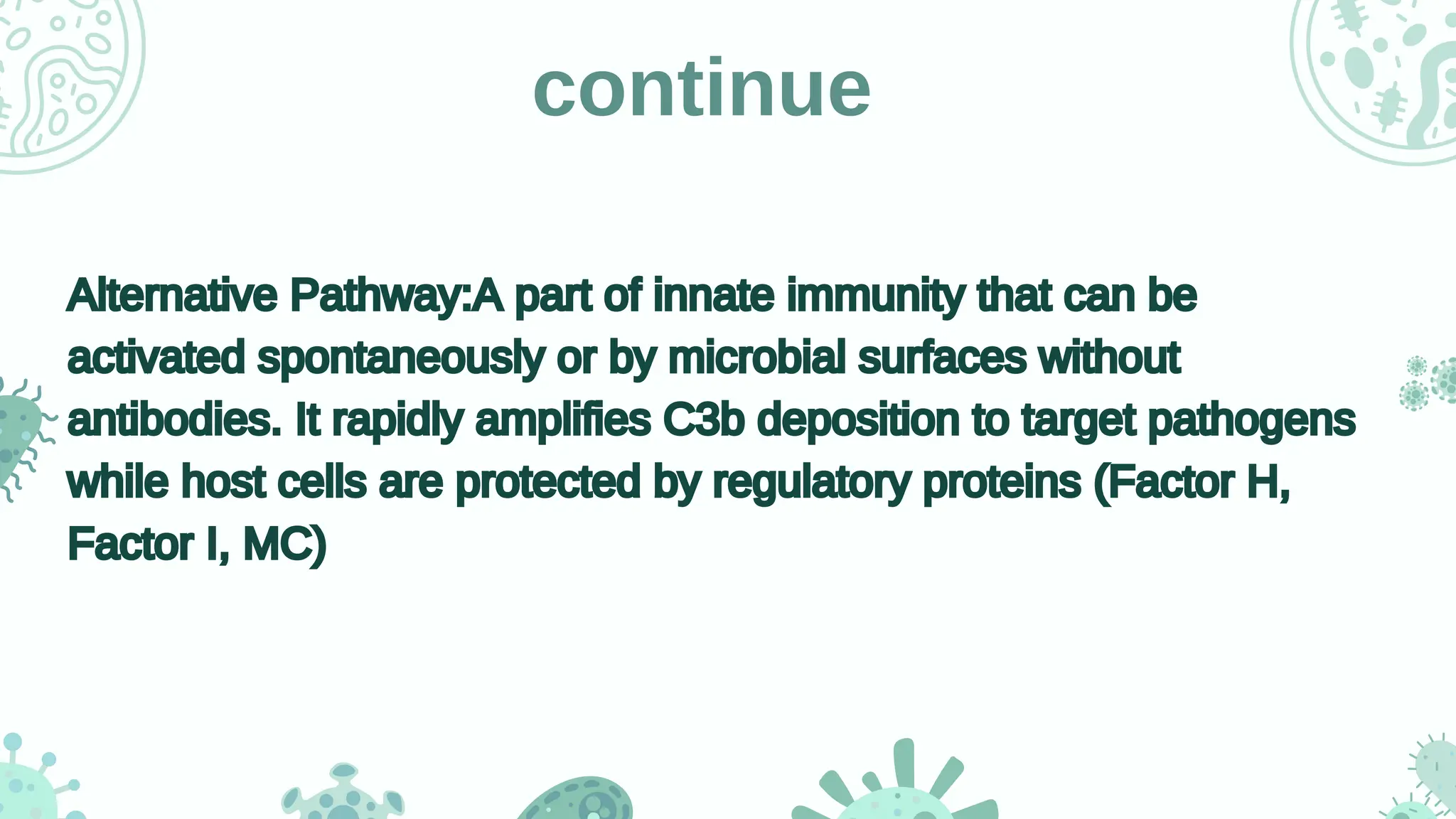
Alternative Pathway:A partof innate immunity that can be
activated spontaneously or by microbial surfaces without
antibodies. It rapidly amplifies C3b deposition to target pathogens
while host cells are protected by regulatory proteins (Factor H,
Factor I, MC)
Alternative Pathway:A part of innate immunity that can be
activated spontaneously or by microbial surfaces without
antibodies. It rapidly amplifies C3b deposition to target pathogens
while host cells are protected by regulatory proteins (Factor H,
Factor I, MC)
18.
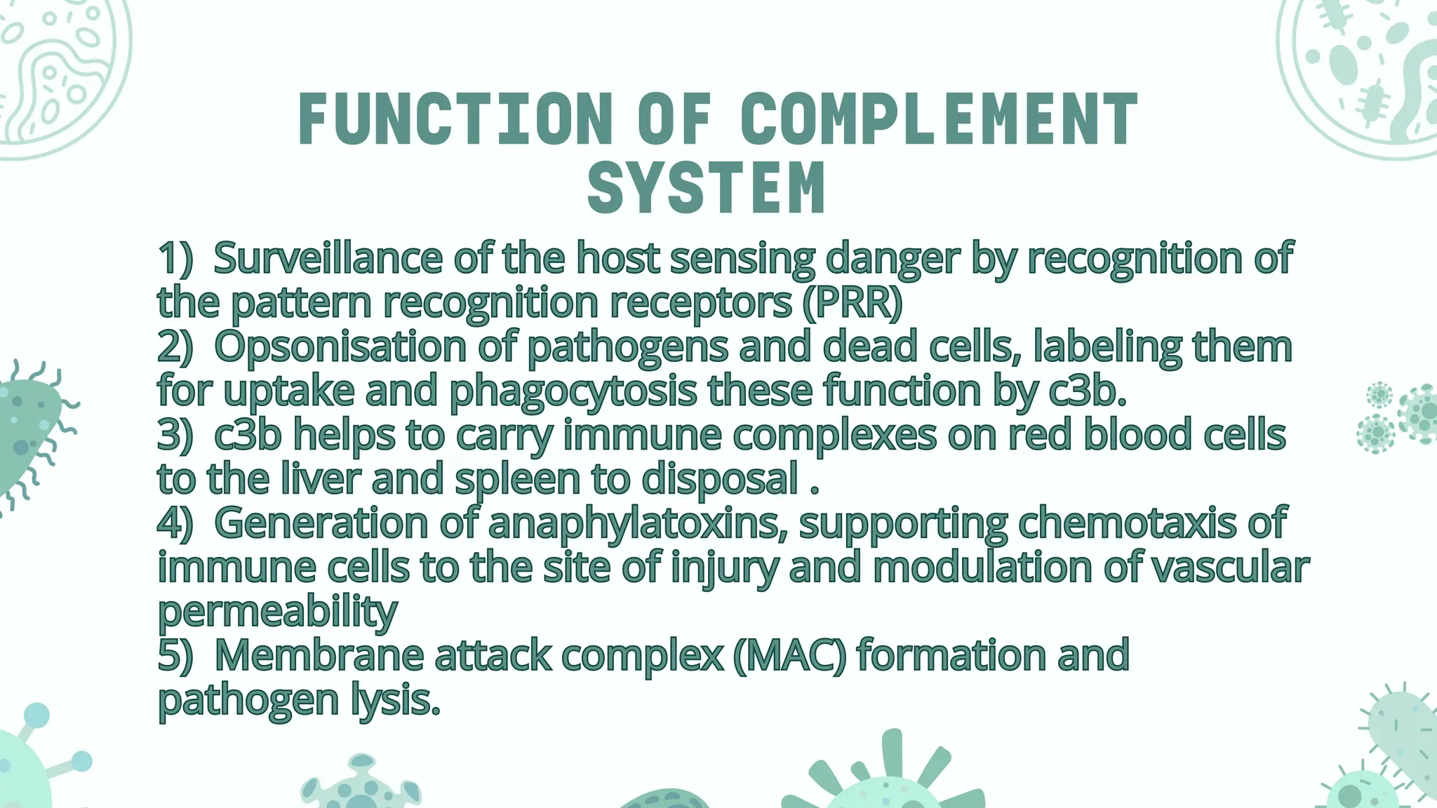
FUNCTION OF COMPLEMENT
SYSTEM
1)Surveillance of the host sensing danger by recognition of
the pattern recognition receptors (PRR)
2) Opsonisation of pathogens and dead cells, labeling them
for uptake and phagocytosis these function by c3b.
3) c3b helps to carry immune complexes on red blood cells
to the liver and spleen to disposal .
4) Generation of anaphylatoxins, supporting chemotaxis of
immune cells to the site of injury and modulation of vascular
permeability
5) Membrane attack complex (MAC) formation and
pathogen lysis.
1) Surveillance of the host sensing danger by recognition of
the pattern recognition receptors (PRR)
2) Opsonisation of pathogens and dead cells, labeling them
for uptake and phagocytosis these function by c3b.
3) c3b helps to carry immune complexes on red blood cells
to the liver and spleen to disposal .
4) Generation of anaphylatoxins, supporting chemotaxis of
immune cells to the site of injury and modulation of vascular
permeability
5) Membrane attack complex (MAC) formation and
pathogen lysis.
19.
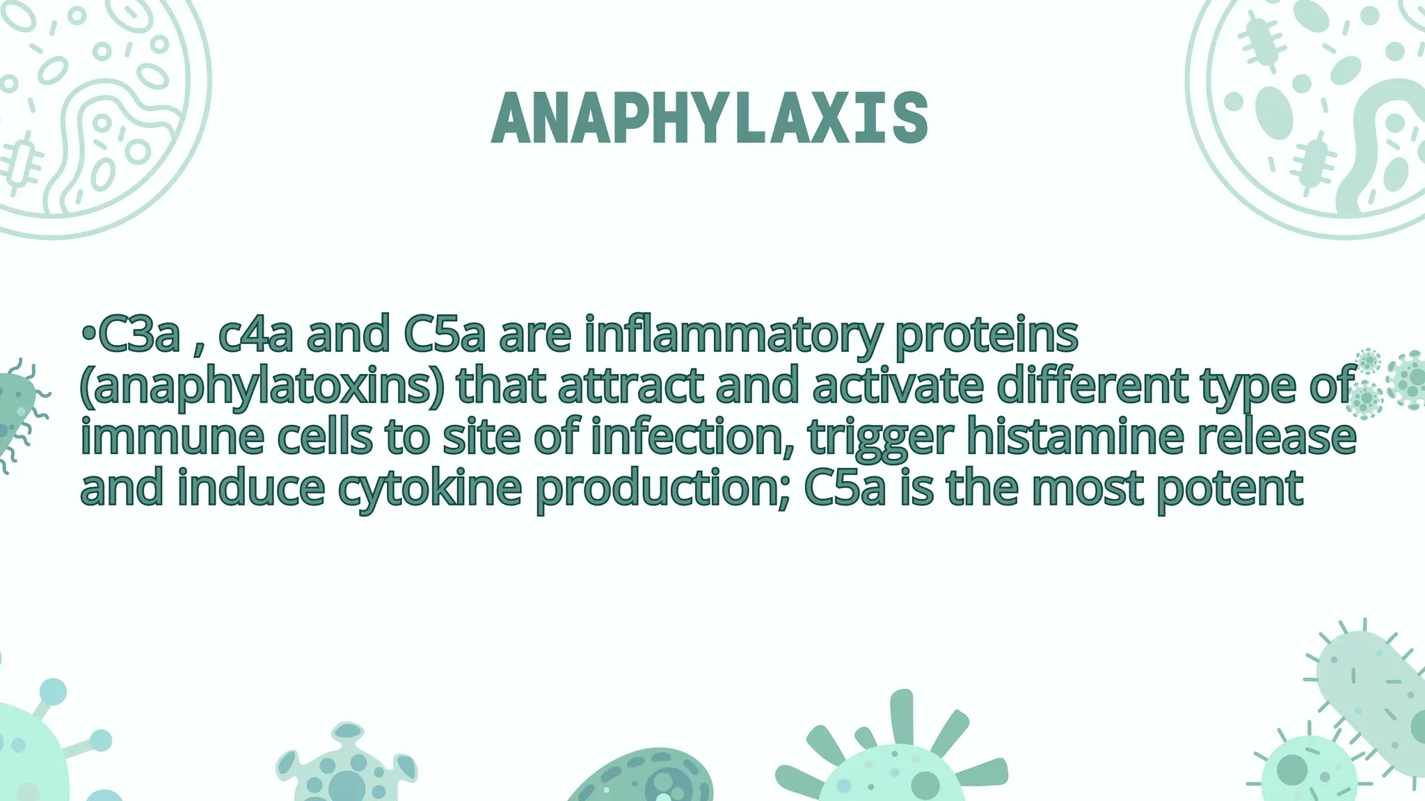
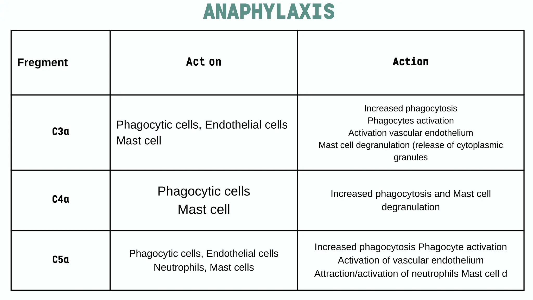
ANAPHYLAXIS
•C3a , c4aand C5a are inflammatory proteins
(anaphylatoxins) that attract and activate different type of
immune cells to site of infection, trigger histamine release
and induce cytokine production; C5a is the most potent
•C3a , c4a and C5a are inflammatory proteins
(anaphylatoxins) that attract and activate different type of
immune cells to site of infection, trigger histamine release
and induce cytokine production; C5a is the most potent
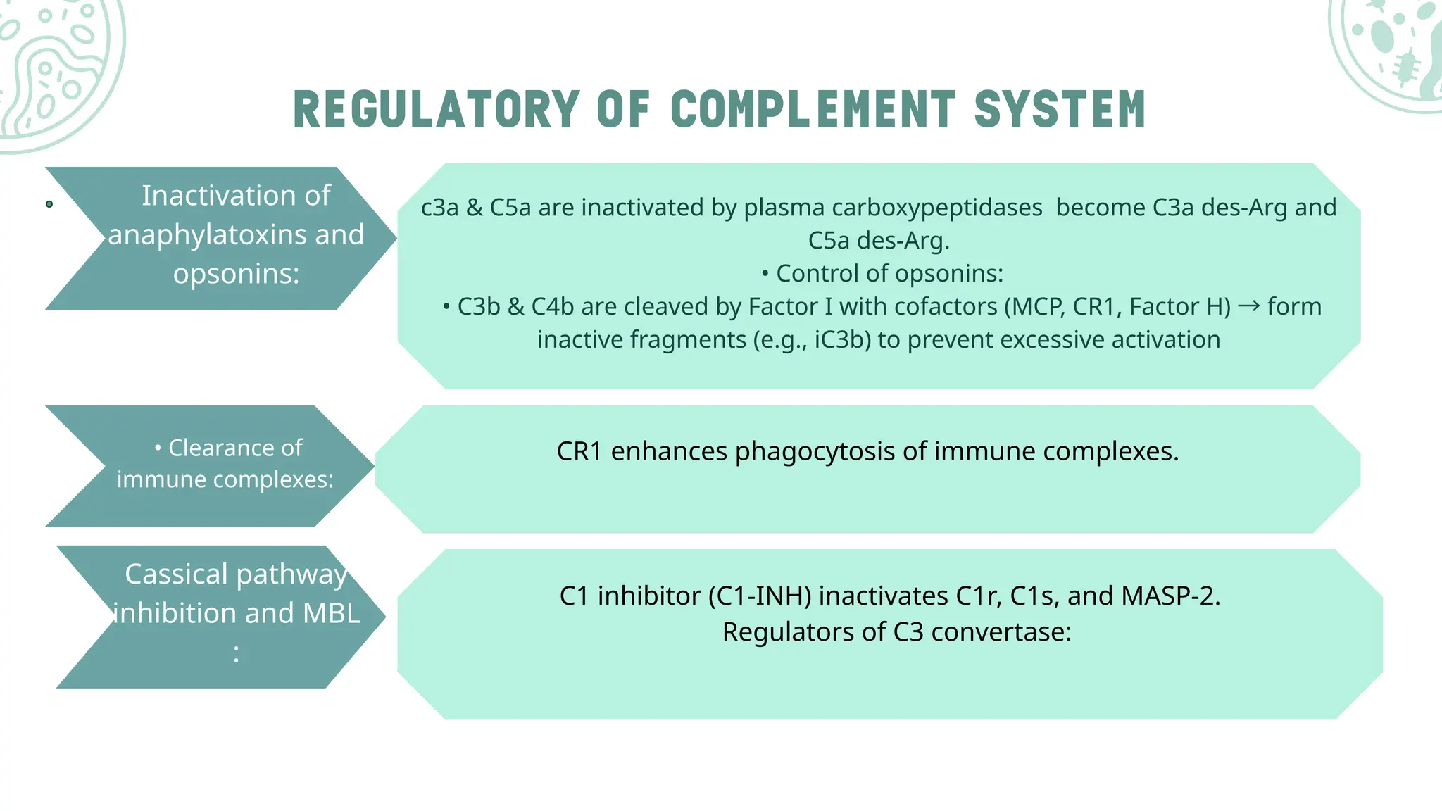
REGULATORY OF COMPLEMENTSYSTEM
.
. Inactivation of
anaphylatoxins and
opsonins:
c3a & C5a are inactivated by plasma carboxypeptidases become C3a des-Arg and
C5a des-Arg.
• Control of opsonins:
• C3b & C4b are cleaved by Factor I with cofactors (MCP, CR1, Factor H) →form
inactive fragments (e.g., iC3b) to prevent excessive activation
• Clearance of
immune complexes:
CR1 enhances phagocytosis of immune complexes.
Cassical pathway
inhibition and MBL
:
C1 inhibitor (C1-INH) inactivates C1r, C1s, and MASP-2.
Regulators of C3 convertase:
22.
• MAC inhibition:
MACassembly is prevented on self-cells by S protein,
vitronectin, and CD59.
23.
COMPLEMENT SYSTEM DISORDE
1.AtypicalHemolytic Uremic Syndrome (aHUS) النمطية غير اليوريمية الدم انحالل متالزمة
Cause: Mutation in Factor H gene (or Factor I, Factor B, CD46).
Effect: Impaired C3 convertase regulation →endothelial cell damage →hemolytic anemia,
thrombocytopenia, and acute renal failure.
.2 Paroxysmal Nocturnal Hemoglobinuria (PNH)االنتيابية الليلية الهيموغلوبين بيلة
Cause: Mutation in PIG-A gene →loss of CD59 and DAF (CD55) on cell surface.
Effect: Uncontrolled complement activation →red blood cell lysis, hemolytic anemia, and
thrombosis.
1.Atypical Hemolytic Uremic Syndrome (aHUS) النمطية غير اليوريمية الدم انحالل متالزمة
Cause: Mutation in Factor H gene (or Factor I, Factor B, CD46).
Effect: Impaired C3 convertase regulation →endothelial cell damage →hemolytic anemia,
thrombocytopenia, and acute renal failure.
.2 Paroxysmal Nocturnal Hemoglobinuria (PNH)االنتيابية الليلية الهيموغلوبين بيلة
Cause: Mutation in PIG-A gene →loss of CD59 and DAF (CD55) on cell surface.
Effect: Uncontrolled complement activation →red blood cell lysis, hemolytic anemia, and
thrombosis.
24.
COMPLEMENT SYSTEM DISORDE
4.Systemic Lupus Erythematosus (SLE) الجهازية الحمامية الذئبة
Cause: Deficiency of C1q, C1r, C1s, C2, or C4.
Effect: Defective clearance of immune complexes and apoptotic cells →autoimmune reactions, leading to
fever, rash, nephritis, and anemia.
REFERENCE
Varela, J. C.,& Tomlinson, S. (2015). Complement: An overview for the clinician. Hematology/Oncology
Clinics of North America, 29(3), 409–427. Available in PMC:
https://www.ncbi.nlm.nih.gov/pmc/articles/PMC4456616/
Nesargikar, P., Spiller, B., & Chavez, R. (2012). The complement system: History, pathways, cascade and
inhibitors. European Journal of Microbiology & Immunology, 2(2), 103–111.
https://doi.org/10.1556/eujmi.2.2012.2.2
Nesargikar, P. N., Spiller, B., & Chavez, R. (2014). The complement system: History, pathways, cascade
and inhibitors. European Journal of Microbiology & Immunology, 2(2), 103–111. Available in PMC:
https://www.ncbi.nlm.nih.gov/pmc/articles/PMC3956958/