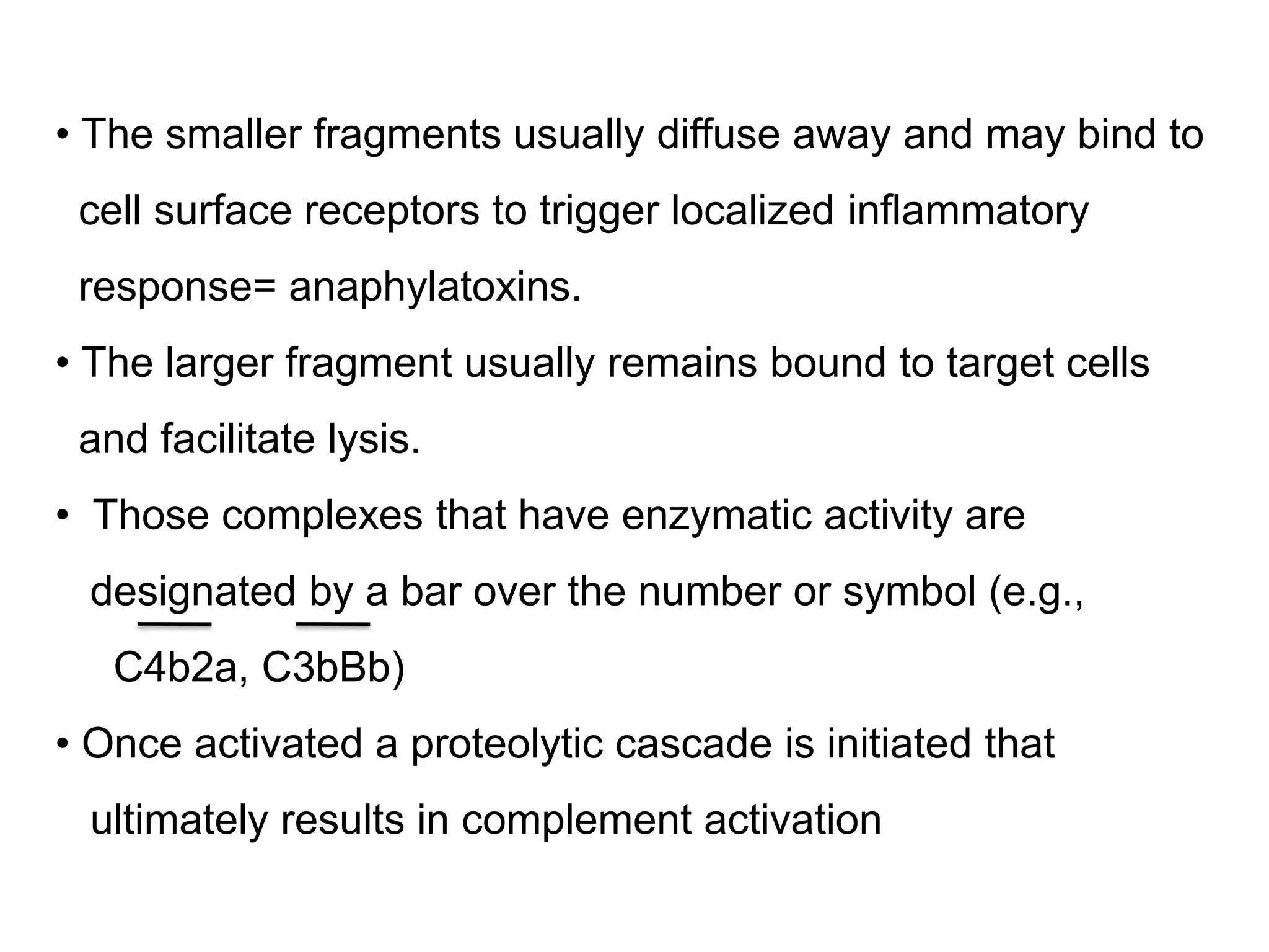
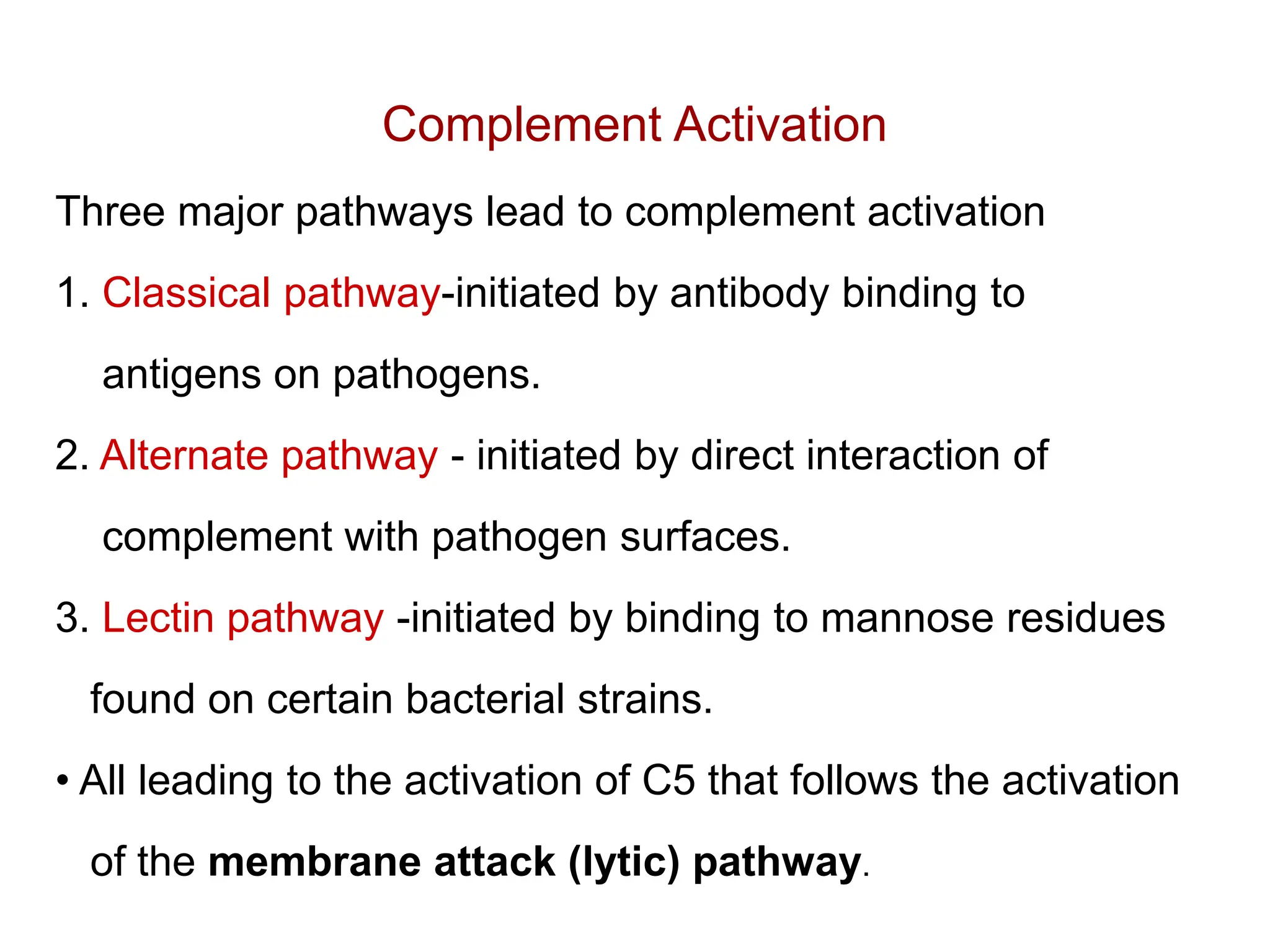
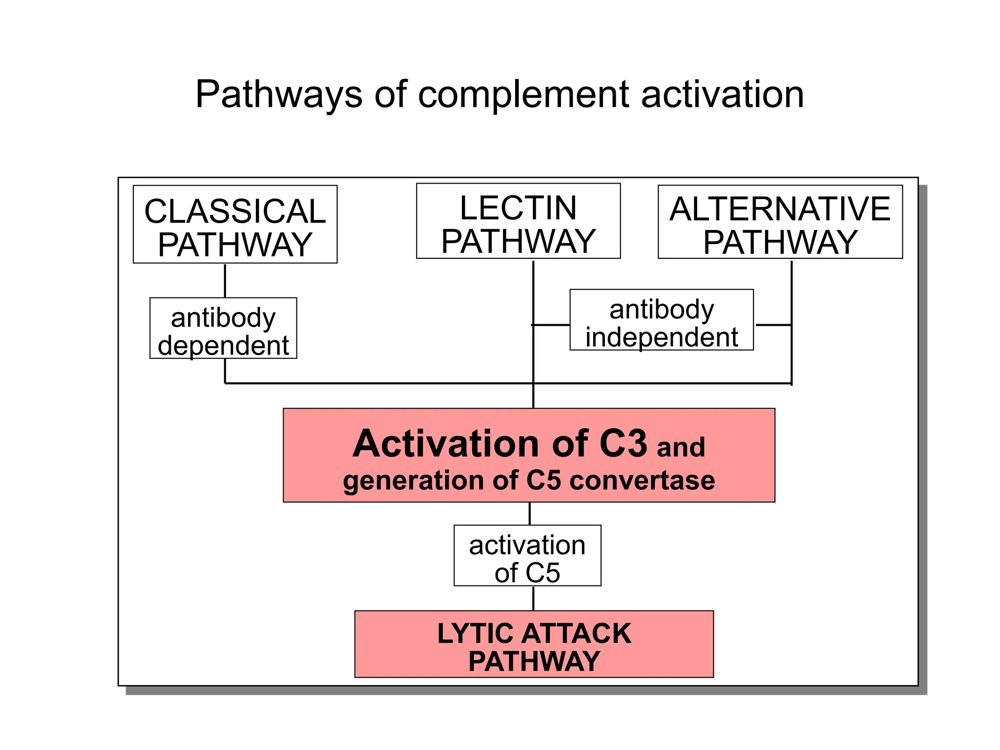
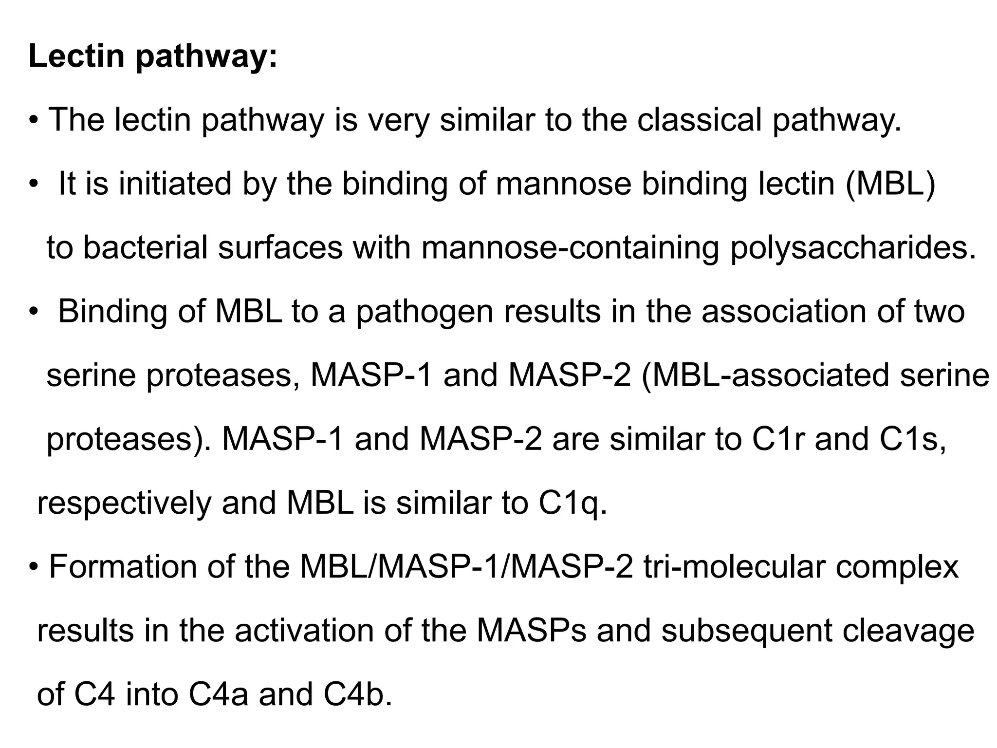
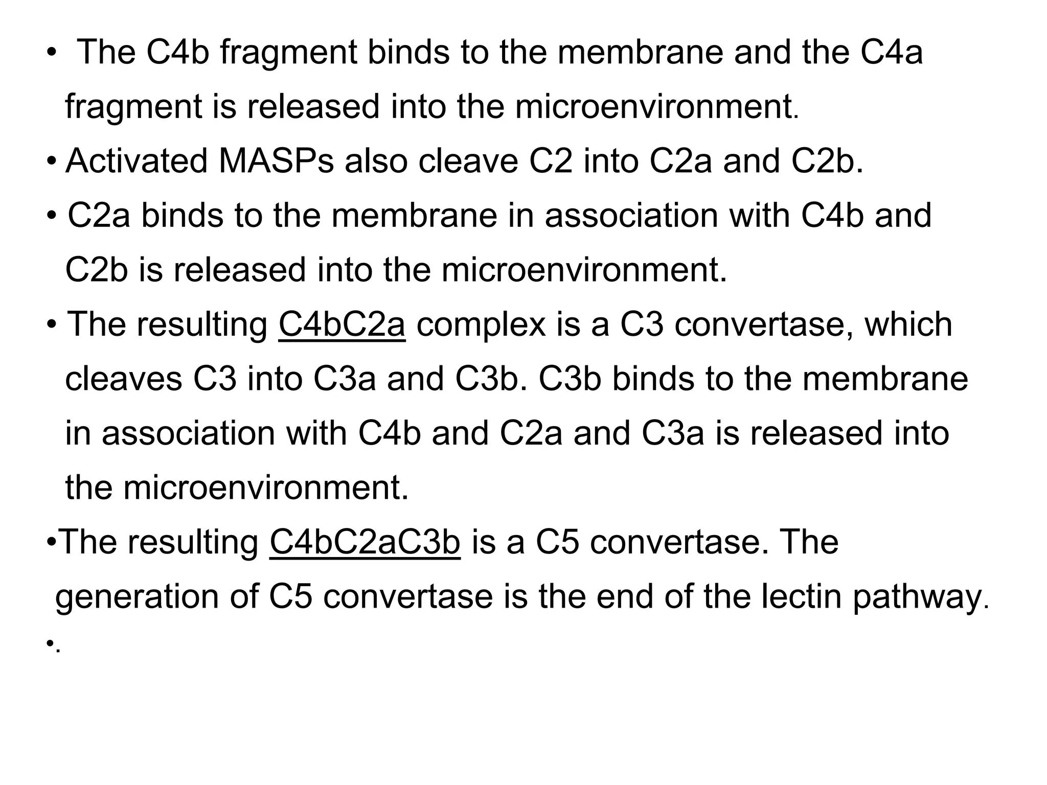
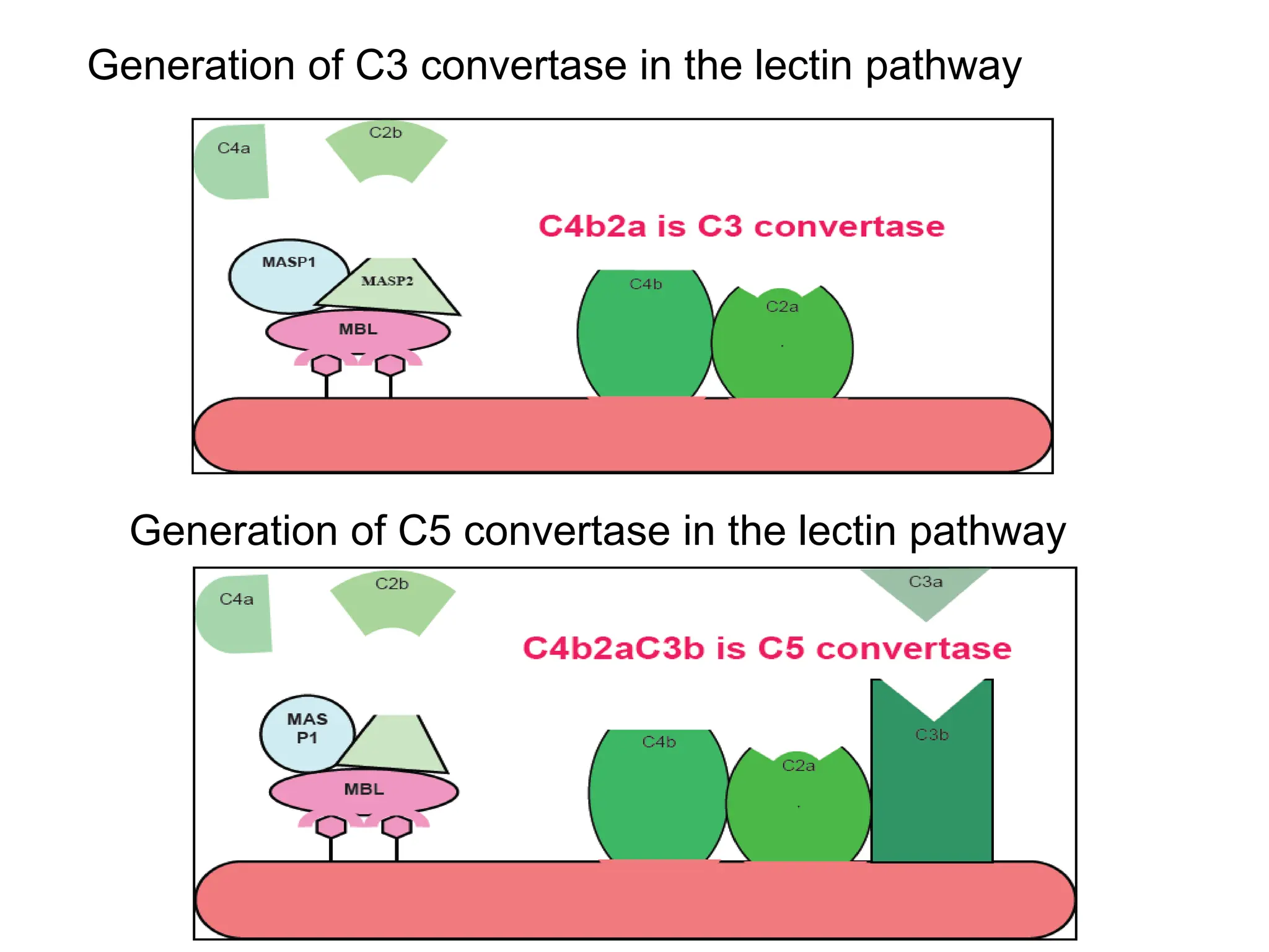
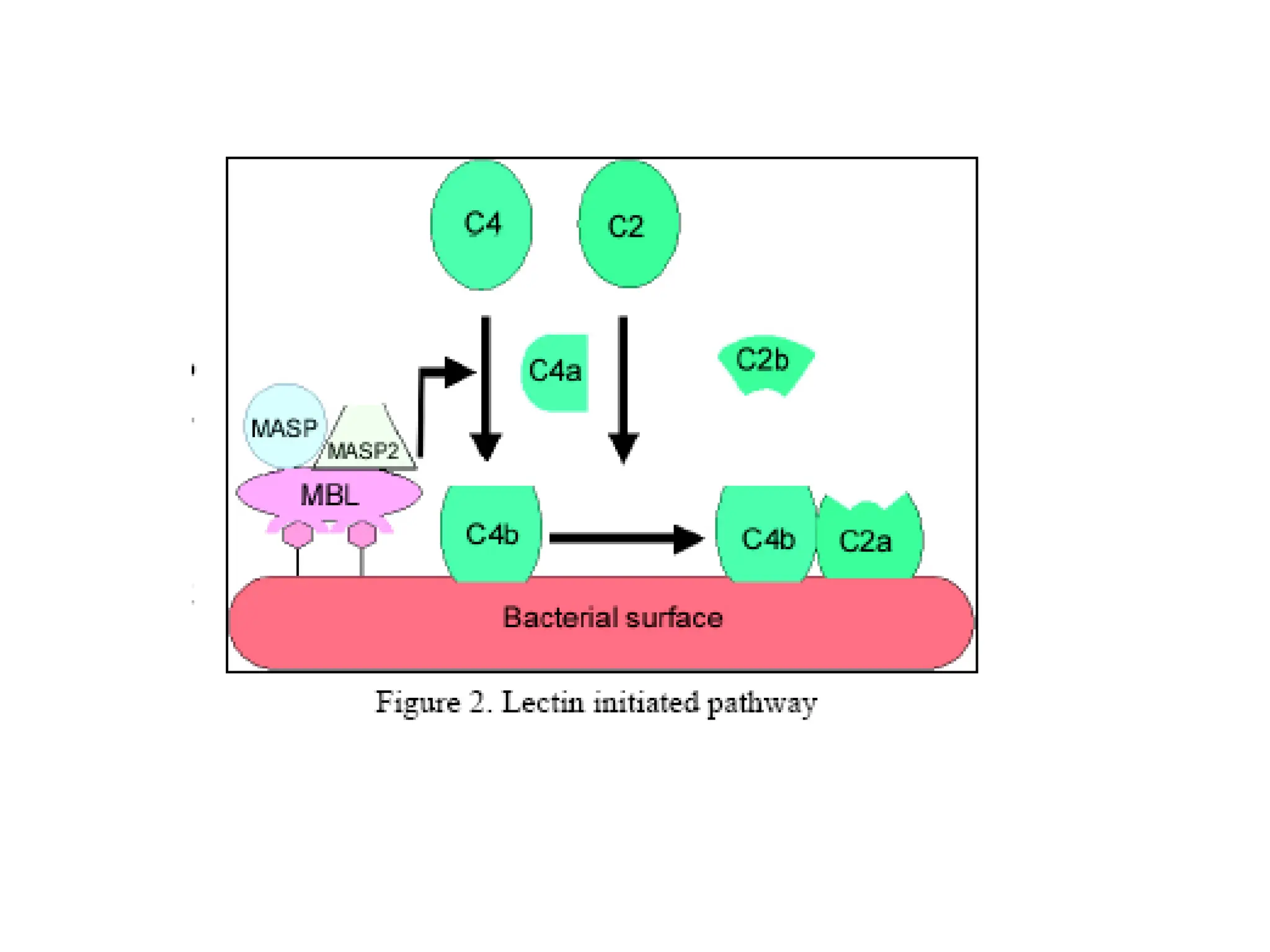
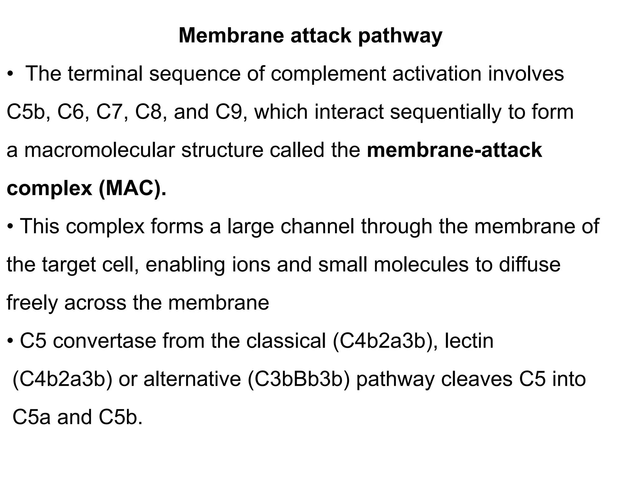
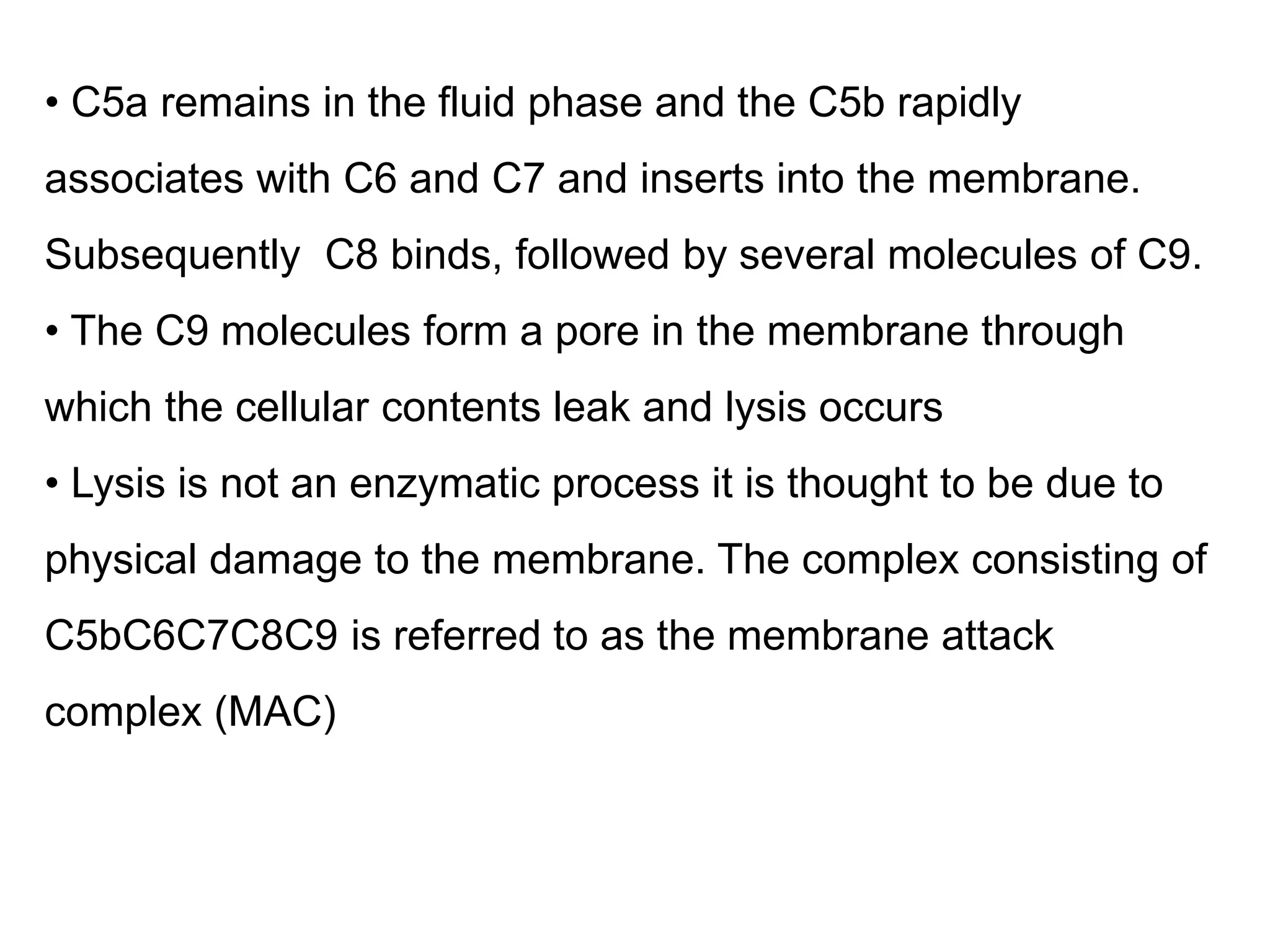
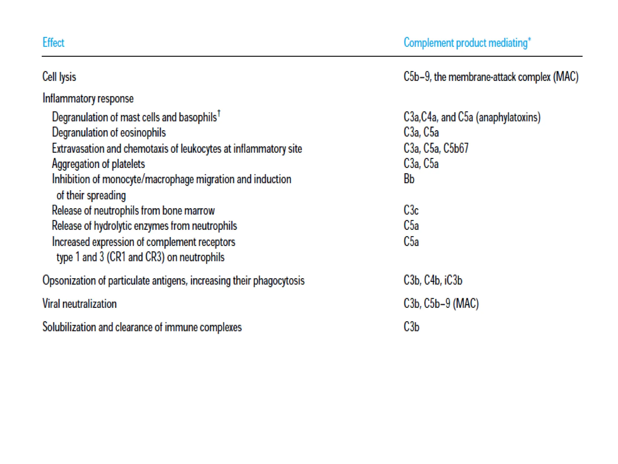
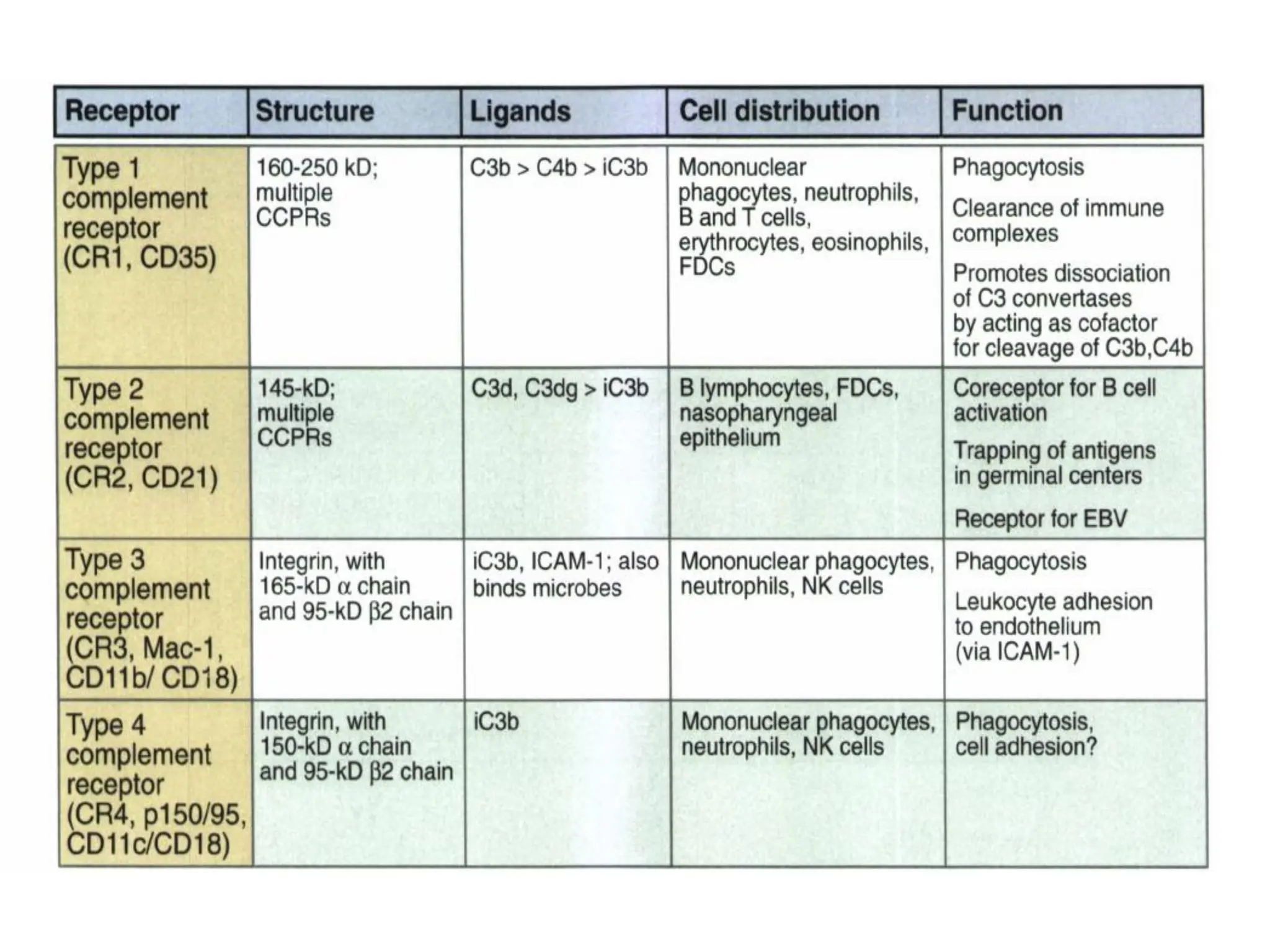
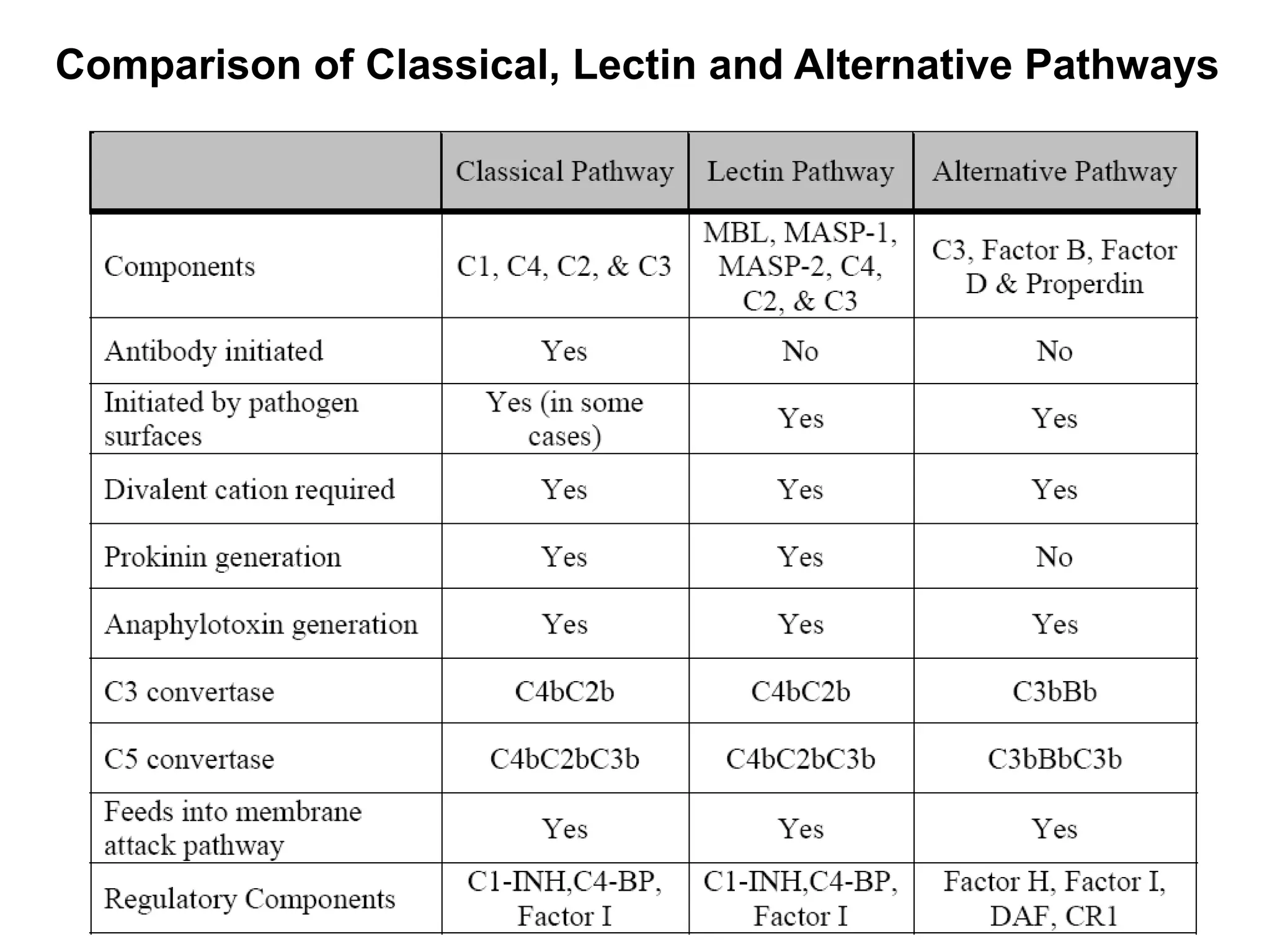
The complement system is a crucial immune response mechanism consisting of over 25 proteins that eliminate microbes through direct lysis, opsonization, and inflammation. It can be activated via three pathways: the classical pathway through antibody-antigen interactions, the lectin pathway via mannose binding lectin, and the alternative pathway initiated by foreign cell surfaces, leading to the formation of the membrane attack complex. Each pathway generates a convertase that facilitates the sequential activation of complement proteins ultimately resulting in pathogen destruction.
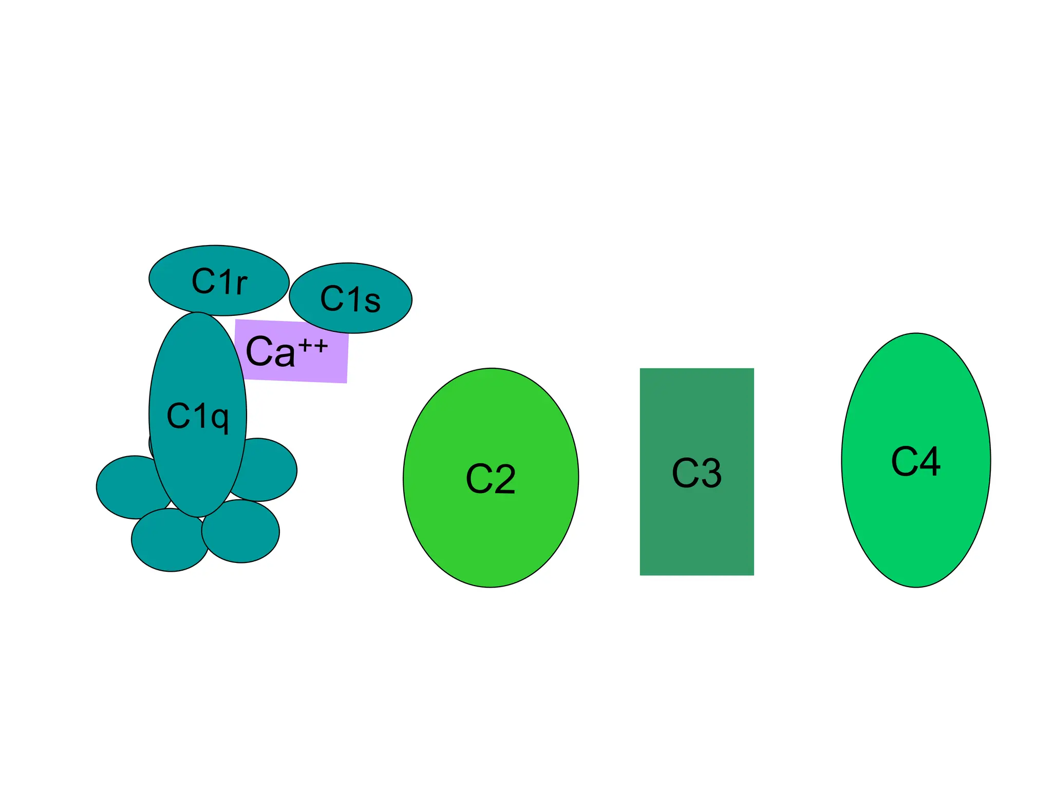
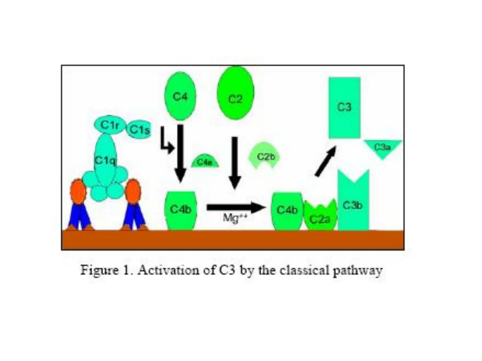
![• C1 a multi-subunit protein containing three different proteins
C1q, C1r and C1s, binds to the Fc region of IgG and IgM
antibody molecules that have interacted with antigen.
• C1 binding does not occur to antibodies that have not
complexed with antigen and binding requires calcium and
magnesium ions.
• In some cases C1 can bind to aggregated immunoglobulin
[e.g. aggregated IgG] or to certain pathogen surfaces in the
absence of antibody).](https://image.slidesharecdn.com/complement-240822053103-c4ad4a9f/75/Complement-System-and-its-activation-pathways-8-2048.jpg)


![COMPLEMENT SYSTEM[immunology]](https://cdn.slidesharecdn.com/ss_thumbnails/rollno05dvncomplementsystem-160328114058-thumbnail.jpg?width=640&height=640&fit=bounds)


