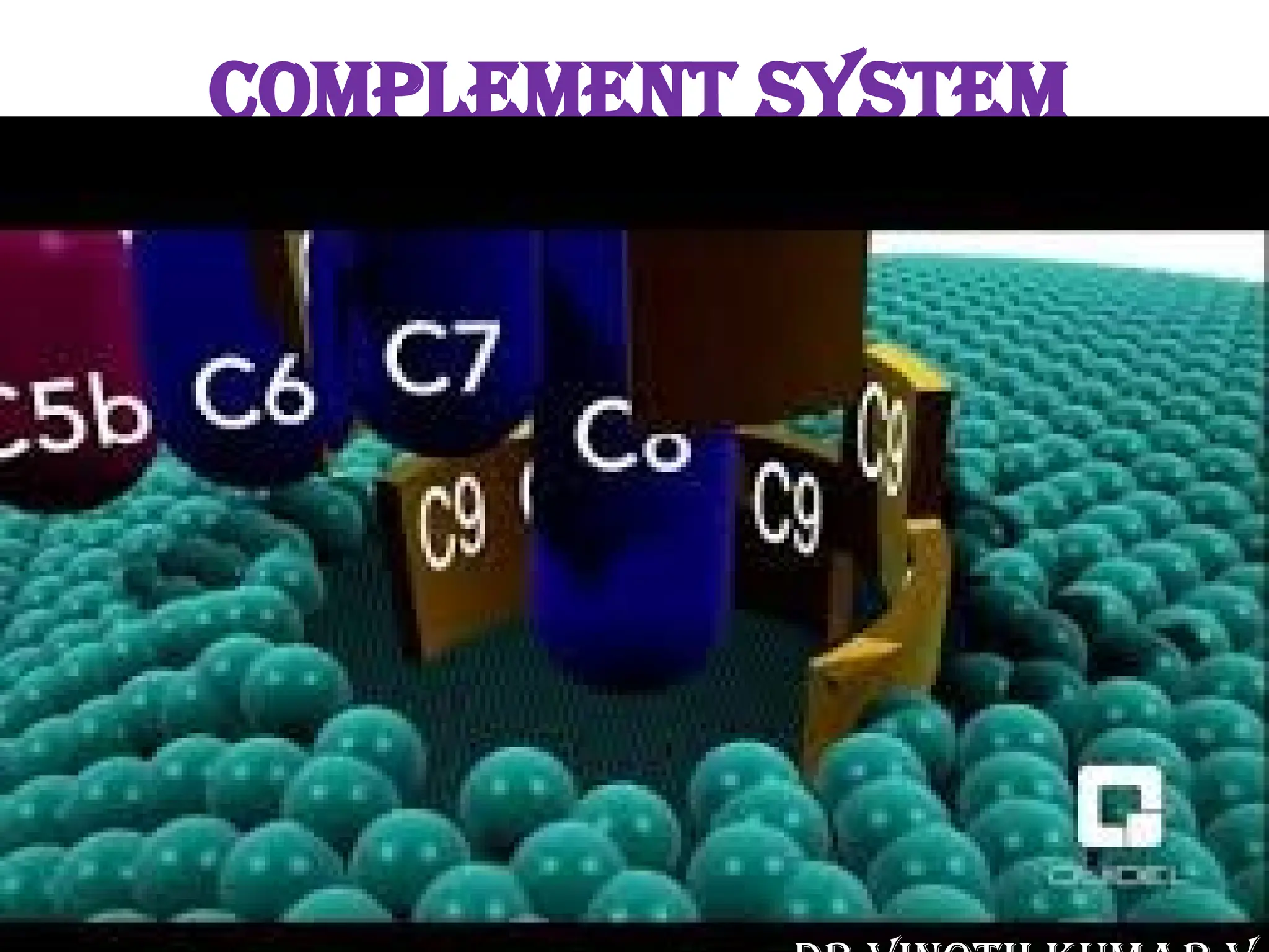
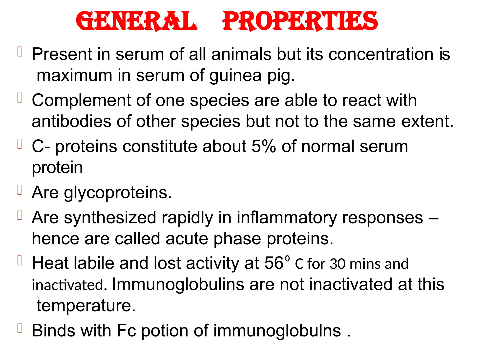
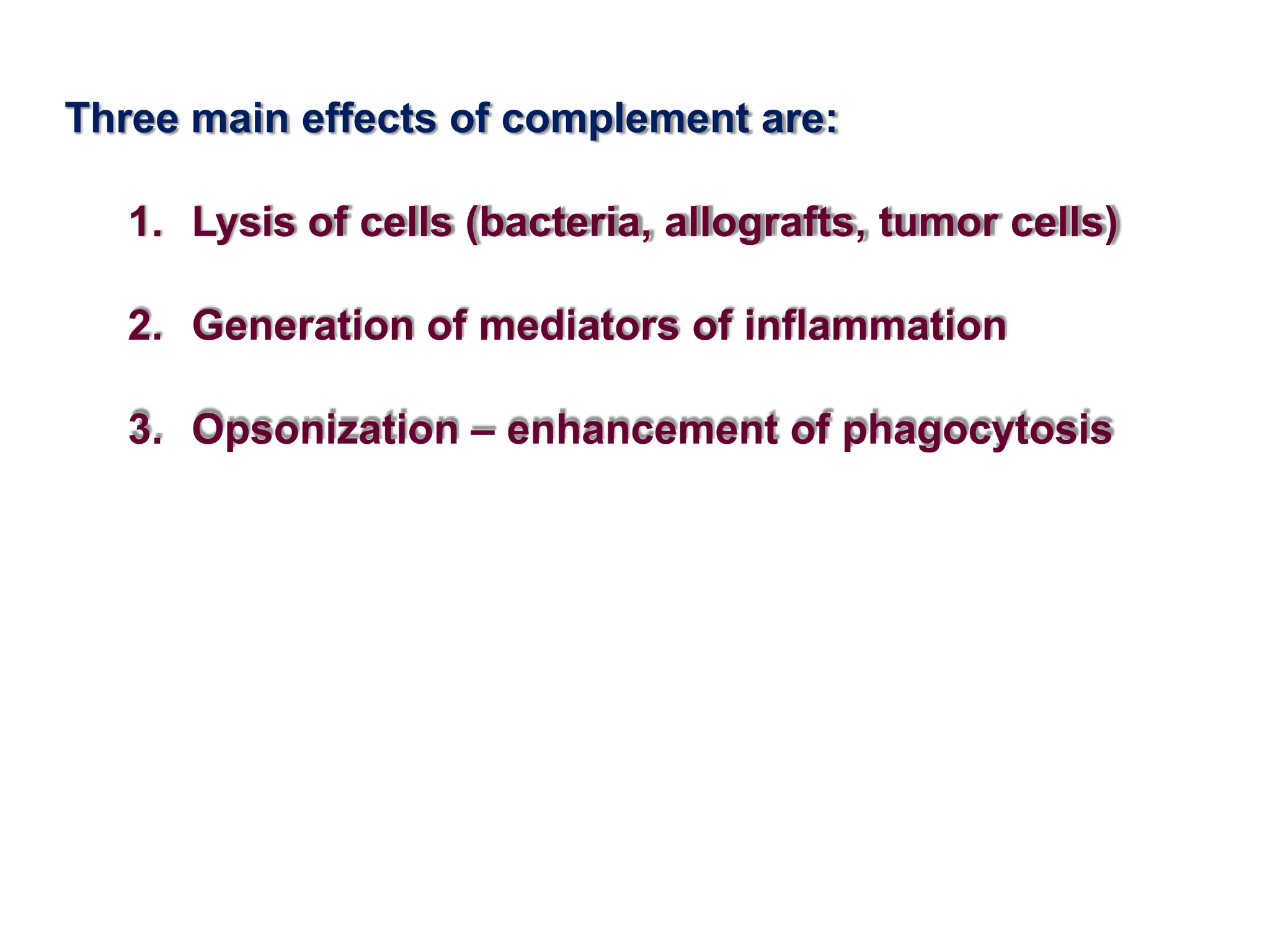
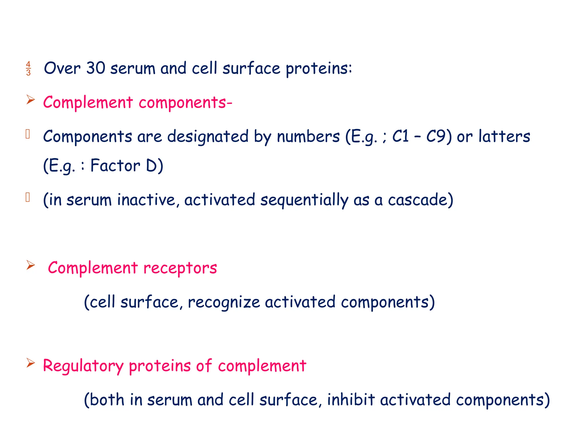
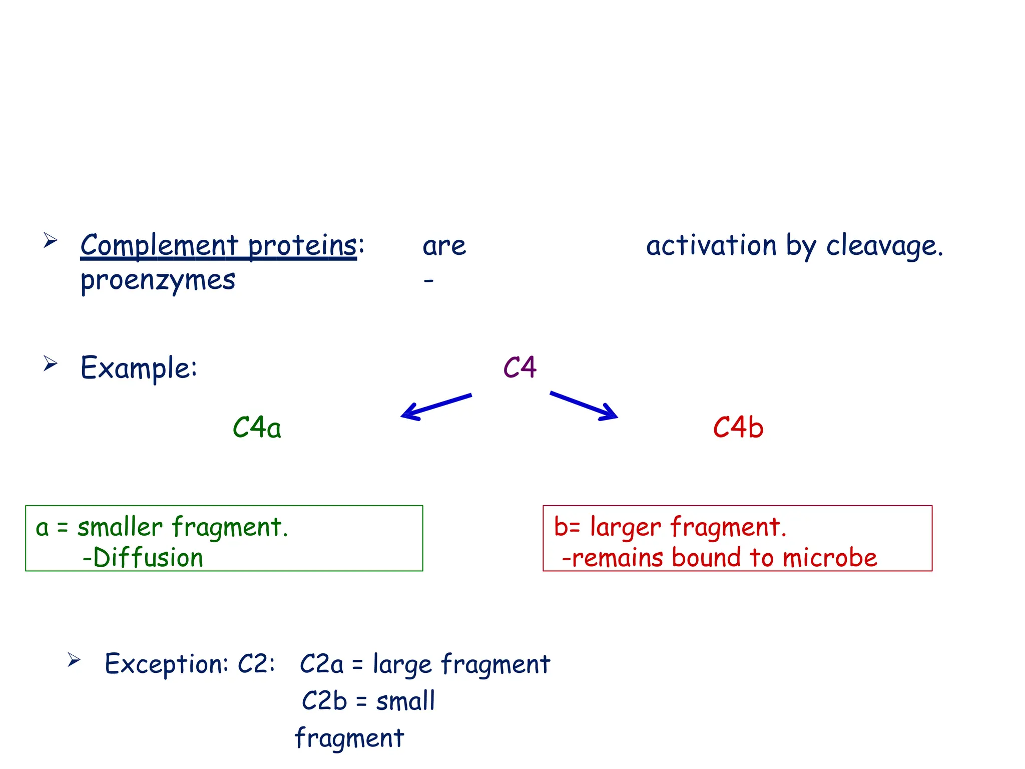
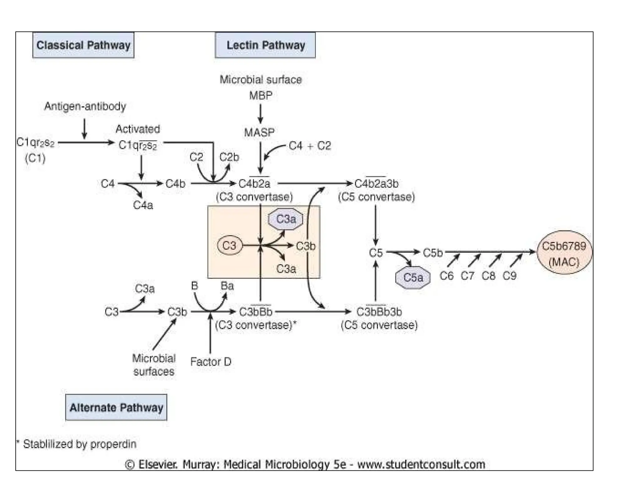
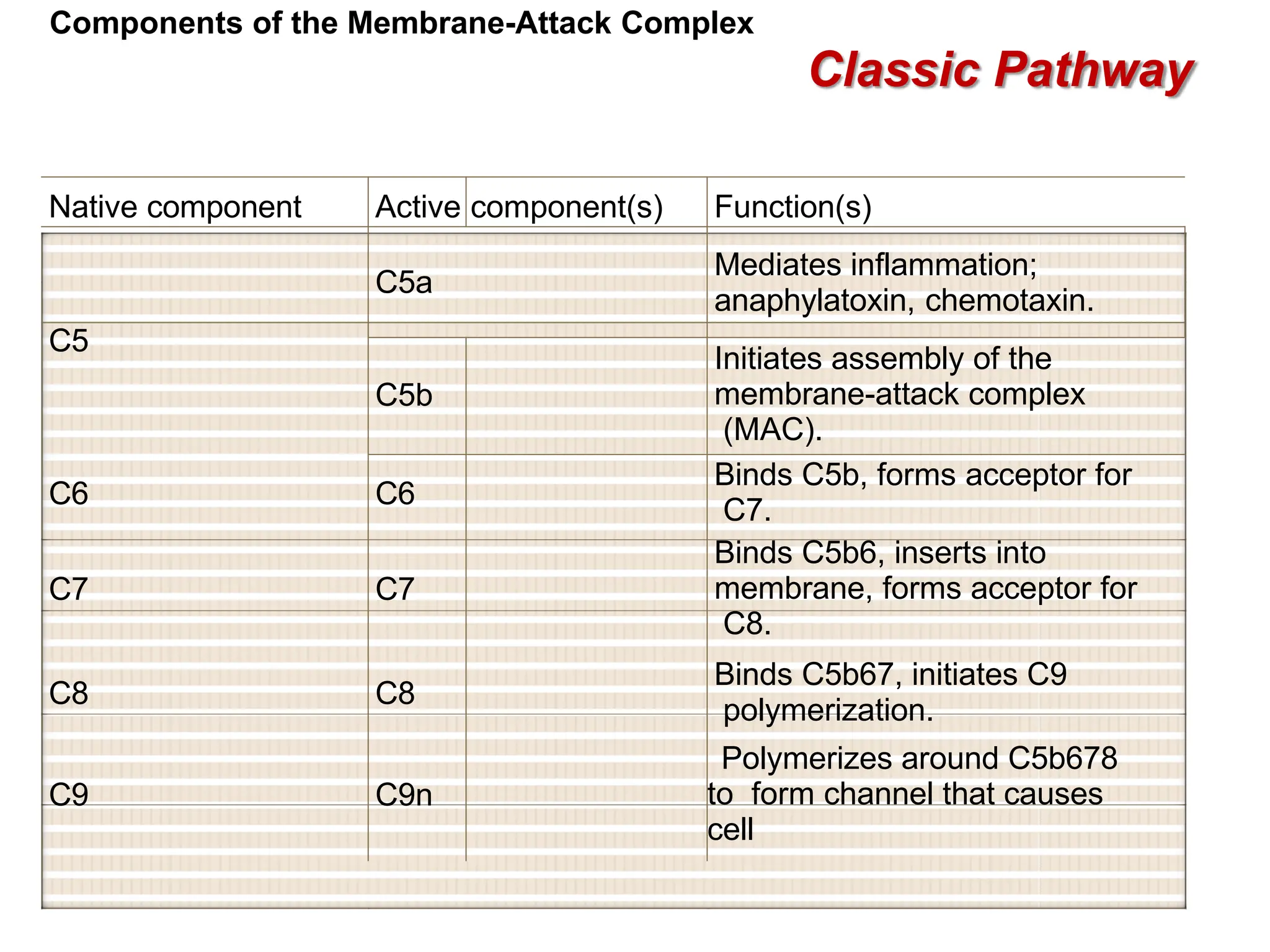
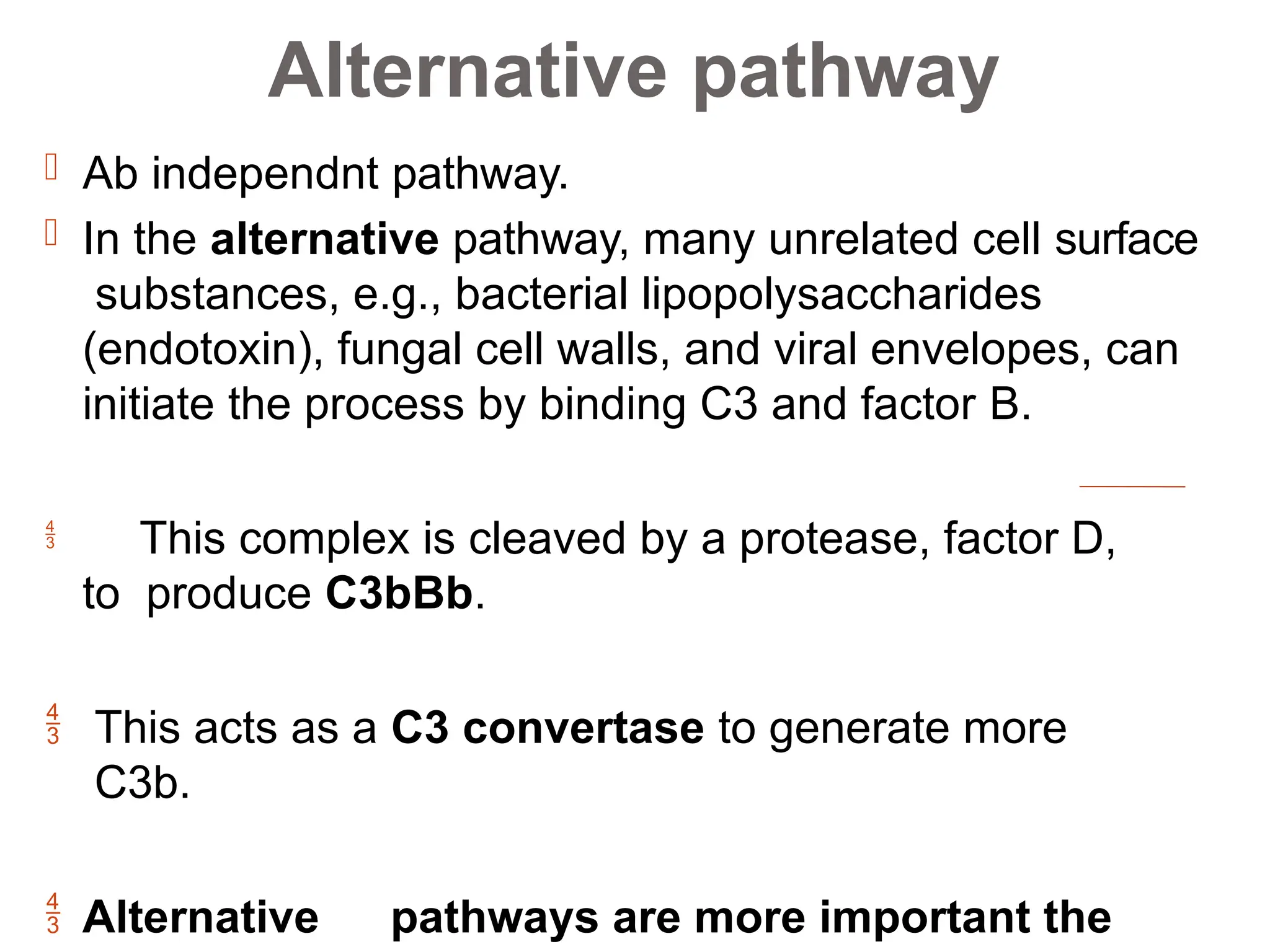
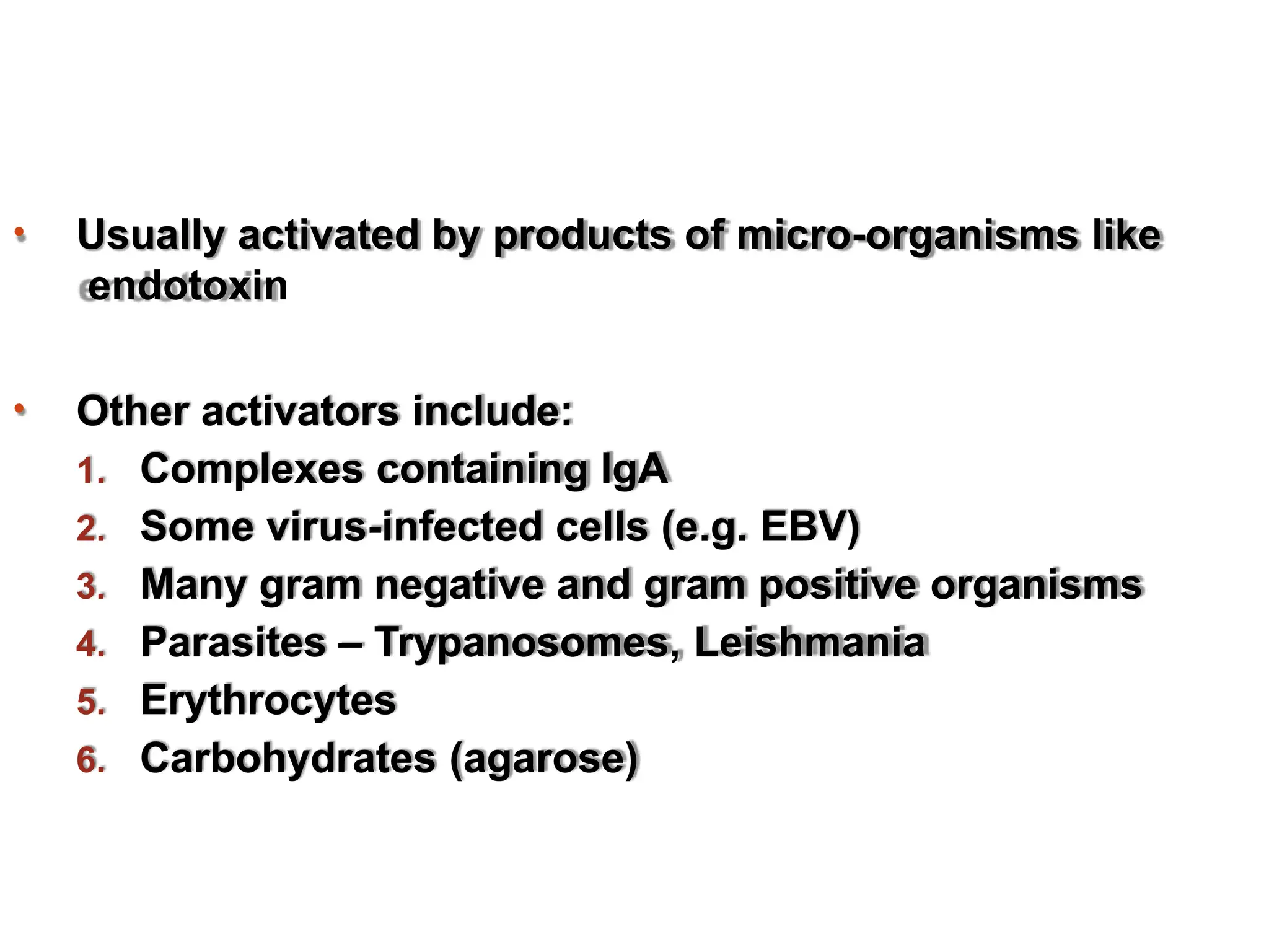
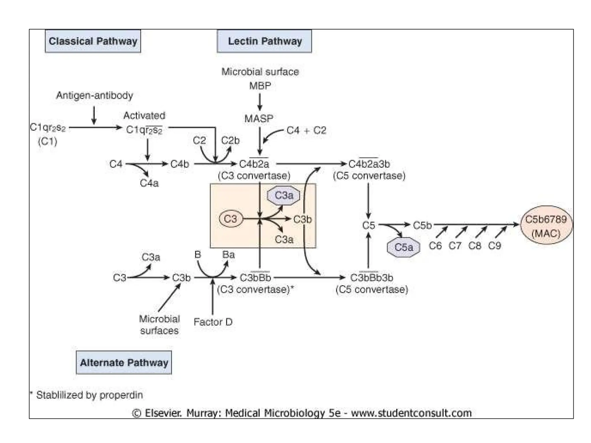
Complement system
 It is named “complement system” because it was first identified as
a heat-labile component of serum that “complemented or augment”
antibodies in the killing of bacteria.
 Consists of serum and cell surface proteins involved in defense
against pathogens and tissue damage mediated by antibodies
 The Complement system is the major effector of cellular and
humoral branch of immune system.
 Plays major role in both innate and adaptive immunity.
INTRODUCTION
 Complement system represents a group of about 30
proteins which augment or complement the immune
response.
 Most of these proteins are found in serum or on cell
surfaces.
 Synthesized in liver as inative precursors and are activated by
proteolysis durig their interaction in a sequential manner.
 Also produced by blood monocytes, tissue macrophages and
epithelial cells of the gastrointestinal and genitourinary
tract.
HISTORY
 Research on complement began in the 1890s, when
Jules Bordet at the Institute Pasteur in Paris
showed that sheep antiserum to the bacterium Vibrio
cholerae caused lysis of the bacteria and that heating
the antiserum destroyed its bacteriolytic activity.
 He named those substances as
Alexins.
 Paul Ehrlich coined the term
complement.
General properties
 Present in serum of all animals but its concentration is
maximum in serum of guinea pig.
 Complement of one species are able to react with
antibodies of other species but not to the same extent.
 C- proteins constitute about 5% of normal serum
protein
 Are glycoproteins.
 Are synthesized rapidly in inflammatory responses –
hence are called acute phase proteins.
 Heat labile and lost activity at 56⁰ C for 30 mins and
inactivated. Immunoglobulins are not inactivated at this
temperature.
 Binds with Fc potion of immunoglobulns .
Three main effects of complement are:
1. Lysis of cells (bacteria, allografts, tumor cells)
2. Generation of mediators of inflammation
3. Opsonization – enhancement of phagocytosis
 Over 30 serum and cell surface proteins:
 Complement components-
- Components are designated by numbers (E.g. ; C1 – C9) or latters
(E.g. : Factor D)
- (in serum inactive, activated sequentially as a cascade)
 Complement receptors
(cell surface, recognize activated components)
 Regulatory proteins of complement
(both in serum and cell surface, inhibit activated components)
 Complement proteins: are
proenzymes -
activation by cleavage.
 Example: C4
 Exception: C2: C2a = large fragment
C2b = small
fragment
a = smaller fragment.
-Diffusion
b= larger fragment.
-remains bound to microbe
C4a C4b
Complement Pathway
Three pathway of complement activation
1.Classical pathway:-
Is antibody dependent pathway and triggered by
formation of soluble antigen-antibody complex or by
binding of the antibody to the antigen present on
the target cell surface.
2.Alternative pathway:-
Is antibody independent pathway stimulated by
antigen directly eg. Bacterial cell surface components.
3.Lectin Pathway:-
Also antibody independent but resembles
classical pathway.
Stages of complement Activation
Three main stages in the activation of complement by
any pathway are
 Formation of C3 convertage
 Formation C5 convertage
 Formation of membrane attack complec(MAC)
• The initiation and formation of C3 convertage are
different in classical and alternative pathway . These
then follow the parralel route to merge at C5 convertage
stage and finally generate the MAC by a common
route.
 Sequential activation of complement components occurs
via one of three pathways:
 the classic pathway,
 the lectin pathway, and
 the alternative pathway .
 Of these pathways, the lectin and the alternative
pathways are more important the first time we are
infected by a microorganism because the antibody
required to trigger the classic pathway is not present.
 The lectin pathway and the alternative pathway are,
therefore, participants in the innate arm of the immune
system.
 All three pathways lead to the production of
C3b, the central molecule of the
complement cascade.
 The presence of C3b on the surface of a
microbe marks it as foreign and targets it for
destruction. C3b has two important
functions:
(1)It combines with other complement
components to generate C5 convertase,
the enzyme that leads to the production of
the membrane attack complex and
(2)it opsonizes bacteria because phagocytes
have receptors for C3b on their surface.
 Part of acquired immunity.
 In the classic pathway, antigen–antibody complexes
activate C1 to form a protease, which cleaves C2
and C4 to form a C4bC2a complex, C2a and C4b split
off.
 The C4bC2a is C3 convertase, which cleaves C3
molecules into two fragments, C3a and C3b.
 C3b forms a complex with C4b,2b, producing a new
enzyme, C5 convertase (C4b2a3b), which cleaves
C5 to form C5a and C5b
 C5b binds to C6 and C7 to form a complex that
interacts with C8 and C9 to produce the membrane
attack complex (C5b,6,7,8,9), which causes
cytolysis.
 Note that the "b" fragment continues in the main
Classical pathway
 Only IgM and IgG fix complement.
 One molecule of IgM can activate complement;
however, activation by IgG requires two cross-linked
IgG molecules.
 Of the IgGs, only IgG1, IgG2, and IgG3 subclasses fix
complement; IgG4 does not.
 C1 is bound to a site located in the Fc region of the
heavy chain.
 C1 is composed of three proteins, C1q, C1r, and
C1s.
 C1q is an aggregate of 18 polypeptides that binds
to the Fc portion of IgG and IgM.
 It is multivalent and can cross-link several
immunoglobulin molecules.
 C1s is a proenzyme that is cleaved to
form an active
protease.
Classic Pathway
Components of the Classical Pathway
Native component Active component(s) Function(s)
C1(q,r,s)
C1q
Binds to antibody that has bound
antigen, activates C1r.
C1r
Cleaves C1s to activate protease
function.
C1s Cleaves C2 and C4.
C2
C2a Unknown.
C2b
Active enzyme of classical pathway;
cleaves C3 and C5.
C3
C3a Mediates inflammation; anaphylatoxin.
C3b
Binds C5 for cleavage by C2b.
Binds cell surfaces for opsonization
and activation of alternate
pathway.
C4
C4a Mediates inflammation.
C4b
Sant
Binds C2 for cleavage by C1s. Binds
cell surfaces for opsonization.
osh
Classic Pathway
Components of the Membrane-Attack Complex
Native component Active component(s) Function(s)
C5a
Mediates inflammation;
anaphylatoxin, chemotaxin.
C5
C5b
Initiates assembly of the
membrane-attack complex
(MAC).
C6 C6
Binds C5b, forms acceptor for
C7.
C7 C7
Binds C5b6, inserts into
membrane, forms acceptor for
C8.
C8 C8
Binds C5b67, initiates C9
polymerization.
C9 C9n
Polymerizes around C5b678
to form channel that causes
cell
 Ab independnt pathway.
 In the alternative pathway, many unrelated cell surface
substances, e.g., bacterial lipopolysaccharides
(endotoxin), fungal cell walls, and viral envelopes, can
initiate the process by binding C3 and factor B.
 This complex is cleaved by a protease, factor D,
to produce C3bBb.
 This acts as a C3 convertase to generate more
C3b.
 Alternative pathways are more important the
Alternative pathway
• Usually activated by products of micro-organisms like
endotoxin
• Other activators include:
1. Complexes containing IgA
2. Some virus-infected cells (e.g. EBV)
3. Many gram negative and gram positive organisms
4. Parasites – Trypanosomes, Leishmania
5. Erythrocytes
6. Carbohydrates (agarose)
Alternative Pathway
Components of the Alternate Pathway
Native component
Active
component(s)
Function(s)
C3
C3a
Mediates inflammation;
anaphylatoxin.
C3b
Binds cell surfaces for
opsonization and activation
of alternate pathway.
Factor B
Factor D
Properdin
B
Binds membrane bound
C3b. Cleaved by Factor D.
Ba Unknown.
Bb
D
P
Cleaved form stabilized by
P produces C3 convertase.
Cleaves Factor B when
bound to C3b.
Binds and stabilizes
membrane bound C3bBb.
Lectin Pathway
 Also known as the MBL Pathway
 In the lectin pathway, mannan-binding lectin (MBL)
(also known as mannose-binding protein) binds to the
surface of microbes bearing mannan (a polymer of
the sugar, mannose).
 Binding causes activation of MASP (MBP- associated
serine proteases)  cleave C2 and C4 and activate
the classic pathway.
 Note that this process bypasses the antibody-requiring
step and so is protective early in infection before
antibody is formed.
Membrane attack complex
 Cleavage of C5 into C5a and C5b.
 C5 (structurally homologous to C3 and C4, lacks
internal thioester bond )
 C5b initiates formation of MAC (complex of C5b,
C6, C7, C8 and multiple C9 molecules ) binds to
C6, and C7 , recruits C8 and complex penetrates
more deeply into the membrane.
 C9, a pore-forming molecule with homology to
perforin. The complex of C5b678 forms a nidus for
C9 binding and polymerization
 Penetrates membrane bilayers to form pores
 Disrupt the osmotic barrier, leading to swelling and
lysis of susceptible cells Abbas et.al.Cellular&Molecular immunology 6th edition
Biologic Effects of complement:
1. Opsonization
• C3b & C1q; enhance phagocytosis
2. Chemotaxis
• C5a and C5,6,7 complex  attract neutrophils
• C5a – enhance adhesiveness of neutrophils to the
endothelium
3. Anaphylatoxin (C3a, C4a, C5a)
• Cause degranulation of mast cells
• Bind directly to smooth muscles of bronchioles 
bronchospasm
4. Cytolysis (MAC)
• Disrupt the membrane & the entry of water and
electrolytes into the cell
5. Enhancement of antibody production
• Binding of C3b to its receptors on the surface of
activated B cells  enhanced antibody production
Regulation of Complement System
1. C1 inhibitor
• Important regulator of classic pathway
• A serine protease inhibitor (serpin)
• Irreversibly binds to and inactivates C1r and
C1s, as well as MASP in lectin pathway
2. Factor H
• Regulate alternative pathway
• Reduce amount of C5 convertase available
• With both cofactor activity for the factor I-
mediated C3b cleavage, and decay accelerating
activity against C3bBb (C3 convertase)
3. Properdin
• Protects C3b and stabilizes C3 convertase
4. Factor I
• Cleaves cell-bound or fluid phase C3b and C4b
 inactivates C3b and C4b
5. Decay accelerating factor (DAF)
• Glycoprotein on surface of human cells
• Prevents assembly of C3bBb or accelerates
disassembly of preformed convertase 
no formation of MAC
• Acts on both classical and alternative
6. C4b-binding protein (C4BP)
• Inhibits the action of C4b in classical pathway
• Splits C4 convertase and is a cofactor for factor I
7. Complement Receptor 1 (CR-1)
• Co-factor for factor I, together with CD46
8. Protectin (CD59) and Vitronectin (S protein)
• Inhibits formation of MAC by binding C5b678
• Present on “self” cells to prevent complement
from damaging them
Clinical Aspects of
complement
1. Deficiency of C5-C8 & Mannan-binding lectin
• Predispose to severe Neisseria bacteremia
2. Deficiency of C3
• Severe, recurrent pyogenic sinus & resp. tract
infections
3. Deficiency of C1 esterase inhibitor
• Angioedema inc. capillary permeability and
edema
4. Deficiency of DAF
• Increased complement-mediated hemolysis

paroxysmal nocturnal hemoglobinuria
5. Transfusion mismatches
• Activation of complement  generate
large amounts of anaphylatoxins & MAC
 red cell hemolysis
6. Autoimmune diseases
• Immune complexes bind complement 
low complement levels + activate
inflammation  tissue damage
7. Severe liver disease
• Deficient complement proteins

predispose to infection with pyogenic
bacteria
8. Factor I deficiency
• Low levels of C3 in plasma due to
unregulated activation of alternative
pathway  recurrent bacterial infections
in children
• Mutations in factor I gene  implicated
in
development of Hemolytic Uremic
Syndrome
THANK U

complement system and characteristics of CS

  • 1.
  • 2.
     It isnamed “complement system” because it was first identified as a heat-labile component of serum that “complemented or augment” antibodies in the killing of bacteria.  Consists of serum and cell surface proteins involved in defense against pathogens and tissue damage mediated by antibodies  The Complement system is the major effector of cellular and humoral branch of immune system.  Plays major role in both innate and adaptive immunity. INTRODUCTION
  • 3.
     Complement systemrepresents a group of about 30 proteins which augment or complement the immune response.  Most of these proteins are found in serum or on cell surfaces.  Synthesized in liver as inative precursors and are activated by proteolysis durig their interaction in a sequential manner.  Also produced by blood monocytes, tissue macrophages and epithelial cells of the gastrointestinal and genitourinary tract.
  • 4.
    HISTORY  Research oncomplement began in the 1890s, when Jules Bordet at the Institute Pasteur in Paris showed that sheep antiserum to the bacterium Vibrio cholerae caused lysis of the bacteria and that heating the antiserum destroyed its bacteriolytic activity.  He named those substances as Alexins.  Paul Ehrlich coined the term complement.
  • 5.
    General properties  Presentin serum of all animals but its concentration is maximum in serum of guinea pig.  Complement of one species are able to react with antibodies of other species but not to the same extent.  C- proteins constitute about 5% of normal serum protein  Are glycoproteins.  Are synthesized rapidly in inflammatory responses – hence are called acute phase proteins.  Heat labile and lost activity at 56⁰ C for 30 mins and inactivated. Immunoglobulins are not inactivated at this temperature.  Binds with Fc potion of immunoglobulns .
  • 6.
    Three main effectsof complement are: 1. Lysis of cells (bacteria, allografts, tumor cells) 2. Generation of mediators of inflammation 3. Opsonization – enhancement of phagocytosis
  • 7.
     Over 30serum and cell surface proteins:  Complement components- - Components are designated by numbers (E.g. ; C1 – C9) or latters (E.g. : Factor D) - (in serum inactive, activated sequentially as a cascade)  Complement receptors (cell surface, recognize activated components)  Regulatory proteins of complement (both in serum and cell surface, inhibit activated components)
  • 8.
     Complement proteins:are proenzymes - activation by cleavage.  Example: C4  Exception: C2: C2a = large fragment C2b = small fragment a = smaller fragment. -Diffusion b= larger fragment. -remains bound to microbe C4a C4b
  • 9.
    Complement Pathway Three pathwayof complement activation 1.Classical pathway:- Is antibody dependent pathway and triggered by formation of soluble antigen-antibody complex or by binding of the antibody to the antigen present on the target cell surface. 2.Alternative pathway:- Is antibody independent pathway stimulated by antigen directly eg. Bacterial cell surface components. 3.Lectin Pathway:- Also antibody independent but resembles classical pathway.
  • 10.
    Stages of complementActivation Three main stages in the activation of complement by any pathway are  Formation of C3 convertage  Formation C5 convertage  Formation of membrane attack complec(MAC) • The initiation and formation of C3 convertage are different in classical and alternative pathway . These then follow the parralel route to merge at C5 convertage stage and finally generate the MAC by a common route.
  • 11.
     Sequential activationof complement components occurs via one of three pathways:  the classic pathway,  the lectin pathway, and  the alternative pathway .  Of these pathways, the lectin and the alternative pathways are more important the first time we are infected by a microorganism because the antibody required to trigger the classic pathway is not present.  The lectin pathway and the alternative pathway are, therefore, participants in the innate arm of the immune system.
  • 12.
     All threepathways lead to the production of C3b, the central molecule of the complement cascade.  The presence of C3b on the surface of a microbe marks it as foreign and targets it for destruction. C3b has two important functions: (1)It combines with other complement components to generate C5 convertase, the enzyme that leads to the production of the membrane attack complex and (2)it opsonizes bacteria because phagocytes have receptors for C3b on their surface.
  • 13.
     Part ofacquired immunity.  In the classic pathway, antigen–antibody complexes activate C1 to form a protease, which cleaves C2 and C4 to form a C4bC2a complex, C2a and C4b split off.  The C4bC2a is C3 convertase, which cleaves C3 molecules into two fragments, C3a and C3b.  C3b forms a complex with C4b,2b, producing a new enzyme, C5 convertase (C4b2a3b), which cleaves C5 to form C5a and C5b  C5b binds to C6 and C7 to form a complex that interacts with C8 and C9 to produce the membrane attack complex (C5b,6,7,8,9), which causes cytolysis.  Note that the "b" fragment continues in the main Classical pathway
  • 14.
     Only IgMand IgG fix complement.  One molecule of IgM can activate complement; however, activation by IgG requires two cross-linked IgG molecules.  Of the IgGs, only IgG1, IgG2, and IgG3 subclasses fix complement; IgG4 does not.  C1 is bound to a site located in the Fc region of the heavy chain.  C1 is composed of three proteins, C1q, C1r, and C1s.  C1q is an aggregate of 18 polypeptides that binds to the Fc portion of IgG and IgM.  It is multivalent and can cross-link several immunoglobulin molecules.  C1s is a proenzyme that is cleaved to form an active protease.
  • 16.
    Classic Pathway Components ofthe Classical Pathway Native component Active component(s) Function(s) C1(q,r,s) C1q Binds to antibody that has bound antigen, activates C1r. C1r Cleaves C1s to activate protease function. C1s Cleaves C2 and C4. C2 C2a Unknown. C2b Active enzyme of classical pathway; cleaves C3 and C5. C3 C3a Mediates inflammation; anaphylatoxin. C3b Binds C5 for cleavage by C2b. Binds cell surfaces for opsonization and activation of alternate pathway. C4 C4a Mediates inflammation. C4b Sant Binds C2 for cleavage by C1s. Binds cell surfaces for opsonization. osh
  • 17.
    Classic Pathway Components ofthe Membrane-Attack Complex Native component Active component(s) Function(s) C5a Mediates inflammation; anaphylatoxin, chemotaxin. C5 C5b Initiates assembly of the membrane-attack complex (MAC). C6 C6 Binds C5b, forms acceptor for C7. C7 C7 Binds C5b6, inserts into membrane, forms acceptor for C8. C8 C8 Binds C5b67, initiates C9 polymerization. C9 C9n Polymerizes around C5b678 to form channel that causes cell
  • 18.
     Ab independntpathway.  In the alternative pathway, many unrelated cell surface substances, e.g., bacterial lipopolysaccharides (endotoxin), fungal cell walls, and viral envelopes, can initiate the process by binding C3 and factor B.  This complex is cleaved by a protease, factor D, to produce C3bBb.  This acts as a C3 convertase to generate more C3b.  Alternative pathways are more important the Alternative pathway
  • 19.
    • Usually activatedby products of micro-organisms like endotoxin • Other activators include: 1. Complexes containing IgA 2. Some virus-infected cells (e.g. EBV) 3. Many gram negative and gram positive organisms 4. Parasites – Trypanosomes, Leishmania 5. Erythrocytes 6. Carbohydrates (agarose)
  • 21.
    Alternative Pathway Components ofthe Alternate Pathway Native component Active component(s) Function(s) C3 C3a Mediates inflammation; anaphylatoxin. C3b Binds cell surfaces for opsonization and activation of alternate pathway. Factor B Factor D Properdin B Binds membrane bound C3b. Cleaved by Factor D. Ba Unknown. Bb D P Cleaved form stabilized by P produces C3 convertase. Cleaves Factor B when bound to C3b. Binds and stabilizes membrane bound C3bBb.
  • 22.
    Lectin Pathway  Alsoknown as the MBL Pathway  In the lectin pathway, mannan-binding lectin (MBL) (also known as mannose-binding protein) binds to the surface of microbes bearing mannan (a polymer of the sugar, mannose).  Binding causes activation of MASP (MBP- associated serine proteases)  cleave C2 and C4 and activate the classic pathway.  Note that this process bypasses the antibody-requiring step and so is protective early in infection before antibody is formed.
  • 25.
    Membrane attack complex Cleavage of C5 into C5a and C5b.  C5 (structurally homologous to C3 and C4, lacks internal thioester bond )  C5b initiates formation of MAC (complex of C5b, C6, C7, C8 and multiple C9 molecules ) binds to C6, and C7 , recruits C8 and complex penetrates more deeply into the membrane.  C9, a pore-forming molecule with homology to perforin. The complex of C5b678 forms a nidus for C9 binding and polymerization  Penetrates membrane bilayers to form pores  Disrupt the osmotic barrier, leading to swelling and lysis of susceptible cells Abbas et.al.Cellular&Molecular immunology 6th edition
  • 26.
    Biologic Effects ofcomplement: 1. Opsonization • C3b & C1q; enhance phagocytosis 2. Chemotaxis • C5a and C5,6,7 complex  attract neutrophils • C5a – enhance adhesiveness of neutrophils to the endothelium 3. Anaphylatoxin (C3a, C4a, C5a) • Cause degranulation of mast cells • Bind directly to smooth muscles of bronchioles  bronchospasm
  • 27.
    4. Cytolysis (MAC) •Disrupt the membrane & the entry of water and electrolytes into the cell 5. Enhancement of antibody production • Binding of C3b to its receptors on the surface of activated B cells  enhanced antibody production
  • 28.
    Regulation of ComplementSystem 1. C1 inhibitor • Important regulator of classic pathway • A serine protease inhibitor (serpin) • Irreversibly binds to and inactivates C1r and C1s, as well as MASP in lectin pathway 2. Factor H • Regulate alternative pathway • Reduce amount of C5 convertase available • With both cofactor activity for the factor I- mediated C3b cleavage, and decay accelerating activity against C3bBb (C3 convertase)
  • 29.
    3. Properdin • ProtectsC3b and stabilizes C3 convertase 4. Factor I • Cleaves cell-bound or fluid phase C3b and C4b  inactivates C3b and C4b 5. Decay accelerating factor (DAF) • Glycoprotein on surface of human cells • Prevents assembly of C3bBb or accelerates disassembly of preformed convertase  no formation of MAC • Acts on both classical and alternative
  • 30.
    6. C4b-binding protein(C4BP) • Inhibits the action of C4b in classical pathway • Splits C4 convertase and is a cofactor for factor I 7. Complement Receptor 1 (CR-1) • Co-factor for factor I, together with CD46 8. Protectin (CD59) and Vitronectin (S protein) • Inhibits formation of MAC by binding C5b678 • Present on “self” cells to prevent complement from damaging them
  • 31.
    Clinical Aspects of complement 1.Deficiency of C5-C8 & Mannan-binding lectin • Predispose to severe Neisseria bacteremia 2. Deficiency of C3 • Severe, recurrent pyogenic sinus & resp. tract infections 3. Deficiency of C1 esterase inhibitor • Angioedema inc. capillary permeability and edema 4. Deficiency of DAF • Increased complement-mediated hemolysis  paroxysmal nocturnal hemoglobinuria
  • 32.
    5. Transfusion mismatches •Activation of complement  generate large amounts of anaphylatoxins & MAC  red cell hemolysis 6. Autoimmune diseases • Immune complexes bind complement  low complement levels + activate inflammation  tissue damage 7. Severe liver disease • Deficient complement proteins  predispose to infection with pyogenic bacteria
  • 33.
    8. Factor Ideficiency • Low levels of C3 in plasma due to unregulated activation of alternative pathway  recurrent bacterial infections in children • Mutations in factor I gene  implicated in development of Hemolytic Uremic Syndrome
  • 34.