Mediterranean Archaeology and Archaeometry, Vol. 15, No 2,(2015), pp. 243-256
Copyright © 2015 MAA
Open Access. Printed in Greece. All rights reserved.
10.5281/zenodo.16612
IDENTIFICATION OF THE BYZANTINE ENCAUSTIC
MURAL PAINTING IN EGYPT
Basem Gehad*1, Mona Foad Aly2 and Hussein Marey2
1The Grand Egyptian Museum, Conservation Center, Cairo, Egypt
2, 3 Faculty of Archeology, Conservation Center, Cairo University, Egypt
Received: 15/12/2014
Accepted: 29/04/2015 Corresponding author: Basem Gehad (basemgehad2013@gmail.com)
ABSTRACT
Encaustic painting uncovered in the hermitage of Apa Apollo at Baouit- Assuit, was studied by
means of spectroscopic, chromatography as well as elemental x- ray fluorescence, the analysis re-
vealed unique information’s about the composition of the organic binding medium, as well as it
deformation and alteration pattern. The elemental analysis highlights also the types of pigments
used in the artistic palette used by the painter in order to execute his paintings.
Beeswax was proved to be used, from bees which feed on sunflower, lead was the major com-
ponent of the orange pigment indicating the usage of minium, arsenic sulphide mixed with hema-
tite was used for the brownish red color, a copper based blue pigment, probably the Egyptian blue
as well as the green earth mixed with Attachmate was also used for green colors.
The results of the study gives a new information’s about a unique paintings executed with a rare
technique, in Egypt.
KEYWORDS: Encaustic, Monastery, Mural Painting, Byzantine, Assuit, Beeswax, FTIR, GC-MS.
�244 B.GEHAD et al.
1. INTRODUCTION was excavated. The monastery was founded at
It is unknown exactly when and where wax the end of the fourth century, and the
was first used as painting medium, but recent excavation showed that it had become
studies highlight a lot of different examples prosperous by the sixth century. The monastery
around the Mediterranean basin, and it is as- was destroyed by the twelfth century.
sumed that the encaustic paintings started as a In 2005-2006 the IFAO mission uncovered a
pure Greek painting tradition. unique encaustic painting inside the walls of the
The Roman writer Pliny the Elder (1st centu- monks’ houses in the northern part of the her-
ry AD) states that from the oldest Greek mas- mitage of the Apa Apollo at Baouit (Benazeth,
ters –such as Polygnotus– onward, encaustic 2010 ) (Fig.1)
was a common painting technique in ancient
Greece and Rome. This text appears in chapters
"on the artists who painted with encaustic by
means of palette knife or brush" –as stated in
the treatise’s index, which is in fact a history of
ancient Greek and Roman painting (Rackman,
1952). Unfortunately most of the mural paint-
ings painted in wax in Egypt cannot now be lo-
cated, only few of them were uncovered in Figure 1 Encaustic mural painting on the northern wall,
building no.1 , Baouit.
Baouit – Assuit as well as the wadi el Natron.
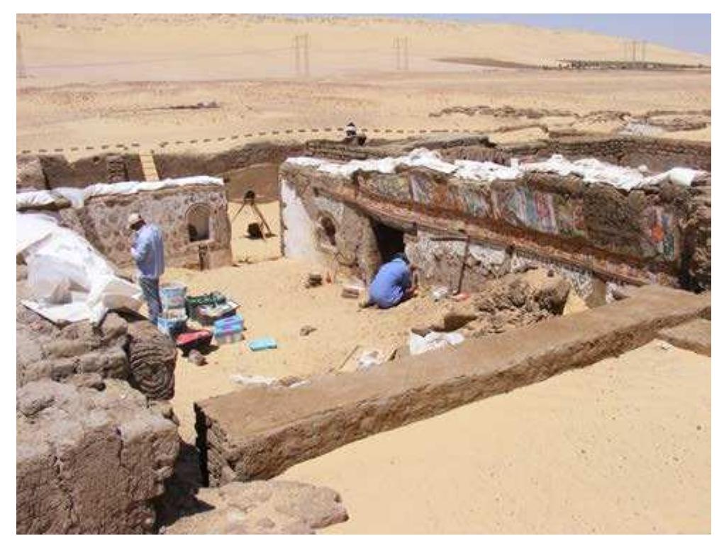
The problem is exacerbated by the lack of pub- A complete painted room known as sale 7
lished scientific reports for this kind of paint- was uncovered in 4 seasons of excavation and
ings. On the other hand, another source of en- given conservation treatment. Unfortunately
caustic paintings should be considered, as it the paintings were destroyed in 2010, The de-
forms a part of the development of encaustic tachment and rescue operation was performed
painting technology in Egypt, which is the by Christophe Guilbaud,Bruno Szkotnicki, Ab-
mummy portraits or the so-called the fayoum ied Mahmoud and Basem Gehad in April
portrait. Studies of these encaustic objects in- 2010,due to the collapsing of the mud brick
form us about the technology of encaustic paint- walls of the house; now they are detached and
ing in Egypt from the beginning of the 1st cen- kept in trays in the store rooms.
tury C.E to the end of the 3rd century C.E . The paintings were uncovered in a construc-
In 1991 the Institut Français d’Archéologie tion used as housing, nominated as Building 1,
Orientale (IFAO) uncovered a hitherto un- excavated by Marie-Hélène Rutschowscaya and
known painting of the annunciation in the Ramez W. Boutros, consisted of several rooms
western semi dome of the church of the holy set around a courtyard. The most southerly of
virgin in Dier el Surian (Innemee , 1999). During these rooms controlled access to the building,
a continuation of the work in 1995 different with a door opening to the outside and another
painting stratifications were identified; the four opening onto the courtyard. On the north side,
layers identified are from different phases of the two rooms were separated by a corridor that led
church. to a kitchen. On the east side stood an ensemble
Another encaustic mural painting was of five vaulted halls. The walls and the vault of
identified in one of the monks’ houses at baouit. the biggest of these (Hall 7) were decorated
The town of Bawit (or Baouit in French) is locat- with mural paintings (Fig.2).
ed between Dayrut and Asyut, 1.25 miles (2km) On the north side of this room, the paintings
west of the village of Bawit and about 17 miles of the vault depicted episodes from the birth of
(28 Km) south of El Ashmunin. Christ: the dream of Joseph, the voyage to Beth-
The site is famous from the excavations lehem, the nativity with the miracle of Salome
carried out there at the beginning of the and the shepherds, the presentation in the
twentieth century. The monastery excavated at Temple and the adoration of the Magi. Below, at
Baouit was dedicated to saint Apollo and called the base of the vault, is a geometric frieze (Bé-
Apa Apollo.Not more than 10 percent of the site nazeth et al, 2008).
Mediterranean Archaeology and Archaeometry, Vol. 15, No 2, (2015), pp. 243-256
� 245
IDENTIFICATION OF THE BYZANTINE ENCAUSTIC MURAL PAINTING IN EGYPT
The hypotheses of painting using molten wax
(or the application of hot instruments for
painting with the wax) or the use of the Punic
wax (saponification wax) mentioned by Pliny
has been argued between different scholars. At
present the most widely accepted hypothesis
states that the ancient technique of encaustic
painting consisted of the application of colored
beeswax in its molten state, a theory which is
based upon ancient literary sources. As the
Roman writer Seneca wrote that the choosing
and applying of the paint was performed
rapidly (Doxiadis 1995), this would be possible
Figure 2 Room no.7 , building no.1 , during excavation, only with the hot or molten wax. Petrie also
photo to the north -west ( Courtesy of IFAO). mentioned that the environment of Egypt
On the south side of the room, the vault bears would keep the wax melted or near melting
the representations of a series of characters in point (Petrie 1911).
which one can recognise two of the founders of But at the same time some scholars have not
Bawit monastery and nine prophets, each of accepted this idea, disagreeing with the idea of
whom holds a scroll inscribed with a passage using molten wax for the following reasons:
from their prophecy. The base of the vault is molten wax cannot produce the long thin and
decorated with a frieze of meanders alternating diluted brush strokes observed, as molten wax
with birds. The wall itself is decorated with a sets and dries very fast, giving only thick and
pattern of diamond shapes each holding a green short brush strokes, the faces indicate not brush
leaf. Another geometric pattern, covered with marks but palette knife marks, which could
large blooming flowers, is painted on the west only be reproduced by saponification of wax,
wall. The east wall, of which only the lower part the terminological meaning of the word
remains, holds three niches. The decoration of encaustic could refer to the method and the
the largest, set in the middle of the wall, was tools but not the molten wax .
found in the debris. It depicted Christ The second hypothesis is the use of water
surrounded by seraphim and the symbols of the soluble encaustic or punic wax. According to
four evangelists. ancient recipe given by Pliny, after bleaching
A great effort is being made to reassemble the the wax using sea water and sun light, the wax
fragments of the encaustic painting, which is a was then boiled with an alkaline solution
representation of the saints and the holy family produced from the burning of ash, , as the
in Egypt in a Coptic and byzantine style. water filtered from burned ash contains high
The precise composition of the medium has proportions of potassium hydroxide and minor
proved to be a continual source of disagree- proportions of sodium hydroxide.
ment. The etymology of the word encaustic in The idea of using saponified wax or punic wax
ancient lexicons and dictionaries is derived has met with great criticism, as there was no
from the Greek word enkaio or enkaiein which direct reference to it, as well as problems con-
means to burn or to be executed by fire (Lieber, cerning the color fading if the saponified wax
1840).Tthe meaning given by the lexicon indi- with its salts was used.
cates the method used in the preparation, which The word sapo, Latin for soap, first appears in
depends mainly on heating the wax before the Pliny the Elder's Historia Naturalis (Rackman,
application. Pliny refers to the impregnation of 1952), which discusses the manufacture of soap
the pigments with the wax by melting and from tallow and ashes, but before this a refer-
preparing cylindrical shapes or color cakes. ence to soap production appears in The Ebers
(Doxiadis, 1995). papyrus from Egypt, 1550 BC, indicating that
the ancient Egyptians bathed regularly and
combined animal and vegetable oils with alka-
Mediterranean Archaeology and Archaeometry, Vol. 15, No 2, (2015), pp. 243-256
�246 B.GEHAD et al.
line salts to create a soap-like substance ( Scholl, (Ramer, 1979; (Meilunas, Bentsen & Steinberg,
2002). 1990; Pitthard, Vak, Griesser, Stanek, &
The pigments used by the encaustic painters Laubenberger, 2007; Kühn, 1960), (Moretto,
were, according to the recent analytical results Orsega, & Mazzocchin, 2011). Infrared spectra
performed in both encaustic mummy portrait were obtained in the mid-range using JASCO-
and paint saucers from the roman period, red 6100 FTIR spectroscopy. About 1 to 3 mg from
lead (minium) for the orange red color, the archeological samples were ground with 99-
Egyptian blue for the blue pigment, white lead 97mg of KBr in an agate mortar, Percentage
or gypsum for the white color, ochre mixed transmittance was collected in the range of
with jarosite and in some cases madder lake for 4000–400 cm−1 with 4 cm−1 resolution.
the rose and the flesh tones, and ochre mixed.
2.5 Gas chromatography–mass spectrometry (gc-ms)
2. MATERIAL AND METHODS Gas chromatography – mass spectrometry is
2.1 X-Ray Fluorescence (XRF) one of the most effective methods widely used
for the identification of organic material and the
Non-destructive chemical- elemental analyses
alteration aspect connected to the aging of these
were collected for twelve samples using Niton
materials. GC-MS has been widely used for the
XLt -793 W portable XRF spectrophotometer
identification of wax in general and beeswax
device, that produced measurements in parts
specifically, the method followed in this re-
per million (ppm). The NITON XLt x-ray tube
search was described in different research as
based analyzer is a completely portable instru-
(White, 1978), (Bonaduce & Colombini, 2004)
ment with a one hand trigger operation and a
and (Maia & Nunes, 2013). Type of GC/MS:
touch screen with full navigation, a complete
thermo scientific trace GC ultra-coupled with
energy spectra view, and an RS232 download
ISQ single Quadra pole MS, type of column TG5
port. All samples were exposed for a minimum
MS, working protocol followed: initial tempera-
of 180 seconds. XRF charts were produced us-
ture 50 0C held for 2 min, 50 0C / min ramp to
ing NITON xrf software. The principle compo-
180 0C, 3 0C / min ramp to 300 o C, 6 0C / min
nents and the cluster analyses were performed
ramp to 320 0C.
using PAST statistical software program, ver-
For sample preparation, approximately 3 mg
sion 2.10.
of beeswax was dissolved in 4 ml of chloroform;
2.2 SEM-EDX the solution was mechanically shaken for 2 min
The scanning electron microscope used was a to complete dissolution of beeswax.
Quanta 3D 200 I) (FEI Philips – Holland) cou- 3. RESULTS
pled with EDX. Column pressure 60 PA, low
3.1 Plaster and paint substrate
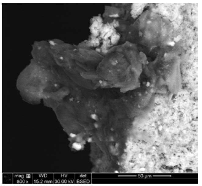
vacuum, In back scattered mode (BSED).
The investigation of the cross sectioned sam-
2.3 X- Ray Diffraction (XRD)
ples, using SEM, indicates six layer in the com-
For the x-ray diffraction samples were analysis position of the mural painting subjected for the
using panlytical X-pert pro with a Cu anode, study showed that the paint layer could be de-
working at 40 mA / 45 kV. The samples were scribed according to the SEM-BSED investiga-
analysed in a nondestructive mode without any tions as following:(Fig.3 ):
sample preparation. An approximately flat sur- (a) Silty sand render with a thickness around
faced archeological sample was attached into 2.5mm, this layer is reinforced with strips of
the sample holder inside the xrd apparatus. The chopped straw, the strip length could be from
data were interpreted using the embedded 300 to 500 µm, according to the SEM investiga-
software. tion, the straw could be from Barley straw
2.4 Fourier Transform IR spectroscopy (FTIR) (Hordeum vulgare L.)
(b) A thick lime based render with a thickness
Fourier transform infrared spectroscopy has about 1.5mm, this layer is also reinforced with
been widely used in the study of wax and pig- chopped straw, and the layer is rich with large
ments, the method used was in accordance with
Mediterranean Archaeology and Archaeometry, Vol. 15, No 2, (2015), pp. 243-256
� 247
IDENTIFICATION OF THE BYZANTINE ENCAUSTIC MURAL PAINTING IN EGYPT
grain sand particles ranging from 100 to 150 3.2.1 Orange paint layer
µm. Two samples from the orange paint layer
(c) Moderate lime based coat, the thickness of were investigated and analysed by means of
this layer is about 150 µm. SEM coupled with EDX,-indicating the presence
(d) Two fine coats with very fine sand particles, of highly back scattered elements in the paint
the inner is about 45 µm while the outer is layer, while elemental analysis using XRF and
about 120 µm. EDX proved the presence of Lead (Pb) as a ma-
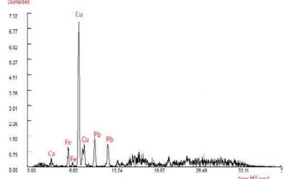
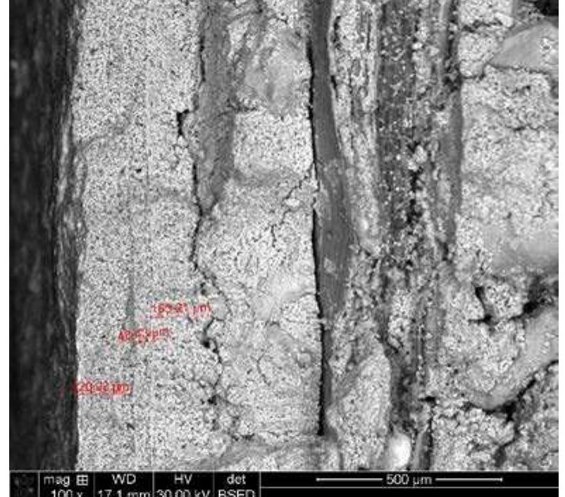
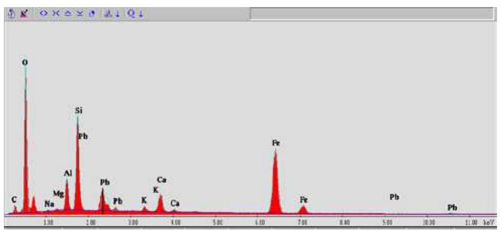
(e) The paint layer which ranges from 5 to 20 jor element as well as Iron (Fe) and Mn,Cu,As.
µm. (Fig. 5,6)
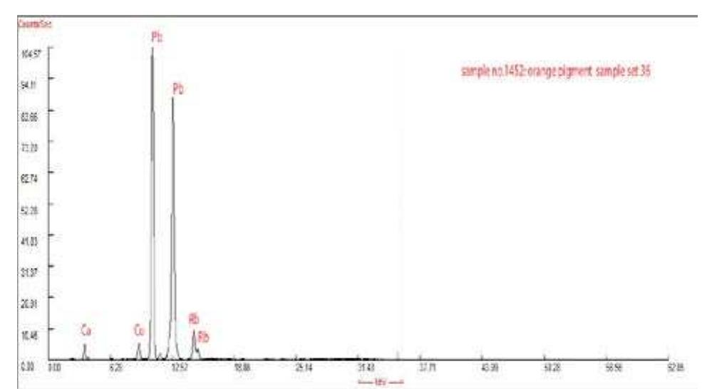
Figure 5 XRF pattern for the orange pigment, lead was
the major element indicated in the sample.
Figure 3 SEM indicating the stratigraphic composition
and the thicknesses of the encaustic paint layer and its
substrate, Baouit, Building no.1.
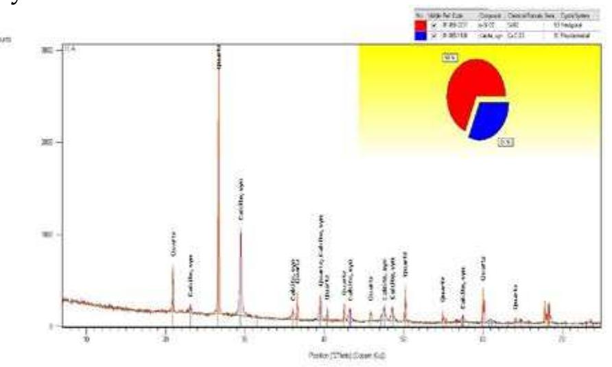
The X-ray diffraction analysis of the paint layer
proved that the white preparation layers are
mainly calcium carbonate (Fig. 4), which could
be interpreted as a lime based plaster, executed
on a silty sand render. The main components of Figure 6 EDX analysis for the orange paint layer indicat-
ing the presence of Pb, Fe. Al, Ca and traces from Mn
the plaster were quartz and calcite, while the and K.
silty sand render represented by albite as one of
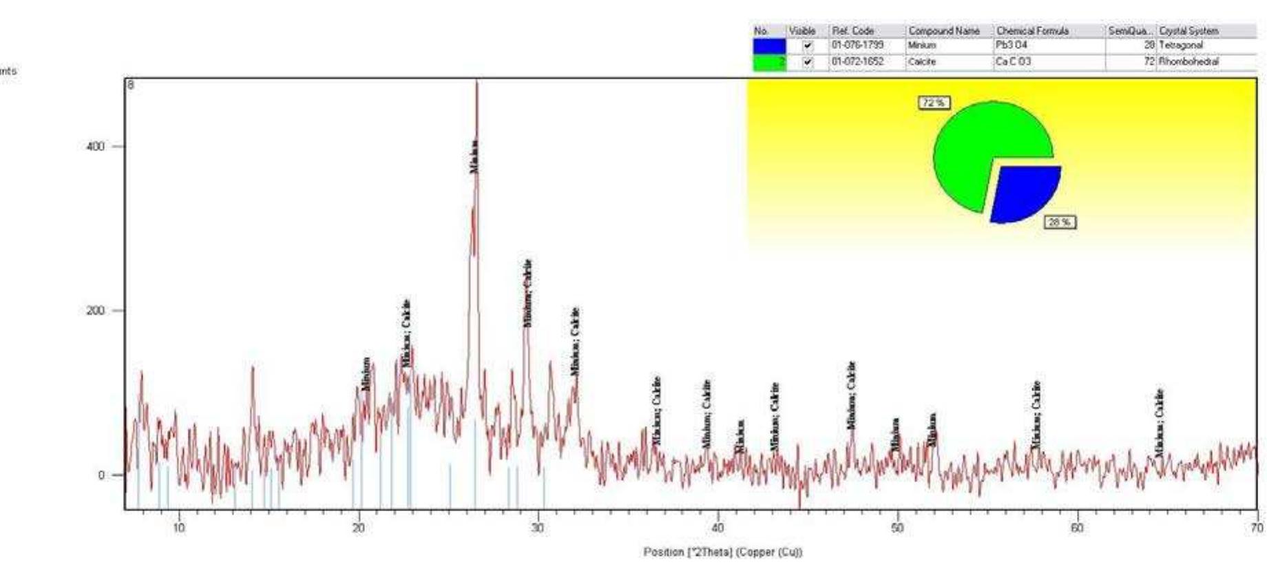
its main components. From the XRD results, no The presence of red lead was confirmed by
alumino-silicate materials were found (ex: Poz- means of XRD analysis, where both minium
zolan) and that confirms that air lime and not and calcite were identified (Fig.7).
hydraulic lime was used.
3.2.2 Yellow paint layer
XRF analysis of three samples analysed from
the yellow paint layer indicated that iron (Fe)
was the most abundant element, with some
traces of Ca, Cu.
Two samples (bulk and powder) were ana-
lysed using XRD. The analysis revealed the
presence of natrojarosite, hydrated sodium iron
sulphate, [NaFe3(SO4)2(OH)] for the lemon yel-
low pigment used (Fig.8), a mineral previously
Figure 4 XRD pattern for the encaustic mural painting reported as a yellow pigment in ancient Egypt
substrate indicating the presence calcite and quartz, in- by Le fur (Le fur , 1994) and Colinart (Colinart,
dicating the presence of lime based plaster , with no 2001).
alumino -silicate materials.
Mediterranean Archaeology and Archaeometry, Vol. 15, No 2, (2015), pp. 243-256
�248 B.GEHAD et al.
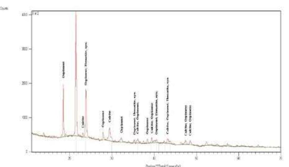
Figure 7 XRD pattern of the orange pigment indicating the presence of minium Pb3O4 and calcite CaCO3.
Figure 8 XRD pattern for the yellow paint layer revealed the presence of natrojarosite, hydrated sodium iron sulphate,
[NaFe3(SO4)2(OH)] for the lemon yellow pigment
The pigment was also identified in different cannot be relied on as a solo analysis technique,
painting examples from the old kingdom (Ak- whether the green pigment was iron based or
hethotp mastaba 6th 6 dyn. Saqqara) towards the copper based was not clear.
middle kingdom period, (ex.Karanak, Luxor), as
well as some other examples from the Ptolemaic
period.
3.2.3 Green paint layer
The olive green pigment used in the monastery
of Baouit was prepared in thin sections, which
allowed a clear observation of large and well
crystallized grains, embedded in a deep green
matrix.
Analysis of the green pigment using XRF indi- Figure 9 XRF result of the green paint layer indicating the
presence of Cu, Ca Fe, and Pb.
cated the presence of copper and iron as major
components (Fig.9), with some traces of magne- EDX analysis revealed the presence of iron
sium and Arsenic. The XRF elemental analysis (Fe), Calcium (Ca), Aluminum (Al), Magnesium
Mediterranean Archaeology and Archaeometry, Vol. 15, No 2, (2015), pp. 243-256
� 249
IDENTIFICATION OF THE BYZANTINE ENCAUSTIC MURAL PAINTING IN EGYPT
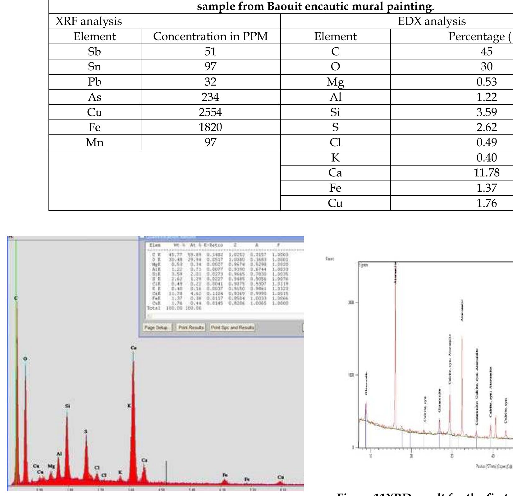
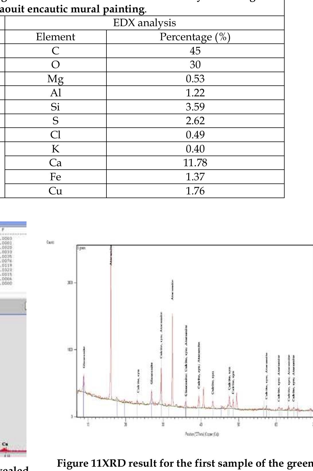
(Mg), potassium (K), Chloride (Cl), Copper (Cu). suggest that glauconite was used rather than
These results confirm the presence of green Celadonite (Figs. 10, Table 1).
earth, while the ratio of Iron (Fe) to (Mg) would
Table no.1: elemental relative percentage resulted fom both XRF and EDX analysis of the green
sample from Baouit encautic mural painting.
XRF analysis EDX analysis
Element Concentration in PPM Element Percentage (%)
Sb 51 C 45
Sn 97 O 30
Pb 32 Mg 0.53
As 234 Al 1.22
Cu 2554 Si 3.59
Fe 1820 S 2.62
Mn 97 Cl 0.49
K 0.40
Ca 11.78
Fe 1.37
Cu 1.76
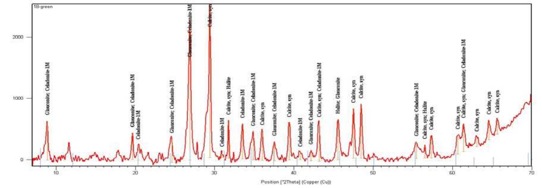
Figure 11XRD result for the first sample of the green
Figure 10 EDX analysis for the green paint layer revealed
paint layer indicating the presence of glauconite, calcite
the presence of Fe, Ca, Al, Mg, K, Cl and Cu.
and attachamite.
Figure 12 XRD pattern for the second green sample indicating the presence of glauconite, calcite and traces of cela-
donite
Mediterranean Archaeology and Archaeometry, Vol. 15, No 2, (2015), pp. 243-256
�250 B.GEHAD et al.
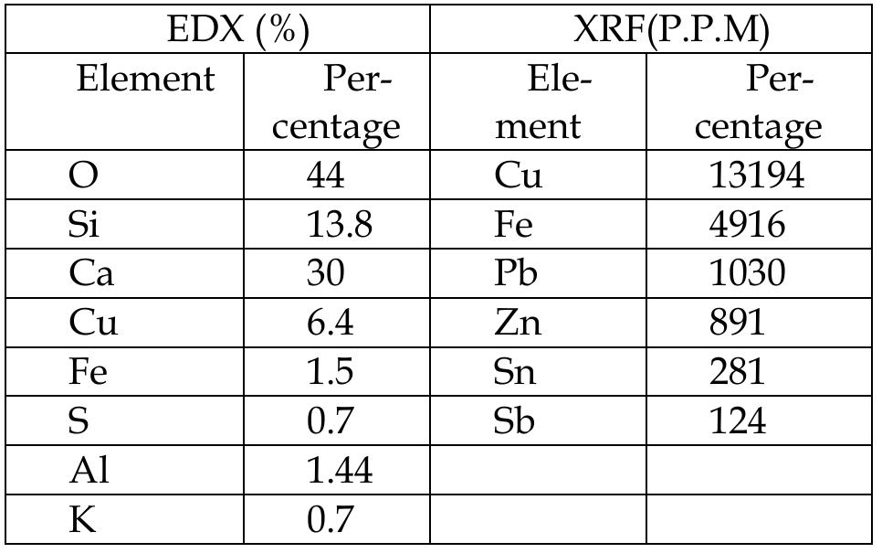
The usage of green earth was conformed us- Table 2 relative percentage elemntal result using xrf and
ing XRD analysis. Two samples were analysed, edx for the blue paint layer.
in one sample glauconite and atacamite were EDX (%) XRF(P.P.M)
present, and in the second sample both glauco- Element Per- Ele- Per-
nite and Celadonite were present (Figs.11, 12). centage ment centage
O 44 Cu 13194
3.2.4 Blue paint layer
Si 13.8 Fe 4916
An investigation of the blue pigment in thin Ca 30 Pb 1030
section showed the presence of anisotropic Cu 6.4 Zn 891
crystals with tubular shape, and refractive in- Fe 1.5 Sn 281
dex less than 1.66.compaed to the canada bal- S 0.7 Sb 124
sam medium used in the sample preparation. Al 1.44
these properties are typical of cuprorivaite, the K 0.7
crystal shape was also observed under sem in-
vestigation (fig no.13) i.e. egyptian blue. ele-
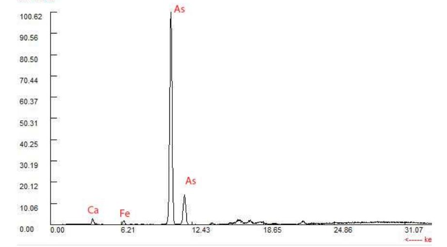
mental analysis using xrf analysis indicated the 3.2.5 BROWNISH RED
presence of copper as a main component; iron The elemental analysis using portable X-ray
also being present. the analysis also indicated fluorescence for the brownish red sample indi-
the presence of lead (fig no.14). the presence of cates the presences of Arsenic (As 26772 ppm)
lead and traces of other elements as tin and and minor amounts of iron (Fe 3974 ppm) and
zinc is of paramount importance as this may calcium (Ca 4251ppm). The XRF result may in-
indicate the reuse of bronze alloy as a source of dicate the use of arsenic sulphide, or orpiment
copper for the fabrication of the egyptian blue. (fig no.15). The xrd pattern of the brownish red
pigment indicates orpiment, calcite and hema-
tite in the composition of the paint layer (fig
no.16). This might explain the dark Chroma of
this color, being due to the mixture of orpiment
and hematite.
Figure 13 SEM image of the blue paint layer revealed the
presence of coarse grains and tubular shaped crystals .
Figure 15 XRF result of the brownish red paint layer in-
dicating the presence of As and traces of Fe.
Figure 14 XRF result for the blue paint layer indicating
Cu as a major elemental component of the blue pigment Figure 16 XRD pattern for the brownish red paint layer
indicating the presence of orpiment, hematite and cal-
cite.
Mediterranean Archaeology and Archaeometry, Vol. 15, No 2, (2015), pp. 243-256
� 251
IDENTIFICATION OF THE BYZANTINE ENCAUSTIC MURAL PAINTING IN EGYPT
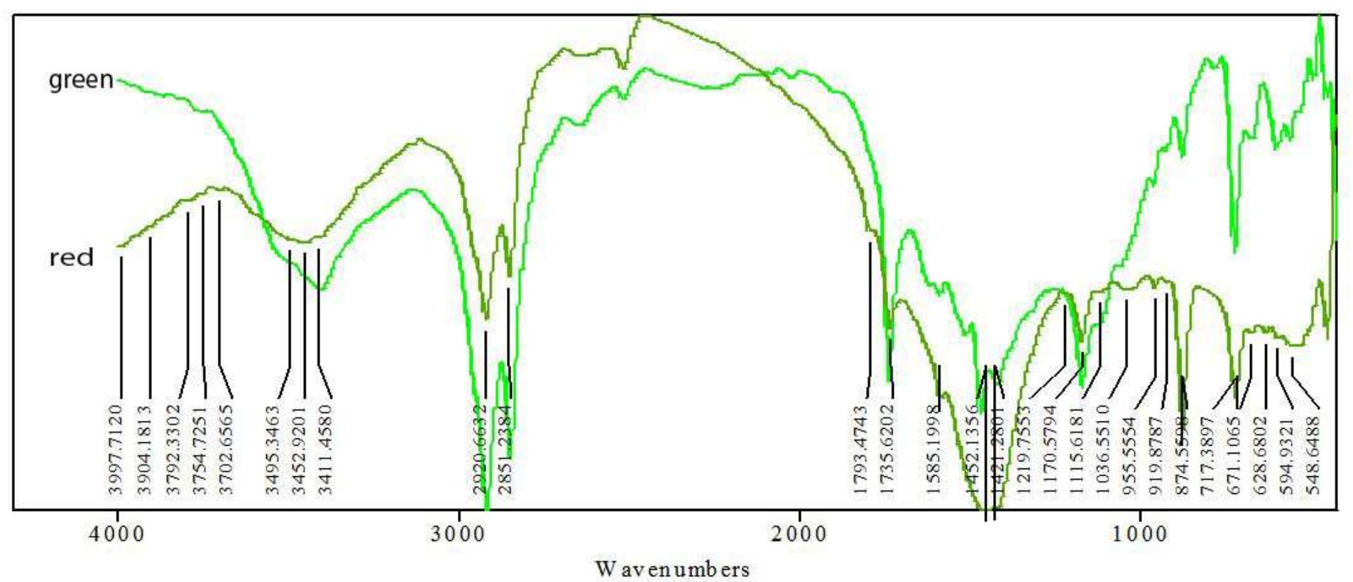
3.3 Organic binding medium ester (Fig.18). The band at 1512 cm-1 could be
The identification of the organic binding me- assigned to carboxylate salts or fatty acid salts
dium was based on the vibrational signature of (Mirghani, Che Man, Jinap, Baharin, & Bakar,
the functional groups for beeswax, using both 2002), while alcohols bands are present in the
Fourier transform infrared analysis, and gas OH stretching at 3407 cm-1 and the alcohols OH
chromatography mass spectrometry analysis. bending at 1122 cm-1 and 1056 cm-1 .
Further information obtained using these two The ftir spectrum of the green sample repre-
methods enabled us to understand the original sents a wax based paint layer, in which a partial
technique used for the execution of the encaus- saponification process in indicated by the pres-
tic technique and the alteration of the material ence of the carboxylate and alcohol functional
over time. groups. This could be due to natural aging of
wax, as the Punic process of saponification will
3.3.1 FTIR analysis result in the complete saponification of wax.
Two samples were analysed by means of The small peak at 958 may indicate the presence
FTIR analysis, from the red and green paint of Si-O, which could be from the green earth
layers. The results were compared to two con- pigment.
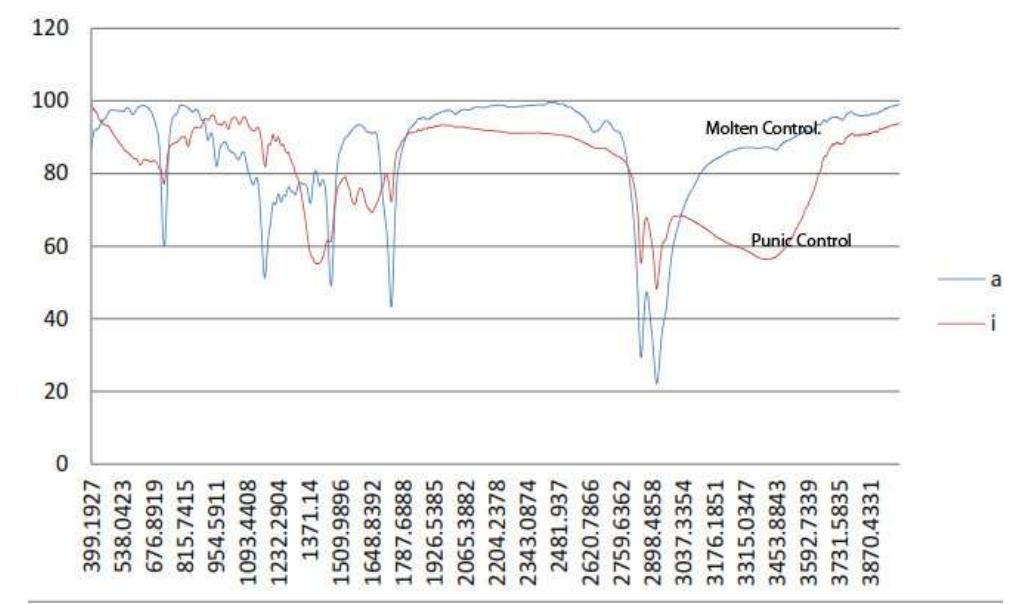
trol samples of bees wax - molten and Punic The FTIR analysis of a sample from a red
beeswax (fig no.17) were prepared- in order to flaking paint layer proved the presence of wax
compare the results with the archeological as a binding medium; this is demonstrated by
samples in order to identify both the presence the presence of the methylene group of the wax
of the wax and the technique used. hydrocarbon, as well as the carbonyl group of
the wax ester.
A significant broad and large band could be
seen in the region of 3450 cm-1 due to the OH
group, probably indicating the presence of tri-
glyceride alcohols. A small band at 1107 and
1028 cm-1 for the long chain alcohols indicates
that small quantities of alcohol could be in the
sample.
Crystalized areas of wax represented by the
small band for C=O at 1735 cm-1 and another
one for the C –O at 1170 cm-1, indicates that cer-
Figure 17 FTIR pattern for two control samples of molten tain alterations could have occurred in the
and Punic ( saponified) wax sample composition.
Inorganic materials were indicated by the
The main functional groups obtained from
presence of a secondary absorption band of the
the contemporary beeswax sample were two
CO3 -2 group of calcium carbonate in the region
main doublets representing stretching in plane
of 2516 cm-1, 1793 cm-1. A broad band at 1458
and out of plane in the region of 2950 and 2850
cm-1 overlapped the CH band in the same re-
cm-1, as well two other sharp doublets at 1460
gion and bending of the CO3-2 is seen at 874 cm-
and 1470 cm-1. The peak in the region of 715 – 1 (Derrick, stulk, & Lundry, 1999).Small peaks in
720 cm-1 indicates the CH2 torsion bend; the C-
the region of 1107 and 1028 cm-1 indicated the
O of the wax ester usually appears in the 1165 –
presence of long chain alcohols (Mirghani, Che
1170 cm-1region; C=O of the organic fatty acids
Man, Jinap, Baharin, & Bakar, 2002), and could
appears in the 1715 -1720 cm-1 region.
be connected to the broad band of the OH at
The FTIR results of the paint layers showed
3454 cm-1.
two strong and sharp methylene stretching
symmetrical and asymmetrical bands at 2919
and 2849 cm-1 respectively, as well as two bands
at 721 cm-1 , 874 cm-1 and 922 cm-1, a strong
C=O at 1735 cm-1 and C-O at 1169 cm-1 of the
Mediterranean Archaeology and Archaeometry, Vol. 15, No 2, (2015), pp. 243-256
�252 B.GEHAD et al.
Figure 18 FTIR pattern of two samples from green and red paint layer, Baouit encaustic mural painting.
The green sample was compared to both a tation and the separation of the main organic
molten bees wax sample and a Punic bees wax components of the wax identified as seen in
sample. The comparison of the ftir charts (Fig.20) mainly hydrocarbons, esters and fatty
(Fig.19) proved that the archeological samples acids, clearly represent the wax alteration. The
were similar to a large extent to the molten con- alteration is shown by:
trolled sample. Some alterations appear in the Presence of long chain alcohols.
archeological paint layer profile, but it does not Depletion of hydrocarbon profile of wax.
resemble totally and intentionally saponified Presence of phenolic compounds.
wax (Punic sample), which means that the pres- Alteration of esters and appearance of gly-
ence of small bands related to long chain alco- ceride esters and fatty acids.
hols or carboxylates, could be interpreted as a
result of natural aging and alteration of wax
due to oxidation and water hydrolysis in an ar-
id and exposed environment.
Figure 19 comparison between FTIR pattern of the green
archaeological paint layer with the two control samples
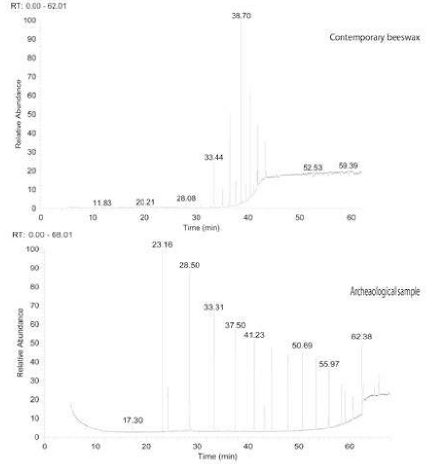
Figure 20 GC-MS result for the beeswax contemporary
of the molten and Punic wax.
sample and the archaeological green sample.
3.3 GC-MS analysis
The four previous aspects are shown in the fol-
The GC-MS analysis for one of the wax based
lowing Table 3, taking into consideration the
paint layers indicates the state of alteration that
relative intensity of the separated compounds:
the binding medium had reached. The fragmen-
Mediterranean Archaeology and Archaeometry, Vol. 15, No 2, (2015), pp. 243-256
� 253
IDENTIFICATION OF THE BYZANTINE ENCAUSTIC MURAL PAINTING IN EGYPT
Table 3 GC-MS analysis results including Retention time and compounds identified from both control beeswax
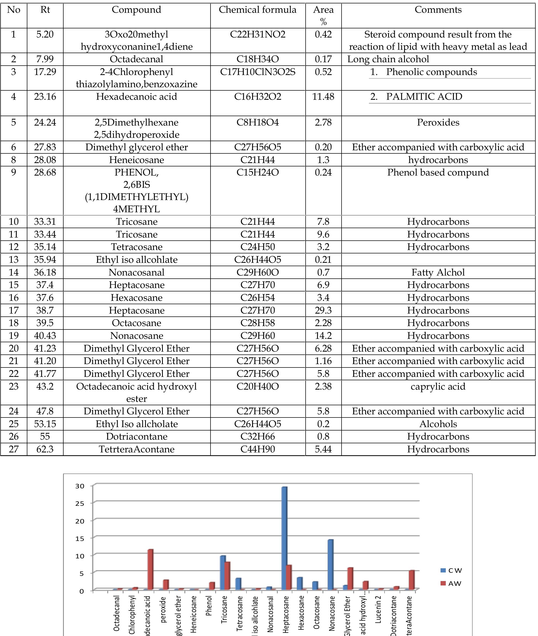
sample and the archaeological green sample.
No Rt Compound Chemical formula Area Comments
%
1 5.20 3Oxo20methyl C22H31NO2 0.42 Steroid compound result from the
hydroxyconanine1,4diene reaction of lipid with heavy metal as lead
2 7.99 Octadecanal C18H34O 0.17 Long chain alcohol
3 17.29 2-4Chlorophenyl C17H10ClN3O2S 0.52 1. Phenolic compounds
thiazolylamino,benzoxazine
4 23.16 Hexadecanoic acid C16H32O2 11.48 2. PALMITIC ACID
5 24.24 2,5Dimethylhexane C8H18O4 2.78 Peroxides
2,5dihydroperoxide
6 27.83 Dimethyl glycerol ether C27H56O5 0.20 Ether accompanied with carboxylic acid
8 28.08 Heneicosane C21H44 1.3 hydrocarbons
9 28.68 PHENOL, C15H24O 0.24 Phenol based compund
2,6BIS
(1,1DIMETHYLETHYL)
4METHYL
10 33.31 Tricosane C21H44 7.8 Hydrocarbons
11 33.44 Tricosane C21H44 9.6 Hydrocarbons
12 35.14 Tetracosane C24H50 3.2 Hydrocarbons
13 35.94 Ethyl iso allcohlate C26H44O5 0.21
14 36.18 Nonacosanal C29H60O 0.7 Fatty Alchol
15 37.4 Heptacosane C27H70 6.9 Hydrocarbons
16 37.6 Hexacosane C26H54 3.4 Hydrocarbons
17 38.7 Heptacosane C27H70 29.3 Hydrocarbons
18 39.5 Octacosane C28H58 2.28 Hydrocarbons
19 40.43 Nonacosane C29H60 14.2 Hydrocarbons
20 41.23 Dimethyl Glycerol Ether C27H56O 6.28 Ether accompanied with carboxylic acid
21 41.20 Dimethyl Glycerol Ether C27H56O 1.16 Ether accompanied with carboxylic acid
22 41.77 Dimethyl Glycerol Ether C27H56O 5.8 Ether accompanied with carboxylic acid
23 43.2 Octadecanoic acid hydroxyl C20H40O 2.38 caprylic acid
ester
24 47.8 Dimethyl Glycerol Ether C27H56O 5.8 Ether accompanied with carboxylic acid
25 53.15 Ethyl Iso allcholate C26H44O5 0.2 Alcohols
26 55 Dotriacontane C32H66 0.8 Hydrocarbons
27 62.3 TetrteraAcontane C44H90 5.44 Hydrocarbons
30
25
20
15
10
5 CW
AW
0
Octadecanoic acid hydroxyl …
Chlorophenyl
Octadecanal
Phenol
Dimethyl glycerol ether
Hexacosane
Octacosane
Heneicosane
Lucenin 2
Hexadecanoic acid
Dotriacontane
Tricosane
TetrteraAcontane
Tetracosane
Heptacosane
Nonacosanal
Dimethyl Glycerol Ether
Nonacosane
Ethyl iso allcohlate
peroxide
Figure 21 Comparison between the main component of contemporary beeswax and the archeological sample from
Baouit
Mediterranean Archaeology and Archaeometry, Vol. 15, No 2, (2015), pp. 243-256
�254 B.GEHAD et al.
4. DISCUSSION light richness of the byzantine palette used for
the encaustic painting in Egypt, during the 6th
The analysis performed on the samples from
century.
the unique encaustic painting from the monas-
The FTIR analysis in the mid-range (400-4000)
tery of Baouit explains the formation of the
for five archaeological samples, proved the
painting palette. The pigments identified were
presence of methylene and ester functional
mainly types identified previously from the
groups identical to those for the wax ftir profile.
roman period, only the brownish red is an ex-
This result confirms the usage of wax and more
ception. The palette shows the variety of
likely beeswax as a binding medium in the mu-
sources where these pigments were obtained;
ral painting of baouit.
both natural and artificial sources were used.
The samples indicate evidence of hydrolysis
The integrated elemental analysis using EDX
or partial saponification of the organic medium,
and XRF confirms the usage of a green earth
the presence of OH vibration bands in the 3400
based pigment in combination with a copper
region as well as 1035 to 1060 cm-1 region
pigment, the XRF shows major amounts of Fe
indicates the presence of long chain alcohols or
and Cu as well as minor amounts of Sb, Sn, Pb
glycerides. One should also mention the ob-
and As, while the EDX gives major Ca peaks
served depletion of the methylene or the
with minor percentage of Al, Fe, Cu, as well
hydrocarbon from the weakness of the peak of
traces of Mn, K, Cl and S. The results match
the characteristic group which may be due to
those of green samples from different periods
the sublimation of the lower molecular
(pharonic and roman period green samples)
hydrocarbons. The same is true for those peaks
from an ancient shrine, analysed by Berry (Ber-
representative of the ester functional group, as
ry, 2002) as well mineralogical identification
they were shifted and weakened, and
and description for green earth (both glauconite
sometimes the intensity of the carbonyl peak
and celadonite) in the literature (Buckley, Bevan,
increased, which indicates the occurrence of
Brown, & Johnson, 1978) (Bearat, 1996).
hydrolysis and formation of carboxylate, for
The presence of both copper based pigment
which some small representative peaks were
and green earth in the XRD pattern, explains the
observed in the archaeological sample charts.
result of the investigation of the thin sectioned
It’s hard to say whether the saponification
sample for the green paint layer , with large
identified on the ftir spectrum for the
green particles embedded in very fine green
archaeological samples was intentionally made
matrix.
by the artist in order to prepare the water
The EDX and the XRF elemental analysis of
soluble encaustic, or that the presence of the
the red pigment indicates lead as a main ele-
saponifaction was due to the water hydrolysis
ment, with traces of iron and calcium, which
as well as other alteration processes, which
could be traces from the white wash. The min-
could have taken place in the wax composition
eralogical composition of the pigment was iden-
during rancidification .
tified using XRD qualitative analysis as Minium
It’s well known that rancidifcation is the de-
(Pb3O4), as well as calcite indicating the lime
caying of unsaturated fatty acids by an oxida-
based render.
tion process; this process could be promoted by
The absence of wallastonite in the qualitative
the presence of trace metals as Ca, Cu, Fe, and
XRD result of the blue pigment indicates the
Zn (Megahed, Nashy , & Ashkar, 2011), which
temperature used in the manufacture of the
already exist in the paint layer from the pig-
Egyptian blue was lower than 950 oC (Riederer,
ments, as well as the presence of heat. These
1997). As no green particles could be observed
two factors existed within the context of the
in the stereo microscopic investigations, the
mural painting at baouit.
theory is supported by well identified sharp
The Chromatogram of the archaeological wax
edge crystals observed under the SEM-BSED
based paint layer shows the highly deteriorated
investigations.
pattern of the wax medium. This was demon-
The identification of pigments like natrojaro-
strated by the presence of long chain alcohols,
site and mixture of orpiment and hematite high-
palimtates and mainly the glycerols of the
Mediterranean Archaeology and Archaeometry, Vol. 15, No 2, (2015), pp. 243-256
� 255
IDENTIFICATION OF THE BYZANTINE ENCAUSTIC MURAL PAINTING IN EGYPT
hydrolysed triglycerides represented by the that the honey and its wax were naturally
ester of fatty acids. formed by bees which fed on sunflowers.
Oxidation or photooxidation may be one of
the main reasons for the alteration of the wax; 5. CONCLUSION
other components found in the archaeological
sample such as phenolic compounds may The painting palette of the encaustic mural
indicate that this process took place. painting at Baouit was identified as: lead red
The profile of the hydrocarbon was also (minium), Egyptian Blue, natroJarosite, a mix-
strongly altered. The main hydrocarbon ture for the brownish red (red ochre and orpi-
heptacosane concentration was 29 % in the ment), and green (glauconite and atacamite
contomperoray wax sample but only 6.9 % in with traces of Celadonite). The mixtures high-
the archaeological wax. This indicates that the light how the ancient artists adapted their mate-
hydrocarbons which represent the main rials to obtain the colors.
backbone of the wax were totally depleted and The identification of beeswax proves the exist-
changed as a result of aging starting with the ence of the encaustic technique as an ongoing
lower molecular weight hydrocarbons. tradition for painting in Egypt during the By-
One of the interesting pieces of information zantium period. The analysis shows also that
that came from the GC-MS analysis, is the the wax was probably not treated with alkali in
presence of lucenin, which is a luteolin flavone order to prepare the Punic; the presence of long
glycoside found in the sunflower family (Bohm chain alcohols could be a result of alteration
& Stussey, 2002) with a molecular composition through natural water hydrolysis and oxidation
(Luteolin 6, 8-di-glucoside), which informs us as a part of the aging process.
ACKNOWLEDGEMENTS
We thank the anonymous reviewers for constructive comments. I would like also thank
Dr.Giselle Hadje, director of the Baouit excavation mission, Dr.Anita Quiles from IFAO, Dr Mo-
hamed Abd el Rohman, from SCA. Kate Fulcher from UCL Institute of Archaeology - British Mu-
seum- department of Conservation Science and Research.
REFERENCES
Bearat,H. (1996) Chemical and mineralogical analyses of * gallo-roman wall painting from dietik-
on, switzerland, Archaeometry 38, 1 81-95.
Bénazeth,D., Herbich,T. (2008) le kôm de baouît : étapes d’une cartographie, BIFAO 108.
Benazeth,D. (2010) Nouvelles Campagnes de fouilles a Baouit(2005, 2006). Etudes Coptes XI,Paris,
pp17-25.
Berry, M. (2002) The Study of Pigments from Shrine I at Ismant el-Kharab, in C. A. Hope and G. E.
Bowen (eds), Dakhleh Oasis Project: Preliminary Reports on the 1994–1995 to 1998– 1999
Field Seasons, 53–60.
Binda, L., Saisi, A., Tiraboschi, C., Valle, S., Colla, C. and Forde, M. C. (2003) Application of sonic
and radar tests on the piers and walls of the Cathedral of Noto. Construction and Building
Materials, vol. 17, 613–627.
Bohm, B. A., & Stussey, T. F. (2002). Flavonoids of sun flower family. Biologia plantarum, 45(2), 226.
Bonaduce, I. and Colombini, M. P. (2004). Charchterisation of beeswax in works of art by gas
chrimatography - mass spectrometery and pyrolysis - gas chromatography- mass
spectrometery procedures. Journal of chromatography, 1028, 297-306.
Buckley, H. A, Bevan, J. C., Brown, K. M., and. Johnson, L. R ( 1978) Glauconite and celadonite"
two separatemineral species Mineralogical Magazine, September, vol. 42, pp. 373-82.
Mediterranean Archaeology and Archaeometry, Vol. 15, No 2, (2015), pp. 243-256
�256 B.GEHAD et al.
Cartwright, C., Middleton, A. (2008) Scientific aspects of ancient faces: mummy portraits from
egypt , in the british museum technical research bulletin, vol2.
Cosentino, P. and Martorana, R. (2001) The resistivity grid applied to wall structures: first results.
Proceedings of the 7th Meeting of the Environmental and Engineering Geophysical Society, Euro-
pean Section, Birmingham, U.K.
Derrick, M., stulk, D., & Lundry, J. (1999). Infra red spectroscopy for conservation science. los angeles:
Getty conservation institute.
Doxiadis, E. (1995). The Mysterious Fayoum Portraits, faces from ancient egypt. Cairo : the American
University in cairo press.
Innemee , K. (1999). Encuastic painting in egypt, painting in Dier el surian. 2, 131-139.
Kühn, H. (1960). Detection and Identification of Waxes, including Punic Wax, by Infra-Red .
Meilunas, R., Bentsen , J., & Steinberg, A. (1990). Analysis of Aged Paint Binders by FTIR
Spectroscopy . Studeis in Conservation , 33-51.
Mirghani, M., Che Man, Y., Jinap, S., Baharin, B., & Bakar, J. (2002). FTIR Spectroscopic
Determination of Soap in Refined Vegetable Oils. JAOCS, 79(2), 113.
Moretto, L. M., Orsega, E. F., & Mazzocchin, G. A. (2011). Spectroscopic methods for the analysis of
celadonite and glauconitein Roman. Journal ofCulturalHeritage, 3-11.
Moretto, L. m., Orsega, E. F., & Mazzocchin, G. A. (2011). Spectroscopic methods fro the analysis of
celadonite and glauconite in roman wall paintings. Journal of cultural Heritage .temperature
gas chromatography and chemometric analysis. food chemistery, 136, 961 - 968.
Megahed, M., Nashy , H., & Ashkar, E. (2011). Evaluation of fried edible oil and determenation of
elemnts content. Agriculture and biology journal of north america, 687-692.
Lieber, F. (1840). Encyclopædia Americana: a popular dictionary of arts and sciences (Vol. IV).
Philadelphia.
Maia, M., & Nunes, F. M. (2013). Authentication of beeswax ( apis mellifera )by high
Petrie, W. (1911). Roman portrait and Memphis (IV). London: school of Archeology in Egypt.
Pitthard, V., Vak, B., Griesser, M., Stanek, S., & Laubenberger, M. (2007). Fayoum Portraits from
the collection of greek and roman antiquites, Kunsthistorisches museum, Vienna.
Technologishe Studien, 4, 11-29.
Rackman , H. (1952). Pliny the elder natural history, chapter XXXV. Cambridge .
Ramer, B. (1979). The Technology, Examination and Conservation of the Fayoum portraits in the
Petrie Museum. Studies in Conservation, 1-13. Spectrography. Studies in Conservation, 5, 71-
81.
Scholl, Reinhold. (2002) Der Papyrus Ebers: die grösste Buchrolle zur Heilkunde Altägyptens.
Walker, A. (2012) The Emperor and the World: Exotic Elements and the Imaging of Middle Byzantine Im-
perial Power, Ninth to Thirteenth Centuries C.E. New York, Cambridge University Press.
White, R. (1978). The application of gas chromatography to the identifcation of waxes. Studies in
conservation, 23(2), 57.
Mediterranean Archaeology and Archaeometry, Vol. 15, No 2, (2015), pp. 243-256
�
 basem gehad
 prof.Mona F O U A D Ali
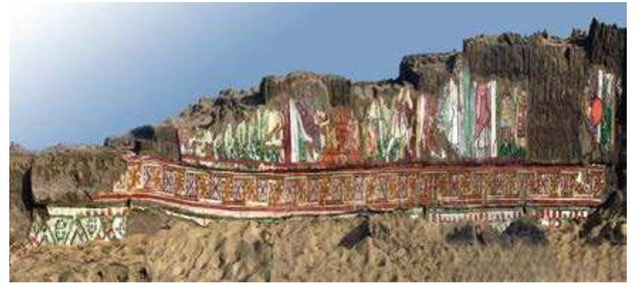
![Figure 8 XRD pattern for the yellow paint layer revealed the presence of natrojarosite, hydrated sodium iron sulphate, [NaFe3(SO,)2(OH)] for the lemon yellow pigment](https://figures.academia-assets.com/50442524/figure_009.jpg)