. 1997 Apr 15;94(8):3920-5.
doi: 10.1073/pnas.94.8.3920.

Functional differentiation of T cells in the intestine of T cell receptor transgenic mice

Affiliations

Functional differentiation of T cells in the intestine of T cell receptor transgenic mice

S D Hurst et al. Proc Natl Acad Sci U S A. .

Abstract

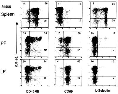
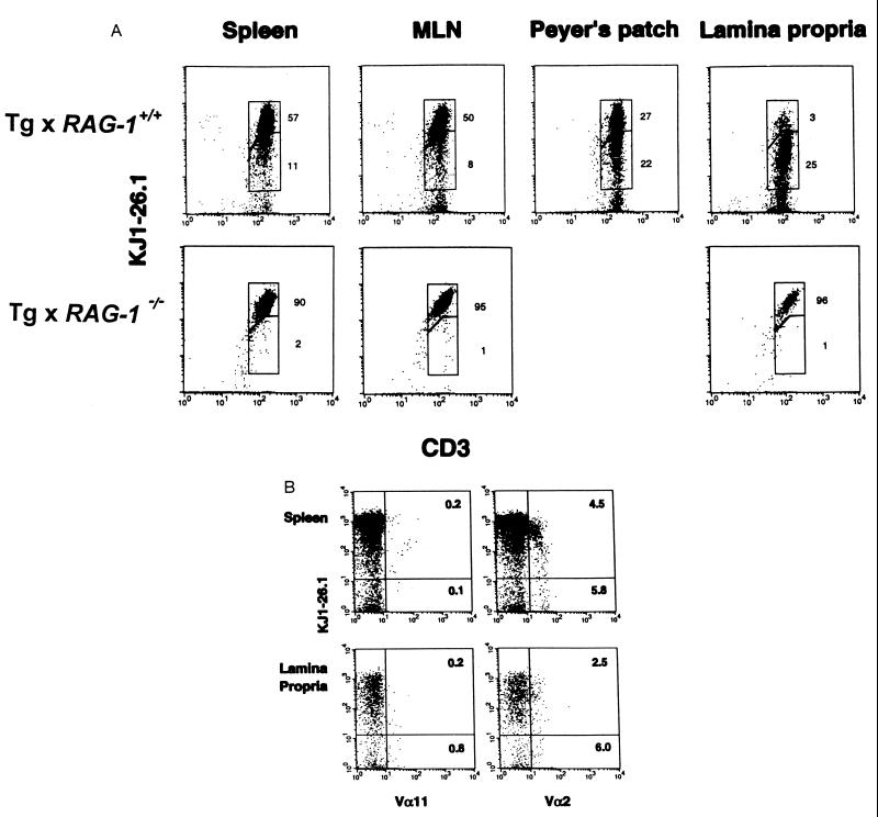
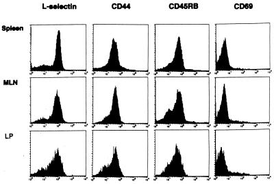
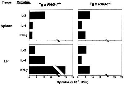
The intestinal lamina propria (LP) is a major effector site of the mucosal immune system where antigen-specific and antigen-nonspecific factors shape the functional responses of CD4+ T helper cells. To study the functional differentiation of LP T helper cells we utilized DO11.10 T cell receptor (TCR) transgenic (Tg) mice that expressed a clonotypic TCR specific for a class II major histocompatibility complex-restricted peptide of chicken ovalbumin. The majority of cells expressing Tg TCR (Tg+) in peripheral lymphoid tissue expressed naive surface phenotypes whereas nearly all Tg+ T cells in the intestinal LP expressed an activated/ memory-like phenotype. Flow cytometric analysis of Tg+ T cell populations revealed that a small proportion of cells in peripheral lymphoid tissue but nearly all cells in the LP expressed dual (Tg plus non-Tg) TCRs. In Tg x recombinase-activating-gene-1-deficient (Tg x RAG-1(-/-)) mice, splenic and LP T cells expressed naive surface phenotypes and produced cytokines equivalent to naive splenic cells from Tg x RAG-1(+/+) mice. In contrast, Tg LP cells from Tg x RAG-1(+/+) mice produced 35-fold greater levels of interferon-gamma and 5-fold greater levels of interleukin 4 compared with naive splenic cells. These findings suggested that activation of Tg+ T cells through endogenous non-Tg TCR had promoted the localization and differentiation of memory-like effector T helper cells in the intestine.


Figures

Figure 1
Figure 1
Surface phenotype of CD4+ T cells in lymphoid and extralymphoid sites. T cells from spleen, PP, and LP of 8-week-old DO11.10 TCR Tg mice were stained with FITC-labeled mAbs specific for CD45RB, CD69, or L-selectin; anti-CD4-PE; and KJl-26.1-BIO followed by Streptavidin-CyChrome. For all populations, lymphoid cells were gated on the basis of forward and 90° angle side scatter and staining for CD4. For each population, the percentage of positively stained cells is shown in quadrants. Quadrants were drawn based on staining with negative control (IgG2a) mAbs. Data shown are representative of more than five experiments.
Figure 2
Figure 2
TCR expression in Tg mice. Spleen, MLN, PP, and LP T cells were harvested from Tg × RAG-1+/+ and Tg × RAG-1−/− mice (PP not detected in Tg × RAG−/− mice) and Tg TCR vs. CD3 expression was analyzed by staining with PE-coupled clonotypic anti-TCR mAb, KJ1–26.1-PE, and FITC-coupled anti-CD3 (A). Results of T helper cells gated on the basis of staining for CD4 are shown. Regions show the distribution of cells with higher (upper region) or lower (lower region) Tg TCR/CD3 ratios within each population. (B) Results are shown for CD4-gated populations of splenic and LP cells from Tg × RAG-1+/+ mice stained with KJ1–26.1-PE and FITC-coupled mAbs specific for endogenous TCR Vα2 and Vα11 chains. Staining for these mAbs in Tg × RAG-1−/− mice was <0.1% on CD4-gated populations (data not shown). Data shown were representative of more than five experiments.
Figure 3
Figure 3
Surface phenotype of CD4+ T cells in Tg × RAG-1−/− mice. Tg T cells from spleen, MLN, and LP were gated on the basis of staining with anti-CD4-PE and KJ1–26.1-BIO followed by Streptavidin-CyChrome and results are shown for staining with FITC-conjugated mAbs specific for L-selectin, CD44, CD45RB, or CD69. Data shown are representative of four experiments.
Figure 4
Figure 4
Induction of cytokine production for LP Tg+ T cells. Purified splenic and LP Tg+ T cells were harvested from Tg × RAG-1+/+ and Tg × RAG-1−/− mice and activated with antigen-presenting cells and Ag. Cytokine levels were determined by ELISA at 48 hr. Data shown are representative of four experiments.

References

    1. Bloom P D, Boedeker E C. In: Seminars in Gastrointestinal Disease. Strober W, Ehrhardt R O, editors. Vol. 7. Philadelphia: Saunders; 1996. pp. 151–166. - PubMed
    1. Tagliabue A, Nencioni L, Caffarena A, Villa L, Boraschi D, Cazzola G, Cavalieri S. Clin Exp Immunol. 1985;62(2):242–247. - PMC - PubMed
    1. Zeitz M, Quinn T C, Graeff A S, James S P. Gastroenterology. 1988;94(2):353–366. - PubMed
    1. Zeitz M, Schieferdecker H L, James S P, Riecken E O. Digestion. 1990;2:280–289. - PubMed
    1. James S P. Gastroenterol Clin N Am. 1991;20:597–612. - PubMed

Publication types

Substances

LinkOut - more resources