. 2022 Nov 9;42(45):8587-8594.
doi: 10.1523/JNEUROSCI.0679-22.2022. Epub 2022 Sep 30.

Integrity of Neuronal Size in the Entorhinal Cortex Is a Biological Substrate of Exceptional Cognitive Aging

Affiliations

Integrity of Neuronal Size in the Entorhinal Cortex Is a Biological Substrate of Exceptional Cognitive Aging

Caren Nassif et al. J Neurosci. .

Abstract

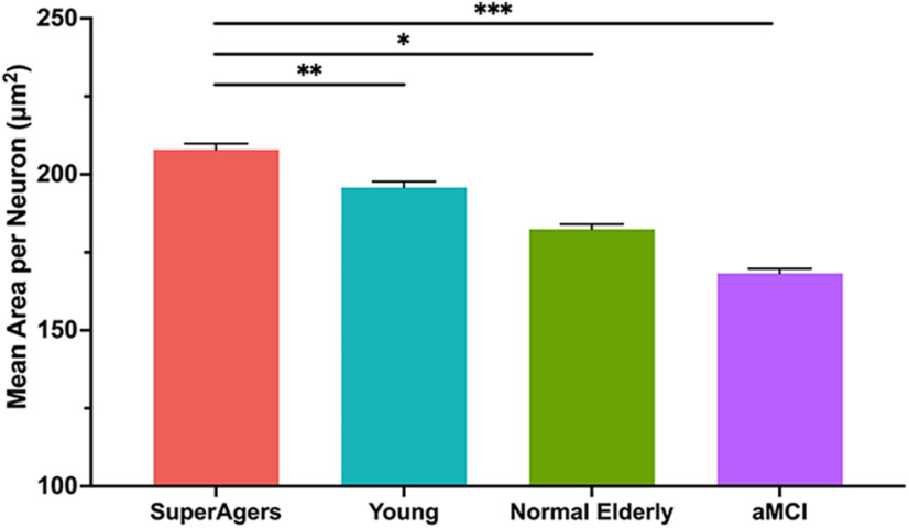
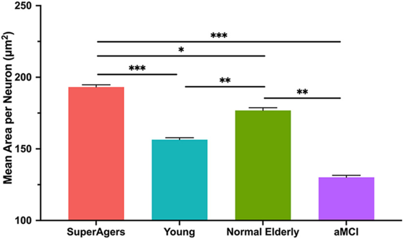
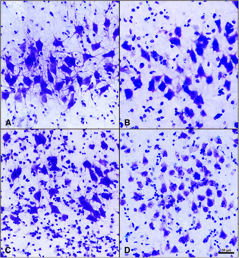
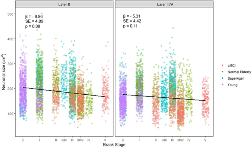
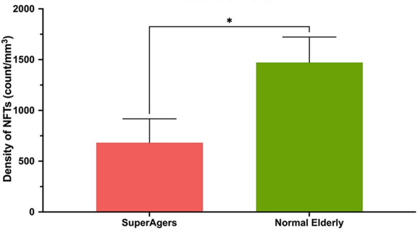
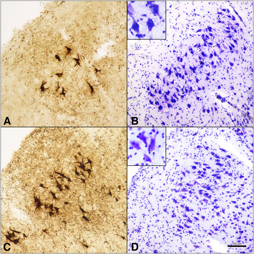
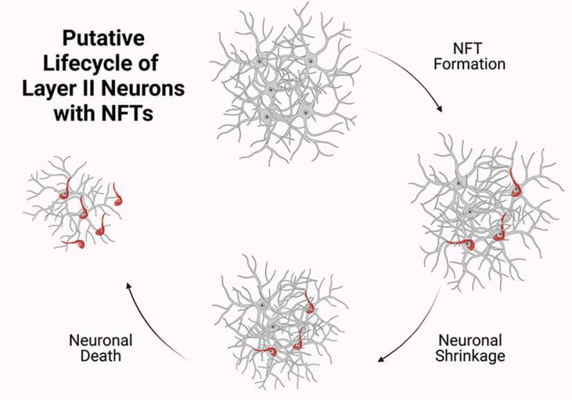
Average aging is associated with a gradual decline of memory capacity. SuperAgers are humans ≥80 years of age who show exceptional episodic memory at least as good as individuals 20-30 years their junior. This study investigated whether neuronal integrity in the entorhinal cortex (ERC), an area critical for memory and selectively vulnerable to neurofibrillary degeneration, differentiated SuperAgers from cognitively healthy younger individuals, cognitively average peers ("Normal Elderly"), and individuals with amnestic mild cognitive impairment. Postmortem sections of the ERC were stained with cresyl violet to visualize neurons and immunostained with mouse monoclonal antibody PHF-1 to visualize neurofibrillary tangles. The cross-sectional area (i.e., size) of layer II and layer III/V ERC neurons were quantified. Two-thirds of total participants were female. Unbiased stereology was used to quantitate tangles in a subgroup of SuperAgers and Normal Elderly. Linear mixed-effect models were used to determine differences across groups. Quantitative measurements found that the soma size of layer II ERC neurons in postmortem brain specimens were significantly larger in SuperAgers compared with all groups (p < 0.05)-including younger individuals 20-30 years their junior (p < 0.005). SuperAgers had significantly fewer stereologically quantified Alzheimer's disease-related neurofibrillary tangles in layer II ERC than Normal Elderly (p < 0.05). This difference in tangle burden in layer II between SuperAgers and Normal Elderly suggests that tangle-bearing neurons may be prone to shrinkage during aging. The finding that SuperAgers show ERC layer II neurons that are substantially larger even compared with individuals 20-30 years younger is remarkable, suggesting that layer II ERC integrity is a biological substrate of exceptional memory in old age.SIGNIFICANCE STATEMENT Average aging is associated with a gradual decline of memory. Previous research shows that an area critical for memory, the entorhinal cortex (ERC), is susceptible to the early formation of Alzheimer's disease neuropathology, even during average (or typical) trajectories of aging. The Northwestern University SuperAging Research Program studies unique individuals known as SuperAgers, individuals ≥80 years old who show exceptional memory that is at least as good as individuals 20-30 years their junior. In this study, we show that SuperAgers harbor larger, healthier neurons in the ERC compared with their cognitively average same-aged peers, those with amnestic mild cognitive impairment, and - remarkably - even compared with individuals 20-30 years younger. We conclude that larger ERC neurons are a biological signature of the SuperAging trajectory.

Keywords: Alzheimer's disease; SuperAging; entorhinal cortex; neurofibrillary tangles; neuronal integrity.


Figures

Figure 1.
Figure 1.
Mean cross-sectional area per neuron in layer II of the entorhinal cortex. Heights of bars represent the difference in mean cross-sectional area per squared micrometer of layer II neurons in the entorhinal cortex between SuperAgers (N = 6), Younger Controls (N = 6), Normal Elderly (N = 7), and individuals with aMCI (N = 5). An overall average of ∼1044 neurons (SD, 229) were measured per group. SuperAgers showed a significantly larger mean area of layer II ERC neurons compared with Normal Elderly, aMCI individuals, and Younger Controls. There were no significant differences in the mean area of layer II neurons between Normal Elderly, aMCI individuals, and Younger Controls. Statistical significance was assessed using a linear mixed-effect model. Error bars represent the SEM. *p < 0.05; **p < 0.01; *** p < 0.001.
Figure 2.
Figure 2.
Mean cross-sectional area per neuron in layer III/IV of the entorhinal cortex. Heights of bars represent the mean cross-sectional area per square micrometer of layer III/V neurons in the entorhinal cortex among SuperAgers (N = 6), Younger Controls (N = 6), Normal Elderly (N = 7), and individuals with aMCI (N = 5). An overall average of ∼1052 neurons (SD, 53) were measured per group. Mean area of layer III/V ERC neurons was significantly larger in SuperAgers compared with Normal Elderly, aMCI individuals, and Younger Controls. Normal Elderly also showed larger neuronal cross-sectional area in layer III/V of the ERC compared with aMCI individuals and Younger Controls. Statistical significance was assessed using a linear mixed-effect model. Error bars represent the SEM. *p < 0.05; **p < 0.01; ***p < 0.001.
Figure 4.
Figure 4.
Layer II neurons of the entorhinal cortex in SuperAgers, Younger Controls, Normal Elderly, and aMCI individuals. A–D, Layer II neurons in SuperAgers, Younger Controls, Normal Elderly, and individuals with aMCI visualized with cresyl violet staining. A, SA 2, a 90-year-old female SuperAger. B, YC 4, a 57-year-old female young control subject. C, NE 6, an 88-year-old male elderly control subject. D, aMCI 1, an 89-year-old female with aMCI. Scale bar: (in D) A–D, 50 µm. A–D, SuperAger (A) shows a significantly larger mean area of layer II ERC neurons compared with Younger Controls (B), Normal Elderly (C), and individual with aMCI (D).
Figure 3.
Figure 3.
Relationship between the cross-sectional area of layer II ERC neurons and Braak stage. Moderate evidence for a negative association of increasing Braak stage on neuronal cross-sectional area (in µm2) was found, yet it did not quite reach statistical significance in ERC layer II (p = 0.08; β = 6.86) and in ERC layer III/V (p = 0.11; β = 5.31). Statistical significance was assessed using a linear mixed-effect model. β, Regression coefficient; SE, SE of the regression coefficient.
Figure 5.
Figure 5.
Density of NFTs in layer II of the entorhinal cortex heights of bars represent the mean density per cubic millimeter of NFTs in layer II neurons in the entorhinal cortex between SuperAgers (N = 5) and Normal Elderly (N = 5). Density of NFTs in layer II ERC neurons was significantly smaller in SuperAgers (mean, 682; SEM, 235) compared with Normal Elderly (mean, 1472; SEM, 251; p < 0.05). This relationship held when controlled for Braak staging. Statistical significance was assessed using a Welch's t test. Error bars represent the SEM.
Figure 6.
Figure 6.
NFTs in layer II ERC neurons compared with neuronal size in SuperAgers and Normal Elderly. A, B, SA 2, a 90-year-old female SuperAger. C, D, NE 6, an 88-year-old male elderly control. SuperAger shows significantly fewer layer II NFTs (A) and larger layer II soma size (B) compared with Normal Elderly (C, D). Scale bar, 100 µm.
Figure 7.
Figure 7.
Putative life cycle of layer II neurons with NFTs. Results suggest that in stellate neurons in layer II of ERC, NFT formation leads to neuronal shrinkage. As previously understood, NFTs undergo biochemical changes and remain as “ghost tangles” after their associated neurons dies. Neuronal shrinkage may be an initial mechanism along the course toward age-related cognitive impairment. Created with BioRender.com.

References

    1. Albert MS, DeKosky ST, Dickson D, Dubois B, Feldman HH, Fox NC, Gamst A, Holtzman DM, Jagust WJ, Petersen RC, Snyder PJ, Carrillo MC, Thies B, Phelps CH (2011) The diagnosis of mild cognitive impairment due to Alzheimer's disease: recommendations from the National Institute on Aging-Alzheimer's Association workgroups on diagnostic guidelines for Alzheimer's disease. Alzheimers Dement 7:270–279. 10.1016/j.jalz.2011.03.008 - DOI - PMC - PubMed
    1. Balasubramanian AB, Kawas CH, Peltz CB, Brookmeyer R, Corrada MM (2012) Alzheimer disease pathology and longitudinal cognitive performance in the oldest-old with no dementia. Neurology 79:915–921. 10.1212/WNL.0b013e318266fc77 - DOI - PMC - PubMed
    1. Braak H, Braak E (1985) On areas of transition between entorhinal allocortex and temporal isocortex in the human brain. Normal morphology and lamina-specific pathology in Alzheimer's disease. Acta Neuropathol 68:325–332. 10.1007/BF00690836 - DOI - PubMed
    1. Braak H, Braak E (1991a) Morphological changes in the human cerebral cortex in dementia. J Hirnforsch 32:277–282. - PubMed
    1. Braak H, Braak E (1991b) Neuropathological stageing of Alzheimer-related changes. Acta Neuropathol 82:239–259. 10.1007/BF00308809 - DOI - PubMed

Publication types