. 2021 Aug 9:12:622737.
doi: 10.3389/fendo.2021.622737. eCollection 2021.

Exenatide Attenuates Obesity-Induced Mitochondrial Dysfunction by Activating SIRT1 in Renal Tubular Cells

Affiliations

Exenatide Attenuates Obesity-Induced Mitochondrial Dysfunction by Activating SIRT1 in Renal Tubular Cells

Yao Wang et al. Front Endocrinol (Lausanne). .

Abstract

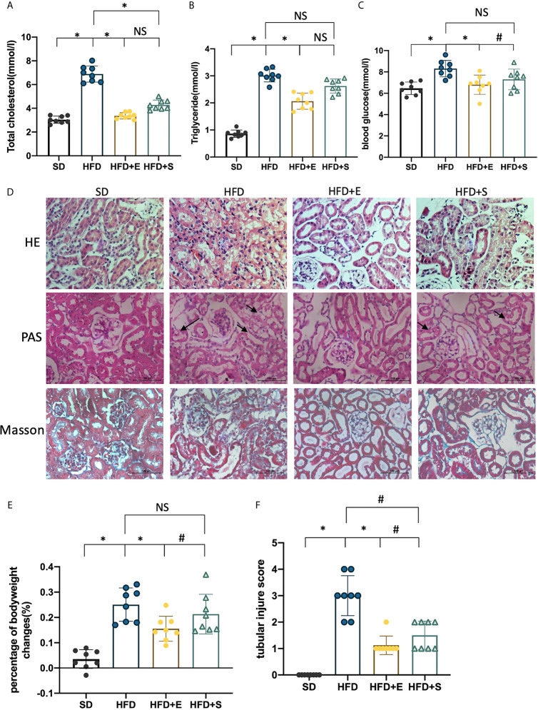
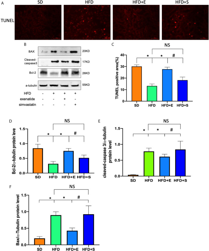
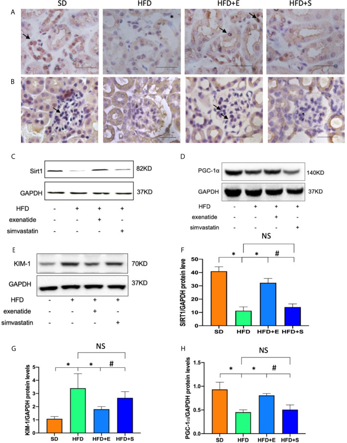
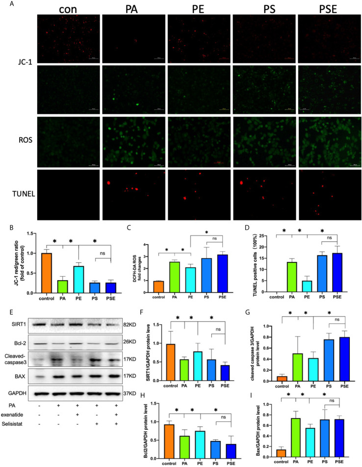
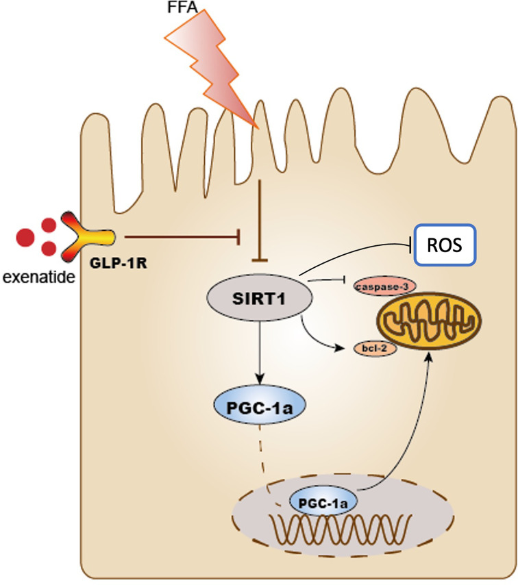
Saturated free fatty acid (FFA)-induced lipotoxicity plays an important role in obesity-induced kidney injury. Exenatide, a Glucagon-like peptide-1 receptor agonist(GLP-1RA), protects against high-fat diet (HFD)-induced kidney injury. The precise mechanism needs to be further explored. This study investigated whether exenatide protects against FFA-induced tubular epithelial cells (TECs) lipotoxicity and elucidated its underlying mechanisms. Here, we show that exenatide treatment reversed HFD induced TECs injuries, including TECs apoptosis and SIRT1 downregulation. The efficacy of exenatide was better than simvastatin. In palmitate (PA)-stimulated HK2 cells, exenatide treatment reversed the downregulation of SIRT1 and prevented an increase in reactive oxygen species (ROS) production, a decrease in mitochondrial membrane potential, and mitochondrial apoptosis. The renal-protective effects of exenatide on the generation of mitochondrial ROS and mitochondrial apoptosis were blocked by inhibiting SIRT1 activation. Collectively, these findings show that exenatide was superior to simvastatin in the treatment of obesity-TECs injuries, the mechanism is partially through SIRT1 restoration, which directly reverses mitochondrial dysfunction and apoptosis.

Keywords: SIRT1; glucagon-like peptide-1 receptor agonist; mitochondrial dysfunction; obesity; renal tubular cells.


Conflict of interest statement

The authors declare that the research was conducted in the absence of any commercial or financial relationships that could be construed as a potential conflict of interest.

Figures

Figure 1
Figure 1
Exenatide improves HFD-induced weight gain, dyslipidemia and renal tubular injury in mice. (A–C, E) Effects of exenatide and simvastatin on TC, TG, blood glucose and body weight changes in HFD mice. (D) H&E, PAS and Masson-trichrome staining of the kidney, arrowheads indicate vacuolation changes in TECs (original image magnification: ×200, bars =100 μm). (F) Quantification of tubular injury. Data are presented as the mean ± SD (n=8). SD, standard diet; HFD, high-fat diet; HFD+E, HFD with exenatide treatment; HFD+S, HFD with simvastatin treatment. *P < 0.01 versus HFD group. #p < 0.01 versus HFD+E group. NS no statistical difference between groups.
Figure 2
Figure 2
Exenatide attenuated renal tubular apoptosis in HFD mice. (A) Kidney apoptosis was detected by TUNEL staining and fluorescence microscopy (original image magnification: ×200, bars=100μm). (B) Western blots of cleaved caspase-3, BAX and Bcl-2 in renal lysates. (C) Quantification of TUNEL-positive cells in the kidney. (D–F) Relative protein expression of cleaved caspase-3, bax and Bcl-2. α-tubulin was used as a control for protein loading. Data are presented as the mean ± SD (n=3). *P < 0.01 versus HFD group. #p < 0.01 versus HFD+E group, NS no statistical difference between groups.
Figure 3
Figure 3
Exenatide restored HFD-repressed SIRT1 expression in mice. (A, B) Immunohistochemistry analysis of SIRT1 expression in kidney tissue (original image magnification: × 200, bars= 100 μm). (C) Western blots of SIRT1 in renal lysates. (D) Western blots of PGC-1α in renal lysates. (E) Western blots of KIM-1 in renal lysates. (F–H) Relative protein expression of SIRT1, PGC-1α, KIM-1. GAPDH was used as a control for protein loading. Data are presented as the mean ± SD (n=3). *P < 0.01 versus HFD group. #p < 0.01 versus HFD+E group. NS no statistical difference between groups.
Figure 4
Figure 4
Exenatide suppresses PA-induced apoptosis via SIRT1 restoration. HK2 cells were treated with 100 nM exenatide plus 10 µM Selisistat and incubated with PA (0.5 mM) for 24 h. (A) Intracellular mitochondrial membrane potential was examined by JC-1 staining, intracellular ROS was examined by DCFH-DA staining and apoptosis was examined by TUNEL staining, all of which were detected by fluorescence microscopy (original image magnification: ×200, bars=100 μm). (B–D) Calculated mean fluorescence intensity of JC-1 and DCFH-DA staining and mean percentages of TUNEL staining-positive cells. (E) Western blots of SIRT1, cleaved caspase-3, Bcl-2 and Bax in cell lysates. (F–I) Relative protein expression of SIRT1, cleaved caspase-3, Bcl-2 and Bax. GAPDH was used as a control for protein loading. CON, control; PA, palmitate; PE, treat with PA and exenatide (100 nM); PS, treat with PA and Selisistat (10 µM); PSE, treat with PA, exenatide (100 nM) and Selisistat (10 µM). Data are presented as the mean ± SD (n=3). *P < 0.01 versus PA group. NS no statistical difference between groups.
Figure 5
Figure 5
Schematic diagram of this study. Exenatide protects HFD induced tubular injury through the preservation of SIRT1. SIRT1 positively regulates the expression of PGC-1α. HFD reduced the expression of SIRT1 and PGC-1α subsequently leads to mitochondria-derived ROS production and mitochondrial apoptosis, which results in the development of renal tubular injury. Treatment with exenatide increased the expression of SIRT1 and PGC-1α and then prevented obesity-induced mitochondrial dysfunction. As a result, exenatide suppressed mitochondria-derived ROS production, alleviated mitochondrial dysfunction, reduced cell apoptosis, and protected against HFD-induced renal tubular injury.

References

    1. Biener A, Cawley J, Meyerhoefer C. The Impact of Obesity on Medical Care Costs and Labor Market Outcomes in the US. Clin Chem (2018) 64(1):108–17. 10.1373/clinchem.2017.272450 - DOI - PubMed
    1. Muntner P, Coresh J, Smith JC, Eckfeldt J, Klag MJ. Plasma Lipids and Risk of Developing Renal Dysfunction: The Atherosclerosis Risk in Communities Study. Kidney Int (2000) 58(1):293–301. 10.1046/j.1523-1755.2000.00165.x - DOI - PubMed
    1. Spalding KL, Bernard S, Näslund E, Salehpour M, Possnert G, Appelsved L, et al. . Impact of Fat Mass and Distribution on Lipid Turnover in Human Adipose Tissue. Nat Commun (2017) 8:15253. 10.1038/ncomms15253 - DOI - PMC - PubMed
    1. Gai Z, Wang T, Visentin M, Kullak-Ublick GA, Fu X, Wang Z. Lipid Accumulation and Chronic Kidney Disease. Nutrients (2019) 11(4):722. 10.3390/nu11040722 - DOI - PMC - PubMed
    1. Nuffer WA, Trujillo JM. Liraglutide: A New Option for the Treatment of Obesity. Pharmacotherapy (2015) 35(10):926–34. 10.1002/phar.1639 - DOI - PubMed

Publication types

MeSH terms