. 2020 Aug 21;295(34):11971-11981.
doi: 10.1074/jbc.RA120.012934. Epub 2020 Jun 22.

MtcB, a member of the MttB superfamily from the human gut acetogen Eubacterium limosum, is a cobalamin-dependent carnitine demethylase

Affiliations

MtcB, a member of the MttB superfamily from the human gut acetogen Eubacterium limosum, is a cobalamin-dependent carnitine demethylase

Duncan J Kountz et al. J Biol Chem. .

Abstract

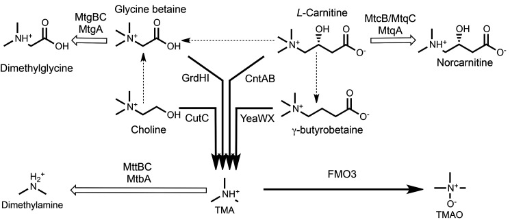
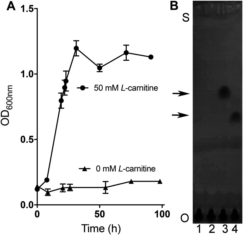
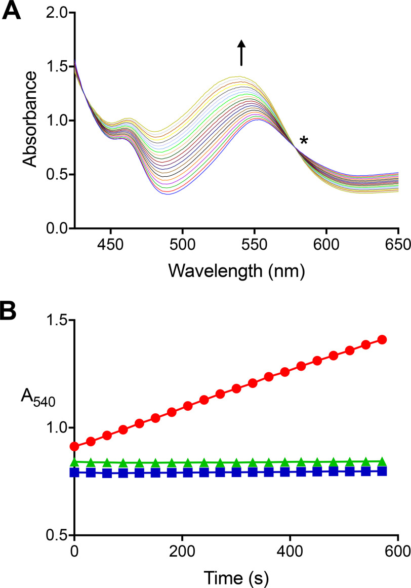
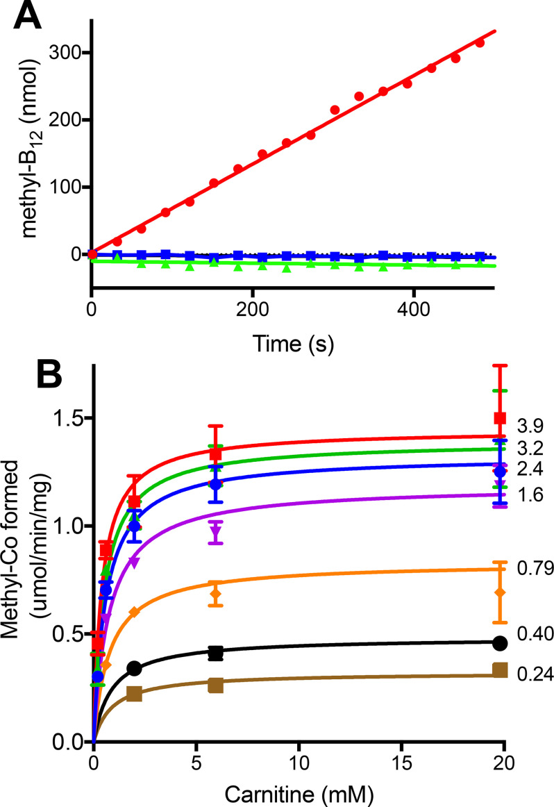
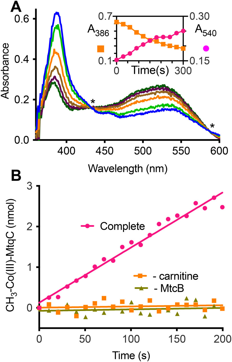
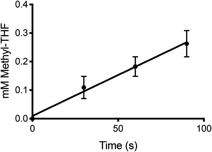
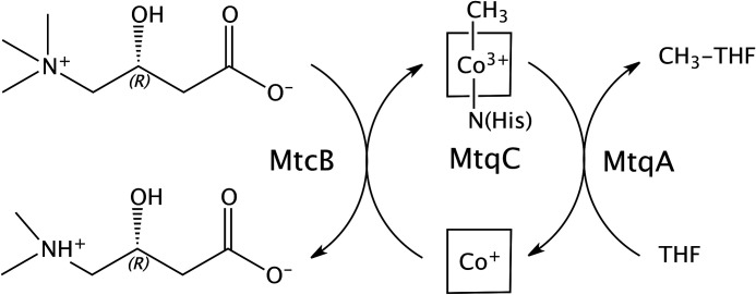
The trimethylamine methyltransferase MttB is the first described member of a superfamily comprising thousands of microbial proteins. Most members of the MttB superfamily are encoded by genes that lack the codon for pyrrolysine characteristic of trimethylamine methyltransferases, raising questions about the activities of these proteins. The superfamily member MtcB is found in the human intestinal isolate Eubacterium limosum ATCC 8486, an acetogen that can grow by demethylation of l-carnitine. Here, we demonstrate that MtcB catalyzes l-carnitine demethylation. When growing on l-carnitine, E. limosum excreted the unusual biological product norcarnitine as well as acetate, butyrate, and caproate. Cellular extracts of E. limosum grown on l-carnitine, but not lactate, methylated cob-(I)alamin or tetrahydrofolate using l-carnitine as methyl donor. MtcB, along with the corrinoid protein MtqC and the methylcorrinoid:tetrahydrofolate methyltransferase MtqA, were much more abundant in E. limosum cells grown on l-carnitine than on lactate. Recombinant MtcB methylates either cob(I)alamin or Co(I)-MtqC in the presence of l-carnitine and, to a much lesser extent, γ-butyrobetaine. Other quaternary amines were not substrates. Recombinant MtcB, MtqC, and MtqA methylated tetrahydrofolate via l-carnitine, forming a key intermediate in the acetogenic Wood-Ljungdahl pathway. To our knowledge, MtcB methylation of cobalamin or Co(I)-MtqC represents the first described mechanism of biological l-carnitine demethylation. The conversion of l-carnitine and its derivative γ-butyrobetaine to trimethylamine by the gut microbiome has been linked to cardiovascular disease. The activities of MtcB and related proteins in E. limosum might demethylate proatherogenic quaternary amines and contribute to the perceived health benefits of this human gut symbiont.

Keywords: acetogenesis; bacterial metabolism; carnitine; cobalamin; energy metabolism; enzyme catalysis; folate; microbiology; microbiome; one-carbon metabolism.


Conflict of interest statement

Conflict of interest—The authors declare that they have no conflicts of interest with the contents of this article.

Figures

Figure 1.
Figure 1.
Microbial quaternary amine metabolism emphasizing reactions known to lead toward (black arrows) or away from (white arrows) TMA and TMAO production as described in the introduction. Pathways with two or more enzymes converting one quaternary amine into another are indicated by dashed arrows. The demethylation of l-carnitine to form norcarnitine is demonstrated for the first time here. Notably, members of the MttB superfamily are involved in all reactions known to lead away from TMA production.
Figure 2.
Figure 2.
E. limosum cultures accumulate norcarnitine during growth on l-carnitine. A, growth dependence of E. limosum upon the addition of 50 mm l-carnitine in LS medium supplemented with 0.1% yeast extract, 0.2% casamino acids, and 10 mm sodium acetate. Error bars, S.D. from an average of three different cultures. B, TLC of culture supernatants demonstrate that l-carnitine is consumed and norcarnitine is produced during growth of E. limosum. Cultures were inoculated into medium containing either no substrate (lanes 1 and 2) or 50 mm l-carnitine (lanes 3 and 4) as described for A. Samples were removed just after inoculation (lanes 1 and 3) and again 24 h after growth had ceased in the l-carnitine–supplemented culture (lanes 2 and 4). Samples were applied at the origin (O) and developed until solvent reached near the top of the plate (S), and then the plates stained were stained with bromocresol green. The top and bottom arrows indicate the migration positions of l-carnitine and norcarnitine standards, respectively (not shown).
Figure 3.
Figure 3.
A carnitine:cob(I)alamin methyltransferase activity is present in extracts of carnitine-grown, but not lactate-grown, E. limosum. A, UV-visible spectra were collected every 30 s during a single carnitine:cob(I)alamin methyltransferase reaction initiated by the addition of extract from carnitine-grown cells. The arrow indicates the direction of increased absorbance at 540 nm with time indicative of methylcob(IIII)alamin formation. The sharp isosbestic point (*) at 578 nm indicates that other cobalamin species did not accumulate appreciably during the reaction. The complete UV-visible spectrum is not shown due to the intense absorbance of Ti(III)citrate and cob(I)alamin below 425 nm. B, absorbance changes at 540 nm (●) in a single reaction containing l-carnitine, cob(I)alamin, and extract of l-carnitine–grown cells. No reaction was observed if l-carnitine was omitted (■) or if l-carnitine–grown cell extract was replaced with lactate-grown cell extract (▴).
Figure 4.
Figure 4.
Purified recombinant MtcB-dependent methylation of cob(I)alamin with l-carnitine. A, methylation was followed by the increase at 540 nm in the complete reaction (●) as described under “Experimental procedures.” Methylation was not observed in the absence of MtcB (▴) or l-carnitine (■). B, kinetic analysis of MtcB in which the l-carnitine concentration was varied at different set cob(I)alamin concentrations. The numbers beside each curve are the millimolar concentration of cob(I)amin used for that data set. Each point is the average of three determinations. Error bars, S.D. Some error bars are not visible due to size of data point markers.
Figure 5.
Figure 5.
MtcB methylates MtqC with l-carnitine. A, UV-visible spectral changes associated with methylation of 50 μm Co(I)-MtqC by MtcB with 40 mm l-carnitine. Spectra shown were taken immediately after initiation (light blue spectrum) of a single reaction and every 60 s thereafter until termination (dark green spectrum). Asterisks indicate locations of isosbestic points for the Co(I)-MtqC and methyl-Co(III)-MtqC spectra. Inset, the progress of the reaction was monitored by increase in absorbance at 540 nm accompanied by the decrease in absorbance at 386 nm. B, methyl-Co(III)-MtqC formation during a complete reaction described under “Experimental procedures” was determined using the calculated extinction coefficient for methyl-Co(III)-MtqC. MtqC methylation was not detectable in the absence of l-carnitine or MtcB. Each curve represents a single representative reaction.
Figure 6.
Figure 6.
In vitro reconstitution of l-carnitine:THF methyl transfer using recombinant MtcB, MtqC, and MtqA. Each time point is the average from three independent reactions and determinations. Error bars, S.D.
Figure 7.
Figure 7.
Schematic of l-carnitine demethylation and methylation of THF by MtcB, MtqC, and MtqA from E. limosum. The ligation of the four nitrogens of the corrin tetrapyrrole ring bound to MtqC is indicated by the square surrounding the cobalt. In the methyl-Co(III) form, the lower axial ligand is a nitrogen provided by a histidine residue that is highly conserved in corrinoid-binding proteins and domains involved in methyltransferase reactions (36).

Comment in

References

    1. Ferguson D. J. Jr., and Krzycki J. A. (1997) Reconstitution of trimethylamine-dependent coenzyme M methylation with the trimethylamine corrinoid protein and the isozymes of methyltransferase II from Methanosarcina barkeri. J. Bacteriol. 179, 846–852 10.1128/jb.179.3.846-852.1997 - DOI - PMC - PubMed
    1. Krzycki J. A. (2004) Function of genetically encoded pyrrolysine in corrinoid-dependent methylamine methyltransferases. Curr. Opin. Chem. Biol. 8, 484–491 10.1016/j.cbpa.2004.08.012 - DOI - PubMed
    1. Paul L., Ferguson D. J., and Krzycki J. A. (2000) The trimethylamine methyltransferase gene and multiple dimethylamine methyltransferase genes of Methanosarcina barkeri contain in-frame and read-through amber codons. J. Bacteriol. 182, 2520–2529 10.1128/jb.182.9.2520-2529.2000 - DOI - PMC - PubMed
    1. Soares J. A., Zhang L., Pitsch R. L., Kleinholz N. M., Jones R. B., Wolff J. J., Amster J., Green-Church K. B., and Krzycki J. A. (2005) The residue mass of l-pyrrolysine in three distinct methylamine methyltransferases. J. Biol. Chem. 280, 36962–36969 10.1074/jbc.M506402200 - DOI - PubMed
    1. Ticak T., Kountz D. J., Girosky K. E., Krzycki J. A., and Ferguson D. J. Jr. (2014) A nonpyrrolysine member of the widely distributed trimethylamine methyltransferase family is a glycine betaine methyltransferase. Proc. Natl. Acad. Sci. U. S. A. 111, E4668–E4676 10.1073/pnas.1409642111 - DOI - PMC - PubMed

Publication types

MeSH terms

Supplementary concepts