. 2020 May 7;5(9):e132055.
doi: 10.1172/jci.insight.132055.

A human-origin probiotic cocktail ameliorates aging-related leaky gut and inflammation via modulating the microbiota/taurine/tight junction axis

Affiliations

A human-origin probiotic cocktail ameliorates aging-related leaky gut and inflammation via modulating the microbiota/taurine/tight junction axis

Shokouh Ahmadi et al. JCI Insight. .

Abstract

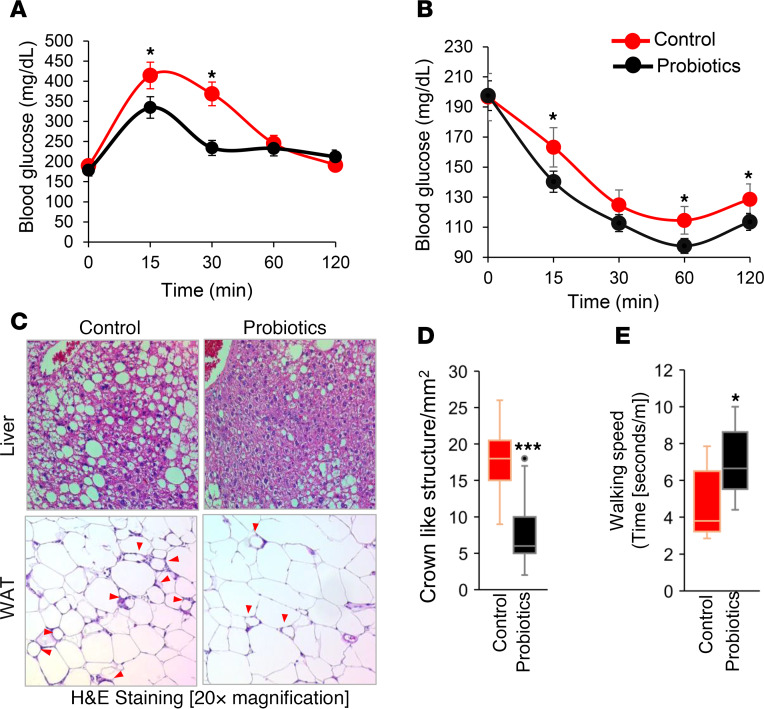
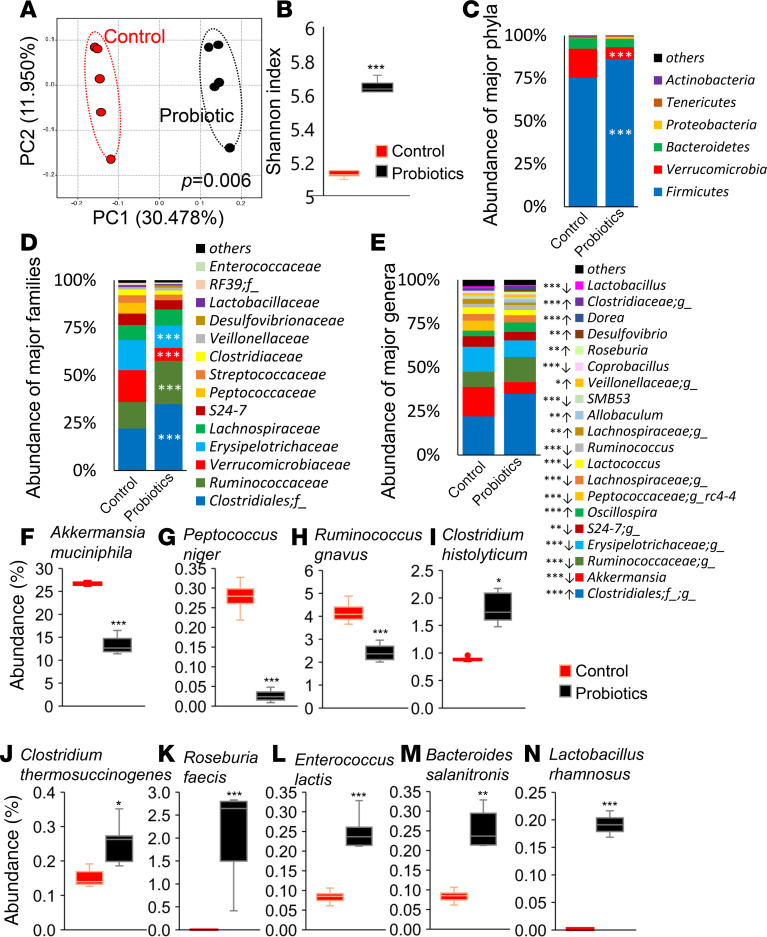
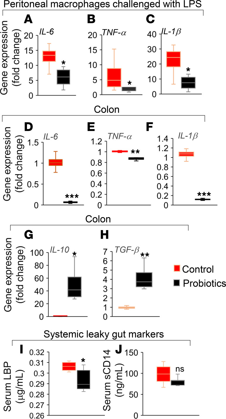
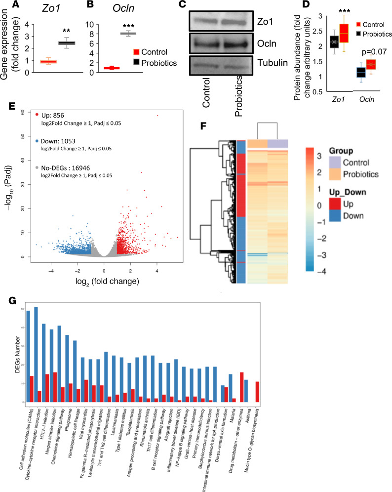
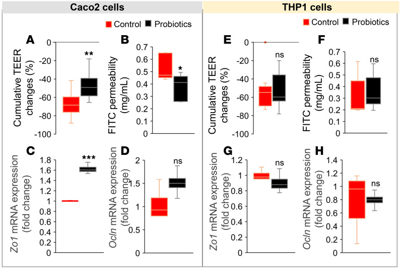
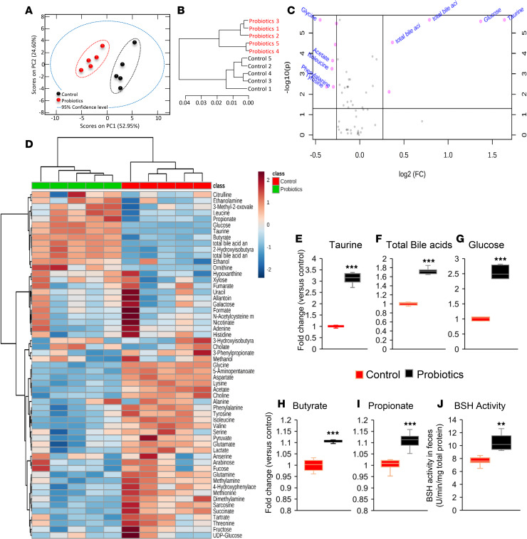
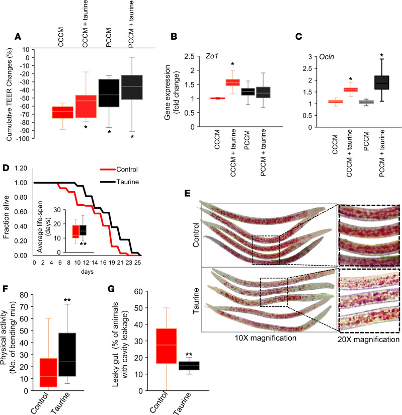
Inflammation is a major risk factor of morbidity and mortality in older adults. Although its precise etiology is unknown, low-grade inflammation in older adults is commonly associated with increased intestinal epithelial permeability (leaky gut) and abnormal (dysbiotic) gut microbiota. The increasing older population and lack of treatments to reduce aging-related microbiota dysbiosis, leaky gut, and inflammation culminates in a rise in aging-related comorbidities, constituting a significant public health concern. Here, we demonstrate that a human-origin probiotic cocktail containing 5 Lactobacillus and 5 Enterococcus strains isolated from healthy infant gut prevented high-fat diet-induced (HFD-induced) microbiota dysbiosis, leaky gut, inflammation, metabolic dysfunctions, and physical function decline in older mice. Probiotic-modulated gut microbiota primarily reduced leaky gut by increasing tight junctions, which in turn reduced inflammation. Mechanistically, probiotics modulated microbiota in a way to increase bile salt hydrolase activity, which in turn increased taurine abundance in the gut that stimulated tight junctions and suppressed gut leakiness. Furthermore, in Caenorhabditis elegans, taurine increased life span, reduced adiposity and leaky gut, and enhanced physical function. The results suggest that such probiotic therapies could prevent or treat aging-related leaky gut and inflammation in the elderly.

Keywords: Gastroenterology; Glucose metabolism; Innate immunity; Macrophages; Microbiology.


Conflict of interest statement

Conflict of interest: The authors have declared that no conflict of interest exists.

Figures

Figure 1
Figure 1. Probiotics feeding prevents HFD-induced metabolic dysfunctions in older mice.
(A and B) Ten weeks of probiotics improved glucose tolerance and enhanced insulin sensitivity in older obese mice, measured by oral glucose tolerance test (A) and insulin tolerance test (B) (n = 6 in control and n = 8 in probiotics groups; *P < 0.05, 2-way ANOVA). (C) Representative images of H&E staining of liver (upper panels) showing reduced fat accumulation and white adipose tissue (WAT; lower panels) showing reduced adipocyte size, along with reduced inflammation (indicated by crown-like structures; red arrows) in probiotics fed mice (n = 8) compared with their controls (n = 6). (D) Crown-like structures are graphed. (E) Probiotic-fed older obese mice (n = 8) exhibited higher physical function presented as walking speed compared with their age- and sex-matched HFD-fed controls (n = 6). Values are mean of n = 6–8 mice in each group, and data are shown as mean ± SEM. *P < 0.05, and ***P < 0.001 by 2-way ANOVA with Bonferroni’s correction (A and B) and Student’s t test (D and E).
Figure 2
Figure 2. Probiotic therapy beneficially modulates gut microbiome in older obese mice.
(A–E) Gut microbiome signature in terms of β-diversity (A), α-diversity (Shannon index) (B), and abundance of major phyla (C), families (D), and genera (E) were significantly changed in probiotic-treated HFD-fed older mice (n = 5) compared with their controls (n = 5). (FN) Specifically, probiotic therapy decreased Akkermansia muciniphila (F), Peptococcus niger (G), and Ruminicoccus gnavus (H) and increased C. histolyticum (I), C. thermosuccinogenes (J), Roseburia faecis (K), Enterococcus lactis (L), Bacteroides salanitronis (M), and Lactobacillus rhamnosus (N). Values are mean of n = 5 in each group, and data are shown as mean ± SEM. *P < 0.05; **P < 0.01, and ***P < 0.001 by PERMANOVA (A), unpaired 2-tailed Student’s t test (F–N), and 1-way ANOVA (B–E).
Figure 3
Figure 3. Probiotics treatment reduces inflammation in peritoneal macrophages and intestine of older obese mice.
(A–C) LPS-induced inflammatory response in terms of mRNA expression of IL-6 (A), TNF-α (B), and IL-1β (C) was reduced in primary macrophages isolated from peritoneal cavity of older HFD-fed mice treated with probiotics (n = 8) compared with their controls (n = 6). (D–H) In addition, the expression of proinflammatory markers such as IL-6 (D), TNF-α (E), and IL-1β (F) were decreased, while antiinflammatory genes like IL-10 (G) and TGF-β (H) mRNA expressions were increased in the colon of probiotic-fed older obese mice (n = 8) compared with their controls (n = 6). (I and J) In addition, systemic leaky gut markers such as LPS binding protein (LBP) (I) and soluble CD14 (J) were reduced in the serum of probiotic-fed older obese mice (n = 7) compared with their controls (n = 6). Values are mean of n = 6–8 in each group, and data are shown as mean ± SEM. *P < 0.05; **P < 0.01, ***P < 0.001 by Student t-test (A–J).
Figure 4
Figure 4. Probiotics treatment increases expression of tight junctions in the intestine of older obese mice.
(A and B) The mRNA expression of tight junction proteins like Zonulin-1 (Zo1) (A) and Occludin (Ocln) (B) were significantly increased in colon of probiotics fed older mice (n = 8) compared with their controls (n = 6). (C and D) Western blot analysis shows that Zo1 protein expression was significantly increased, while Ocln showed marginal increase in the colon tissues of probiotic-treated older mice (n = 7) compared with their controls (n = 6). (E and F) Global gene expression using RNAseq analysis revealed that probiotic feeding significantly increased around 856 genes while it decreased 1053 genes that were distinctly clustered in the probiotic-treated (n = 7) group versus controls (n = 6). (G) Pathway analysis of deferentially expressed genes (DEGs) shows that cell adhesion and cytokine (immune) pathways were more affected by probiotics treatment compared with their controls. Values are mean of n = 6–7 each group, and data are shown as mean ± SEM. **P < 0.01 and ***P < 0.001. Student t test (A, B, D) and random forest analysis (E) were used, as well as hierarchical clustering between samples using hclust, with diagrams drawn with ggplot2 (F) and differential expression of genes (DEGs) (G) were completed using R programs.
Figure 5
Figure 5. Probiotics primarily act on intestinal tight junctions versus immune cells.
(A–C) Treatment of Caco-2 cell monolayers (cultured with THP1 cells) with cecal conditioned media (CCM) from probiotic-fed mice shows significantly less changes in transepithelial electrical resistance (TEER) (A) with reduced FITC-dextran (4 kDa) (B) and increased Zo1 (C) and Ocln (D) mRNA and protein expressions compared with control CCM–treated cells. (E–H) No significant changes were observed in these measures when cocultured THP-1 cells were treated with CCM prepared from probiotics and control mouse cecal contents. Values are mean of 2–3 repeated experiments done in triplicate, and CCM was prepared from cecal contents of n = 6 controls and n = 7 probiotic-treated mice. Data are shown as mean ± SEM. **P < 0.01 ***P < 0.001 by ANOVA with Bonferroni’s corrections and Student’s t test.
Figure 6
Figure 6. Probiotic therapy modulates gut metabolome and increases taurine production by enhancing bile salt hydrolase (BSH) activity in the gut of older HFD-fed mice.
(A–C) Untargeted-unbiased metabolomics analyses show that the production of distinct metabolites shown by principal component analysis (PCA) (A), group clustering (B), and fold change abundance enrichment (C) were significantly changed in probiotic-fed old mice (n = 5) compared with their controls (n = 5). (D) Similarly, metabolite heatmap shows differential clusters that are significantly changed in probiotic-fed older mice feces compared with controls. (E–I) Abundance of taurine (E) and total bile acids (F), glucose (G), butyrate (H) and propionate (I) was significantly increased in the feces of probiotic-fed older mice (n = 5) compared with their controls (n = 5). (H) Probiotics feeding significantly enhanced bile salt hydrolase (BSH) activity in the gut of older mice. Values are mean of n = 5 control and n = 5 probiotics group, and data are shown as mean ± SEM. **P < 0.01 and ***P < 0.001. PLS-tool box in MatLab (A and B) and Welch t-test (E–J) were used.
Figure 7
Figure 7. Taurine significantly decreases epithelial permeability, increases tight junction proteins, reduces fat accumulation, and reduces gut leakiness in C. elegans.
(A–C) Taurine supplementation in cecal conditioned media (CCM) in control significantly reduced changes in TEER of Caco-2 cells monolayer (A) and significantly increased expression of tight junction protein Zo1 (B) and Ocln (C) expression. Values are mean of 2–3 repeated experiments done in triplicate. (D) Taurine treatment increased the life span of C. elegans. Kaplan-Meier curves were generated using survival data in days for each worm (n = 150–195 worms in each group), and statistical significance was calculated using the log-rank test. (E) Composite image of multiple worms. (F and G) Taurine treatment also increased physical activity (F) and intestinal permeability (leaky gut) using Smurf assay (G) in n = 120-150 worms in each group. Values are mean of 2–3 trials of cell culture experiments done in triplicate (A–C) and n = 120-195 worms (D–G). Data are shown as mean ± SEM. *P < 0.05, **P < 0.01 by 2-way ANOVA with Bonferroni’s corrections (A), Student’s t test (B, C, F, G), and log-rank (D).

References

    1. Harper S. Economic and social implications of aging societies. Science. 2014;346(6209):587–591. doi: 10.1126/science.1254405. - DOI - PubMed
    1. Sato Y, Yanagita M. The unprecedented era of aging. Inflamm Regen. 2019;39:15. - PMC - PubMed
    1. Nagpal R, et al. Gut microbiome and aging: Physiological and mechanistic insights. Nutr Healthy Aging. 2018;4(4):267–285. doi: 10.3233/NHA-170030. - DOI - PMC - PubMed
    1. Chung HY, et al. Molecular inflammation: underpinnings of aging and age-related diseases. Ageing Res Rev. 2009;8(1):18–30. doi: 10.1016/j.arr.2008.07.002. - DOI - PMC - PubMed
    1. Kavanagh K, Hsu FC, Davis AT, Kritchevsky SB, Rejeski WJ, Kim S. Biomarkers of leaky gut are related to inflammation and reduced physical function in older adults with cardiometabolic disease and mobility limitations. Geroscience. 2019;41(6):923–933. doi: 10.1007/s11357-019-00112-z. - DOI - PMC - PubMed

Publication types