. 2019 Oct;574(7776):57-62.
doi: 10.1038/s41586-019-1570-z. Epub 2019 Sep 18.

FPR1 is the plague receptor on host immune cells

Affiliations

FPR1 is the plague receptor on host immune cells

Patrick Osei-Owusu et al. Nature. 2019 Oct.

Abstract

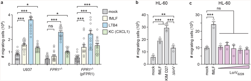
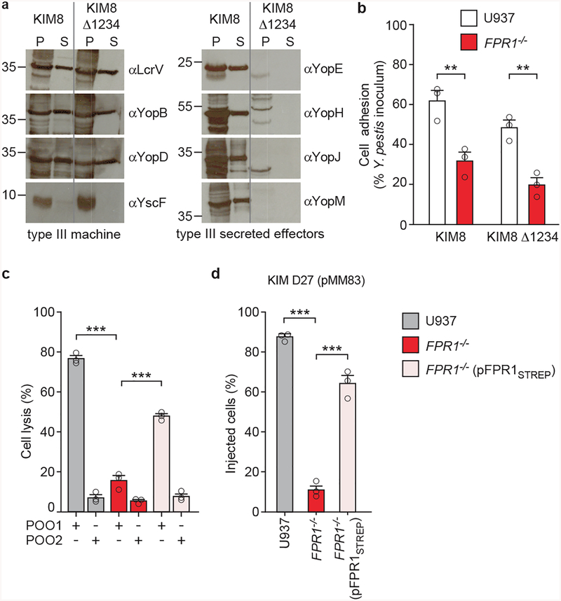
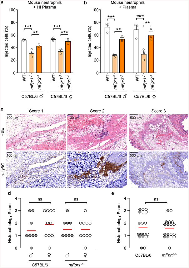
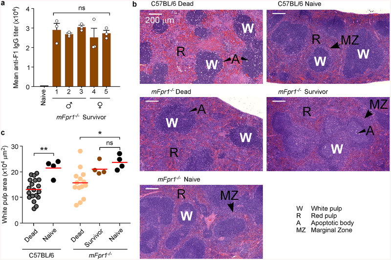
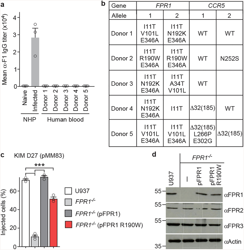
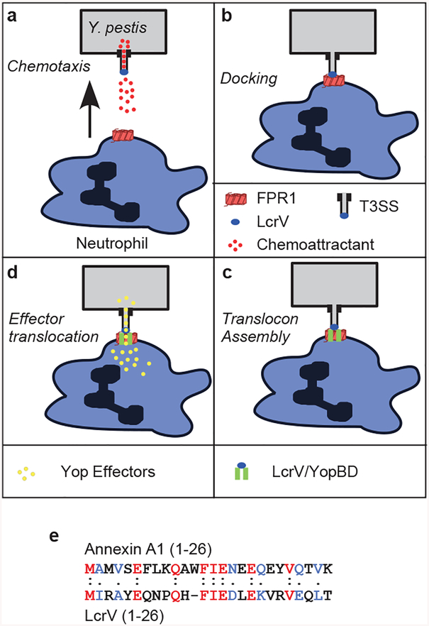
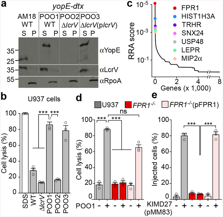
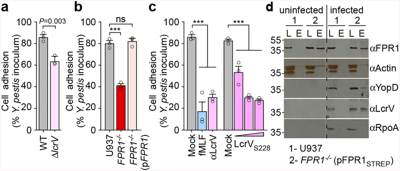
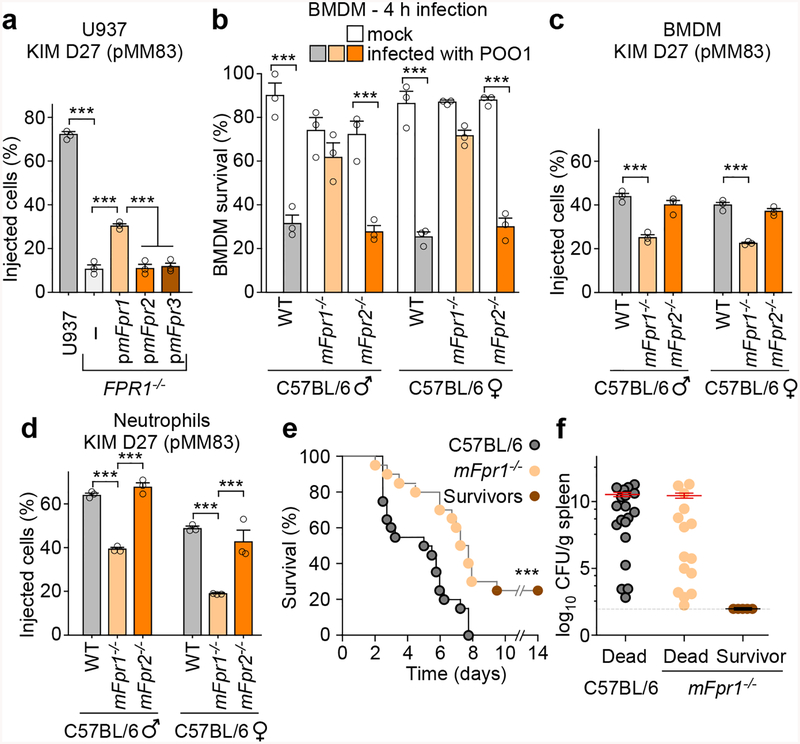
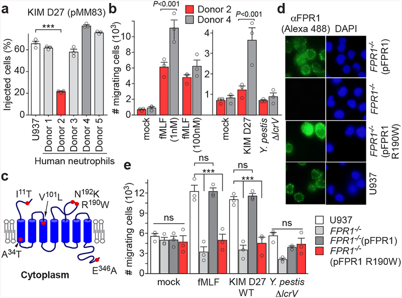
The causative agent of plague, Yersinia pestis, uses a type III secretion system to selectively destroy immune cells in humans, thus enabling Y. pestis to reproduce in the bloodstream and be transmitted to new hosts through fleabites. The host factors that are responsible for the selective destruction of immune cells by plague bacteria are unknown. Here we show that LcrV, the needle cap protein of the Y. pestis type III secretion system, binds to the N-formylpeptide receptor (FPR1) on human immune cells to promote the translocation of bacterial effectors. Plague infection in mice is characterized by high mortality; however, Fpr1-deficient mice have increased survival and antibody responses that are protective against plague. We identified FPR1R190W as a candidate resistance allele in humans that protects neutrophils from destruction by the Y. pestis type III secretion system. Thus, FPR1 is a plague receptor on immune cells in both humans and mice, and its absence or mutation provides protection against Y. pestis. Furthermore, plague selection of FPR1 alleles appears to have shaped human immune responses towards other infectious diseases and malignant neoplasms.


Conflict of interest statement

The authors declare no competing financial interests.

Figures

Extended Data Figure 1 |
Extended Data Figure 1 |. Generation of FPR1−/− U937 cells using CRISPR-Cas9 and genetic complementation.
a, Sequencing results for alleles cloned from CRISPR-Cas9 derived FPR1−/− U937 macrophages. b, Immunoblot analysis for the production of FPR1, FPR2 and FPR3 in U937 macrophages, CRISPR-Cas9 derived FPR1−/− cells and FPR1−/− cells transfected with pFPR1. Numbers to the left of blots indicate migration of molecular weight markers. One of three repeats is shown (a, b).
Extended Data Figure 2 |
Extended Data Figure 2 |. Contribution of CCR5 to Yersinia spp. intoxication and injection by the T3SS.
a, Sequencing results for the alleles of two CCR5−/− U937 isolates obtained using CRISPR-Cas9 mutagenesis. b, Cell survival following incubation with strains Y. pestis KIM D27 and POO1 was measured using the Trypan blue exclusion assay; error bars represent the s.e.m. (n = 4 biological replicates). T3SS injection into U937, FPR1−/−, and CCR5−/− by Y. pestis KIM D27 (c) and Y. pseudotuberculosis YPIII carrying pMM83 (YopM-Bla) (d); error bars represent the s.e.m. (n = 3 biological replicates). One of three repeats is shown (b-d). Statistical analysis was performed using one-way ANOVA with Bonferroni Correction: ***, P <0.001; **, P <0.01.
Extended Data Figure 3 |
Extended Data Figure 3 |. Antibodies and ligands of N-formylpeptide receptor (FPR1) inhibit Y. pestis type III secretion into human neutrophils.
Human neutrophils were stained with the β-lactamase substrate CCF2-AM, infected with wild-type Y. pestis KIM D27 (WT, pMM83) or KLD29 (ΔlcrV, pMM83) variant defective for the type III secretion system (T3SS), and analyzed for (a) blue fluorescence (YopM-Bla translocation into neutrophils and CCF2-AM cleavage) or (b) green fluorescence to derive the percent of stained cells injected with T3SS effector. c, Inhibition of Y. pestis T3SS into human neutrophils by monoclonal (αFPR1m) and polyclonal antibodies against FPR1 (αFPR1p), bacterial LcrV, N-formylpeptide (fMLF), annexin A1 peptide, staphylococcal CHIPS and cyclosporin H. d, Differential interference contrast (DIC) and fluorescence microscopy of mock or fMLF treated differentiated U937 cells infected with green-fluorescent Y. pestis KIM D27 (pEGFP) and stained with F1-specific antibody (red) to reveal extracellular bacteria in merged images of mock, but not in merged images of fMLF-treated cells. Orange and green arrows point to extra- and intracellular bacteria, respectively. e, Antibodies and ligands of FPR1 inhibit Y. pestis T3SS of YopM-Bla into U937 macrophages. One of three repeats is shown (a-e). Error bars represent the s.e.m. (n = 3 biological replicates) (c, e). One-way ANOVA with Bonferroni Correction was used to identify significant differences (c, e): ***, P <0.001; **, P <0.01.
Extended Data Figure 4 |
Extended Data Figure 4 |. Screening 45 monoclonal antibodies for inhibition of Y. pestis T3SS into human neutrophils.
Human neutrophils were stained with the β-lactamase substrate CCF2-AM (green fluorescence), and infected with wild-type Y. pestis KIM D27 (pMM83). Translocation of YopM-Bla into neutrophils results in CCF2-AM cleavage (blue fluorescence) allowing for quantification of percent of stained cells injected with T3SS effector in the absence (Mock) or presence of specific monoclonal antibody. Error bars represent the s.e.m. (n = 3 biological replicates). A representative of three independent experiments is shown.
Extended Data Figure 5 |
Extended Data Figure 5 |. FPR1−/− cells migrate towards chemoattractants other than formylated peptides and differentiated HL-60 cells migrate towards Y. pestis.
a, Numbers of migrating immune cells were quantified in a transwell assay primed with mock, 10 nM fMLF, 10 nM LTB4 or 100 ng ml−1 KC (CXCL1) for U937, FPR1−/− and FPR1−/− (pFPR1) cells. b, Numbers of migrating HL-60 cells were quantified in a transwell assay primed with mock, Y. pestis KIM D27 (WT) or KLD29 (ΔlcrV) (107 CFU/ml). Chemotaxis toward fMLF is shown as a control. c, Increasing concentrations of LcrVS228 (10−1–103 ng/ml) were added to the transwell assay and number of migrating HL-60 cells recorded. Error bars represent the s.e.m. (n = 3 biological replicates); one-way ANOVA with Bonferroni Correction was used to identify significant differences: ***, P<0.001; **, P <0.01; *, P<0.05; ns, not significant. A representative of three independent experiments is shown.
Extended Data Figure 6 |
Extended Data Figure 6 |. Adhesion of Y. pestis to human macrophages does not require effector Yops and loss of FPR1 in human macrophages can be complemented with pFPR1STREP.
a, Y. pestis KIM 8 and its variant KIM 8 Δ1234 (Yop-less) were grown at 37°C in TMH to induce T3SS. Cultures were centrifuged to separate the supernatant (S) from the bacterial pellet (P) and extracts were analyzed by immunoblotting with antibodies specific for YopB (αYopB), YopD (αYopD), YopE (αYopE), LcrV (αLcrV), YscF (αYscF), YopH (αYopH), YopM (αYopM) and YopJ (αYopJ). One of three repeats is shown. b, Wild-type Y. pestis KIM 8 or its Yop-less variant, KIM8 Δ1234, were added to U937 and FPR1−/− cells (MOI of 10) and adherence quantified as percent inoculum. Error bars represent the s.e.m. (n = 3 biological replicates). One-way ANOVA with Bonferroni Correction was used to identify significant differences: **, P<0.01. A representative of three independent experiments is shown. c-d, Genetic complementation in FPR1−/− cells using pFPR1STREP. c, Y. pestis POO1 (yopE-dtx) but not POO2 (ΔlcrV, yopE-dtx) induced cell lysis is restored in FPR1−/− cells transfected with plasmid expressing C-terminal Strep-II tag FPR1 (pFPR1STREP). d, Y. pestis KIM D27 (pMM83) mediated YopM-Bla translocation into U937, FPR1−/− and FPR1−/− (pFPR1STREP) cells. One of three repeats is shown and error bars represent the s.e.m. (n = 3 biological replicates) (c, d). Significant differences were measured with one-way ANOVA with Bonferroni post-hoc analysis: ***, P<0.001.
Extended Data Figure 7 |
Extended Data Figure 7 |. T3SS injection of mFpr1−/− neutrophils in vitro and in vivo.
a-b, Murine plasma does not affect mFpr1−/− neutrophil resistance to T3SS injection. Y. pestis KIM D27 (pMM83) translocation of YopM-Bla into mouse neutrophils, incubated in murine plasma with (a) or without (b) prior heat inactivation (HI). One of three repeats is shown. Error bars represent the s.e.m. (n = 3 biological replicates); one-way ANOVA with Bonferroni Correction was used to identify significant differences: ***, P<0.001; **, P<0.01. c-e, The mFpr1−/− mutation does not abolish the influx of neutrophils into Y. pestis infected tissues. Age and sex matched C57BL/6J and mFpr1−/− mice (4 groups, n = 5 per group, 6- to 8-week old, 5 males and 5 females, 2 experimental replicates) were anesthetized and infected by injection of 1,000 CFU Y. pestis CO92 into the inguinal region. Four hours post-challenge, euthanized animals were necropsied, the dermis surrounding the injection site was removed and fixed in formalin for histopathological analysis. Consecutive thin sectioned slides were stained with hematoxylin and eosin (H&E), or stained by immunohistochemistry with the neutrophil marker anti-Ly6G (α-Ly6G). (c) Slides were analyzed by a blinded investigator and assigned one of four pathology scores: 0 = no neutrophil influx, 1 = local infiltration, 2 = moderate local infiltration and 3 = widespread infiltration. Analysis of neutrophil influx was performed in male (n=8) and in female (n= 10) mice (d) or in all mice (male and female, n=18; 2 animals were excluded from the analysis owing to unclear histology)(e). One-way ANOVA with Bonferroni Correction was used to analyze differences: ns, not significant (b-c).
Extended Data Figure 8 |
Extended Data Figure 8 |. Contribution of mouse N-formylpeptide receptor 1 to plague disease.
a, Serum derived from naïve or Y. pestis infected mFpr1−/− mice (experiment shown in Fig. 4e) was analyzed for IgG specific for capsular fraction 1 antigen (αF1) via ELISA. One of three repeats is shown and error bars represent the s.e.m. (n = 3 biological replicates). b, Representative spleen sections stained with H&E from naïve or Y. pestis infected animals shown in Fig. 4e. c, Measurements of white pulp areas in spleens. Each dot represents the average white pulp surface area in each animal (n=19–101 per animal, data quantified using ImageJ). Horizontal bars denote the average. Significant differences were determined using one-way ANOVA and Bonferroni’s post-hoc analysis (a) and the Mann-Whitney and Kruskal-Wallis tests (c): **, P <0.01; *, P<0.05; ns, not significant.
Extended Data Figure 9 |
Extended Data Figure 9 |. Genetic and functional analyses of FPR1 and CCR5 genes from five human donors.
a, Serum derived from the blood of naïve or Y. pestis infected non-human primates (NHP, Cynomolgous macaque) or human blood donors described in Fig. 5a (donors 1–5) was analyzed for IgG specific for F1 antigen (αF1) via ELISA. b, List of amino acids changes deduced following cloning and sequencing of FPR1 and CCR5 alleles from human blood neutrophils (donors 1–5). c, Quantification of Y. pestis KIM D27 (pMM83) translocation of YopM-Bla into U937 or FPR1−/− macrophages transfected with plasmids pFPR1 and pFPR1 R190W. d, Immunoblot analysis for the production of FPR1, FPR2, FPR3 and actin in U937 macrophages, and derived FPR1−/− cells un-transfected or transfected with pFPR1 and pFPR1 R190W, respectively. One of three repeats is shown (a, c). Error bars represent the s.e.m. (n = 3 biological replicates) (a, c). One-way ANOVA and Bonferroni’s post-hoc analyses (c) was used to identify significant differences: ***, P<0.001.
Extended Data Figure 10 |
Extended Data Figure 10 |. Model summarizing Y. pestis interactions with the plague receptor on human immune cells.
a, Y. pestis releases N-formylpeptides via its type III secretion system (T3SS) in order to attract human neutrophils by activating N-formylpeptide receptor (FPR1) signaling and chemotaxis. b, The Y. pestis T3SS docks on the plague receptor (FPR1) via the LcrV needle cap protein. c, Docking promotes assembly of the membrane translocon (including LcrV, YopD and YopB), which provides a conduit for low-calcium signaling to the bacterial T3SS. d, Low-calcium signaling activates T3SS transport of Yop effectors into the cytoplasm, thereby killing host immune cells. e, LcrV shares homology with the annexin A1 peptide. Alignment performed using Clustal Omega.
Figure 1 |
Figure 1 |. FPR1 is essential for Y. pestis T3SS into U937 macrophages.
a, Y. pestis AM18 (ΔyfeAB, Δpgm) and its variants POO1 (yopE-dtx), POO2 (ΔlcrV, yopE-dtx) and POO3 (ΔlcrV(plcrV), yopE-dtx) were grown at 37°C with 5 mM EGTA to induce T3SS. Cultures were centrifuged to separate the supernatant (S) from the bacterial pellet (P) and extracts analyzed by immunoblotting with antibodies specific for YopE (αYopE), LcrV (αLcrV) and cytoplasmic RNA polymerase subunit A (αRpoA). b, Y. pestis cells (AM18, POO1, POO2 or POO3) were added at MOI of 10 to U937 for 4 hours at 37°C. Cell lysis was measured as LDH activity in centrifuged supernatants. SDS was used to generate a control sample. c, CRISPR-Cas9 mutagenesis of U937 cells was performed to select for variants resistant to Y. pestis POO1 intoxication as compared to Y. pestis POO2 control. Candidate genes were identified by next generation sequencing and data which are representative of three independent replicates were analyzed using the MaGeCK-based robust rank aggregation (RRA) score analysis. d, Y. pestis POO1 induced cell lysis in U937, FPR1−/− and FPR1−/− (pFPR1) cultures. e, Y. pestis KIM D27 (pMM83) mediated YopM-Bla translocation into U937, FPR1−/− and FPR1−/− (pFPR1) cells. Error bars represent the s.e.m. (n = 3 biological replicates) (b,d,e). One-way ANOVA with Bonferroni Correction was used to identify significant differences: ***, P<0.001; ns= not significant. One of three repeats is shown (a-e).
Figure 2 |
Figure 2 |. Immune cell chemotaxis towards Y. pestis is mediated by the T3SS and FPR1.
Numbers of migrating immune cells were quantified in a transwell assay primed with mock, Y. pestis KIM D27 (WT) or KLD29 (ΔlcrV) (107 CFU/ml). Chemotaxis toward fMLF is shown as a control: U937, FPR1−/− and FPR1−/− (pFPR1) cells (a); human neutrophils (b); granulocytes from wild-type (C57BL/6) and mFpr1−/− mice (c). One of three repeats is shown and error bars represent the s.e.m. (n = 3 biological replicates). One-way ANOVA with Bonferroni Correction (a, b) and un-paired Student’s t-test (c) were used to identify significant differences: ***, P<0.001; **, P <0.01; *, P <0.05; ns, not significant.
Figure 3 |
Figure 3 |. Adhesion of Y. pestis to human macrophages involves LcrV binding to N-formylpeptide receptor.
a, Wild-type Y. pestis KIM D27 or its ΔlcrV variant (KLD29) were added to U937 cells (MOI of 10) and adherence quantified as percent inoculum. b, Adherence of Y. pestis KIM D27 to U937, FPR1−/− and FPR1−/− (pFPR1) cells. c, Treatment of U937 cells with 10 μM fMLF or αLcrV or LcrVS228 (10–103 ng/ml) reduces Y. pestis KIM D27 adherence. One of three repeats is shown and error bars represent the s.e.m. (n = 3 biological replicates) (a-c). Significant differences were measured with the two tailed t-test (a), and one-way ANOVA with Bonferroni post-hoc analysis (b, c): ***, P<0.001; ns= not significant. d, Cleared detergent lysates of U937 or FPR1−/− (pFPR1STREP) cells were incubated without (left panel) or with 10 MOI Y. pestis KIM D27 (right panel) and subjected to affinity chromatography on StrepTactin-sepharose. Load (L) and eluate (E) samples were analyzed by immunoblotting with IgG specific for FPR1, actin, YopD, LcrV, and RpoA. One of three repeats is shown.
Figure 4 |
Figure 4 |. Contribution of mouse N-formylpeptide receptors to plague disease.
a, Transfection with pmFpr1, but not pmFpr2 or pmFpr3, restores Y. pestis KIM D27 (pMM83) translocation of YopM-Bla into U937 FPR1−/− cells. One of three repeats is shown and error bars represent the s.e.m. (n = 3 biological replicates). b, Compared to C57BL/6 mice, BMDM from mFpr1−/−, but not mFpr2−/− mice, exhibit increased survival when infected with Y. pestis POO1 (yopE-dtx); one of three repeats is shown and error bars represent the s.e.m (n = 3 biological replicates). Y. pestis KIM D27 (pMM83) translocation of YopM-Bla in mouse BMDM (c) and neutrophils (d) requires mFpr1; one of three repeats is shown and error bars represent the s.e.m. (n = 3 biological replicates). e, Survival of wild-type C57BL/6 and mFpr1−/− mice (n=10 males and 10 females) following subcutaneous inoculation with (600 CFU) Y. pestis CO92. f, Bacterial loads (CFU) in spleen tissues of dead and surviving animals. Horizontal bars denote the mean and error bars represent the s.e.m. One of two repeats is shown (e, f). Significant differences were determined using one-way ANOVA and Bonferroni’s post-hoc analysis (a-d) and the log-rank (Mantel-Cox) test (e): ***, P<0.001.
Figure 5 |
Figure 5 |. Single-nucleotide polymorphism in human FPR1 associated with neutrophil resistance to Y. pestis T3SS.
a, Quantification of Y. pestis KIM D27 (pMM83) translocation of YopM-Bla into neutrophils from five different donors (1–5) as compared to U937 cells. b, Quantification of migrating neutrophils from donors 2 and 4 following addition of fMLF (1–100 nM) or priming with mock, Y. pestis KIM D27 (WT) or KLD29 (ΔlcrV) (2×10⁷ CFU/ml). c, Model illustrating the position of amino acid substitutions in human FPR1. d, Surface display of FPR1 revealed by immunofluorescence microscopy using Alexa 488 labeled anti-FPR1 antibodies (αFPR1) (left). DAPI staining showing nuclei of U937 cells and variants (right). One of three repeats is shown. e, Quantification of migrating U937, FPR1−/−, FPR1−/− (pFPR1) and FPR1−/− (pFPR1 R190W) cells in a transwell assay primed with mock, fMLF, Y. pestis KIM D27 (WT) or KLD29 (ΔlcrV). One of three repeats is shown (a, b, d, e). Error bars represent the s.e.m. (n = 3 biological replicates) (a, b, e). One-way ANOVA and Bonferroni’s post-hoc analyses (a, e) and two-tailed t-test (b) were used to identify significant differences: ***, P<0.001; ns, not significant.

References

    1. Andrades Valtueña A et al. The stone age plague and its persistence in Eurasia. Curr Biol. 27, 3683–3691 (2017). - PubMed
    1. Stenseth NC et al. Plague: past, present, and future. PLoS Med. 5, e3(2008). - PMC - PubMed
    1. Benedictow OJ The Black Death 1346–1353: The Complete History. (Boydell Press, 2004).
    1. Samson M et al. Resistance to HIV-1 infection in Caucasian individuals bearing mutant alleles of the CCR-5 chemokine receptor gene. Nature 382, 722–725 (1996). - PubMed
    1. Liu R et al. Homozygous defect in HIV-1 coreceptor accounts for resistance of some multiply-exposed individuals to HIV-1 infection. Cell 86, 367–377 (1996). - PubMed

Publication types

MeSH terms