. 2018 Apr 4;13(4):e0195463.
doi: 10.1371/journal.pone.0195463. eCollection 2018.

Ranking of osteogenic potential of physical exercises in postmenopausal women based on femoral neck strains

Affiliations

Ranking of osteogenic potential of physical exercises in postmenopausal women based on femoral neck strains

Pim Pellikaan et al. PLoS One. .

Abstract

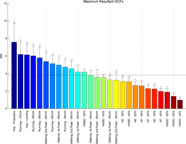
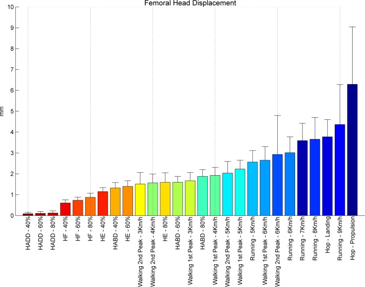
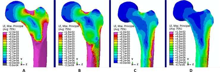
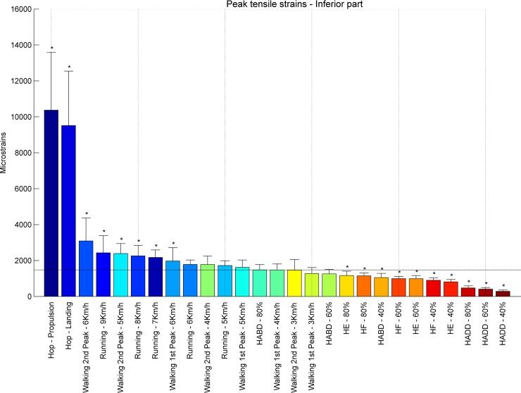
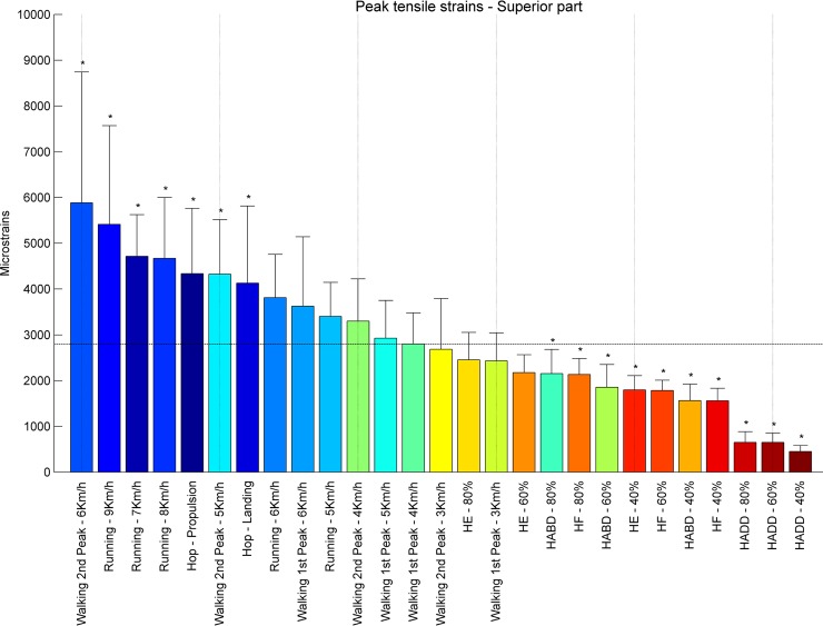
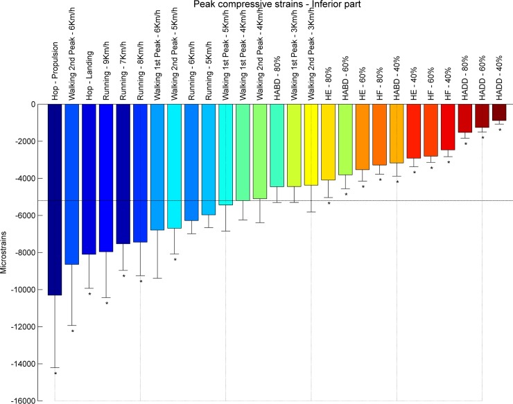
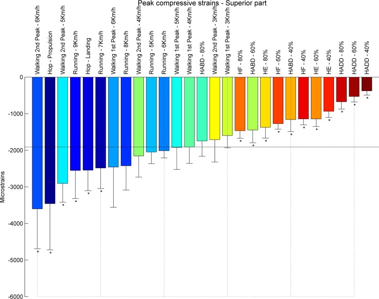
The current study aimed to assess the potential of different exercises triggering an osteogenic response at the femoral neck in a group of postmenopausal women. The osteogenic potential was determined by ranking the peak hip contact forces (HCFs) and consequent peak tensile and compressive strains at the superior and inferior part of the femoral neck during activities such as (fast) walking, running and resistance training exercises. Results indicate that fast walking (5-6 km/h) running and hopping induced significantly higher strains at the femoral neck than walking at 4 km/h which is considered a baseline exercise for bone preservation. Exercises with a high fracture risk such as hopping, need to be considered carefully especially in a frail elderly population and may therefore not be suitable as a training exercise. Since superior femoral neck frailness is related to elevated hip fracture risk, exercises such as fast walking (above 5 km/h) and running can be highly recommended to stimulate this particular area. Our results suggest that a training program including fast walking (above 5 km/h) and running exercises may increase or preserve the bone mineral density (BMD) at the femoral neck.


Conflict of interest statement
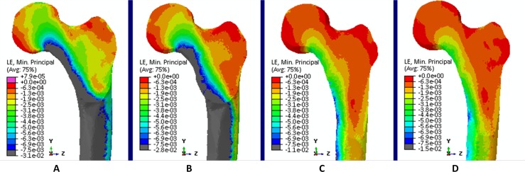
Competing Interests: The authors have declared that no competing interests exist.

Figures

Fig 1
Fig 1. Marker set.
Reflective markers were placed on bone anatomical landmarks of the body. Cluster markers were placed on the upper and lower leg for better motion tracking for these specific body segments. During walking, running and hopping the medial markers of the knee and ankle were removed to avoid any adaptation of the natural movement in case of marker contact.
Fig 2
Fig 2. Ranking hip contact forces.
Average peak HCFs expressed in body weight of each subject [BW] ranked from left to right for the highest (blue) to the lowest HCF’s (red). Asterisks denote the exercises with significantly different peak HCFs compared to walking at 4 km/h (1st peak) indicated by the horizontal line.
Fig 3
Fig 3. Ranking femoral head displacements.
Femoral head displacements in millimeters [mm] during peak HCFs ranked from right to left for the highest (blue) to the lowest displacement (red).
Fig 4
Fig 4. Tensile strains.
Tensile strains at the proximal femur for (A) hopping (propulsion), (B) walking 6 km/h (second peak), (C) walking 4 km/h (first peak) and (D) Hip Abduction at 80% RM.
Fig 5
Fig 5. Ranking tensile strains inferior part.
Average peak tensile strains in μstrains (εμ) in the inferior part of the femoral neck ranked from left to right for the highest (blue) to the lowest strain (red). Asterisks denote the exercises with significantly different peak tensile strains compared to walking at 4 km/h (1st peak) indicated by the horizontal line.
Fig 6
Fig 6. Ranking tensile strains superior part.
Average peak tensile strains in μstrains (εμ) in the superior part of the femoral neck ranked from left to right for the highest (blue) to the lowest strain (red). Asterisks denote the exercises with significantly different peak tensile strains compared to walking at 4 km/h (1st peak) indicated by the horizontal line.
Fig 7
Fig 7. Compressive strains.
Compression strains at the proximal femur for (A) hopping (propulsion), (B) walking 6 km/h (second peak), (C) walking 4 km/h (first peak) and (D) Hip Abduction at 80% RM.
Fig 8
Fig 8. Ranking compression strains inferior part.
Average peak compressive strains in μstrains (εμ) in the inferior part of the femoral neck ranked from left to right for the highest (blue) to the lowest strain (red). Asterisks denote the exercises with significantly different peak compressive strains compared to walking at 4 km/h (1st peak) indicated by the horizontal line.
Fig 9
Fig 9. Ranking compression strains superior part.
Average peak compressive strains in μstrains (εμ) in the s part of the femoral neck ranked from left to right for the highest (blue) to the lowest strain (red). Asterisks denote the exercises with significantly different peak compressive strains compared to walking at 4 km/h (1st peak) indicated by the horizontal line.

References

    1. Hernlund E, Svedbom A, Ivergård M, Compston J, Cooper C, Stenmark J, et al. Osteoporosis in the European Union: medical management, epidemiology and economic burden. Arch Osteoporos. 2013. January;8(1–2):136. - PMC - PubMed
    1. Boonen S, Dejaeger E, Vanderschueren D, Venken K, Bogaerts A, Verschueren S, et al. Osteoporosis and osteoporotic fracture occurrence and prevention in the elderly: a geriatric perspective. Best Pract Res Clin Endocrinol Metab. Elsevier Ltd; 2008;22(5):765–85. doi: 10.1016/j.beem.2008.07.002 - DOI - PubMed
    1. Frost HM. From Wolff’s law to the Utah paradigm: Insights about bone physiology and its clinical applications Vol. 262, Anatomical Record; 2001. p. 398–419. - PubMed
    1. Frost HM. Bone’s mechanostat: A 2003 update. Anat Rec. Wiley Online Library; 2003;275A(2):1081–101. - PubMed
    1. Bassett CAL, Becker RO. Generation of electric potentials by bone in response to mechanical stress. Science (80-). American Association for the Advancement of Science; 1962;137(3535):1063–4. - PubMed

Publication types

LinkOut - more resources