. 2018 May;32(5):2878-2890.
doi: 10.1096/fj.201700919RRR. Epub 2018 Jan 17.

MMP14 is a novel target of PTH signaling in osteocytes that controls resorption by regulating soluble RANKL production

Affiliations

MMP14 is a novel target of PTH signaling in osteocytes that controls resorption by regulating soluble RANKL production

Jesus Delgado-Calle et al. FASEB J. 2018 May.

Abstract

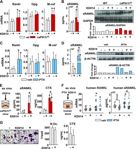
Parathyroid hormone (PTH) affects the skeleton by acting on osteocytes (Ots) in bone through yet unclear mechanisms. We report that matrix metalloproteinase 14 (MMP14) expression/activity are increased in bones from mice with genetic constitutive activation (ca) of the PTH receptor 1 (PTH1R) in Ots (caPTH1ROt) and in bones from mice exposed to elevated PTH levels but not in mice lacking [conditional knockout (cKO)] the PTH1R in Ots (cKOPTH1ROt). Furthermore, PTH upregulates MMP14 in human bone cultures and in Ot-enriched bones from floxed control mice but not from cKOPTH1ROt mice. MMP14 activity increases soluble receptor activator of NF-κΒ ligand production, which in turn, stimulates osteoclast differentiation and resorption. Pharmacologic inhibition of MMP14 activity reduced the high bone remodeling exhibited by caPTH1ROt mice or induced by chronic PTH elevation and decreased bone resorption but allowed full stimulation of bone formation induced by PTH injections, thereby potentiating bone gain. Thus, MMP14 is a new member of the intricate gene network activated in Ots by PTH1R signaling that can be targeted to adjust the skeletal responses to PTH in favor of bone preservation.-Delgado-Calle, J., Hancock, B., Likine, E. F., Sato, A. Y., McAndrews, K., Sanudo, C., Bruzzaniti, A., Riancho, J. A., Tonra, J. R., Bellido, T. MMP14 is a novel target of PTH signaling in osteocytes that controls resorption by regulating soluble RANKL production.

Keywords: antiresorptive; osteoblasts; osteoclasts; osteoporosis.


Conflict of interest statement

The authors thank Drs. Keith Condon, Gretel Pellegrini, and Sumana Posritong and Meloney Cregor (all from Indiana University School of Medicine) for their assistance in tissue and data collection. The authors also thank Drs. David Burr and Munro Peacock (both from the Indiana University School of Medicine) for critical reading of the manuscript. This research was supported by the U.S. Department of Veterans Affairs (1 I01 BX002104-01 to T.B.); the U.S. National Institutes of Health (NIH) National Institute of Arthritis and Musculoskeletal and Skin Diseases (Grant R01-AR059357 to T.B.; R01-AR060332 to A.B.); NIH National Heart, Lung, and Blood Institute (Grant T35 HL1 10854-01); a Scholar Award from the American Society of Hematology (to J.D-C.); and a grant from Instituto de Salud Carlos III (PI12/615), cofunded by the European Union through Fonds Européen de Développement Économique et Régional (FEDER) funds (to J.A.R.). The KD014 neutralizing antibody was obtained under a Materials Transfer Agreement with Kadmon Corp., LLC. J.R.T. was an employee of Kadmon Corp., LCC. The remaining authors declare no conflicts of interest.

Figures

Figure 1.
Figure 1.
Genetic and hormonal activation of PTH receptor signaling in Ots upregulates MMP14 gene expression in murine and human bone. AC) Mmp14 mRNA expression in whole bones (L4) from caPTH1ROt mice (n = 7) and WT control littermates (n = 7) (A), from fl/fl and cKOPTH1ROt mice receiving daily injections of vehicle (veh)/PTH (vehicle/PTH injections, n = 10/8 fl/fl; n = 9/8, cKOPTH1ROt mice) (B), or fed a normal/low-calcium diet (normal/deficient calcium diet, n = 9/9 fl/fl; n = 7/6, cKOPTH1ROt mice) (C). D) MMP14 mRNA expression in human bone fragments treated with vehicle (n = 8) or PTH (n = 8) for 24 h. E, F) Ex vivo bone organ cultures established from normal and Ot-enriched bones were treated with vehicle or PTH for 24 h (n = 4 mice/genotype/treatment). Bars represent means ± sd. *P ≤ 0.05 vs. WT/fl/fl (A), vs. vehicle (B, DF), or vs. a normal diet (C) by Student’s t test (A, D, E) or 2-way ANOVA, Tukey’s post hoc test (B, C, F).
Figure 2.
Figure 2.
Inhibition of MMP14 activity reduces the increase in bone gain coupled to resorption induced by ca of PTH1R signaling in Ots. A, B) Mmp14 mRNA expression (A) MMP14 and MMP2 protein levels (B) (n = 2/group) and band quantification in whole bones (L4) from WT and caPTH1ROt receiving control or KD014 for 8 wk. Fold changes relative to WT/saline-treated mice are shown. Proteins of interest were assayed on different gels. C) Total, spinal, and femoral BMD percentage change calculated over initial BMD measurements in WT and caPTH1ROt mice receiving control/KD014 injections. D) microCT analysis of cortical bone in mice receiving control/KD014 for 8 wk. Control/KD014 injections: n = 10/13, WT; n = 11/10, caPTH1ROt mice. Bars represent means ± sd. *P ≤ 0.05 vs. WT mice of each group, unless otherwise indicated by the lines, by 2-way ANOVA, Tukey’s post hoc test. Ct.Ar, cortical area; Ct.Th, cortical thickness; Ma.Ar, marrow area; Tt.Ar, tissue area.
Figure 3.
Figure 3.
Inhibition of MMP14 activity decreases the elevated bone resorption and bone remodeling displayed by caPTH1ROt mice. A) Longitudinal analysis of serum CTX and TRAP5b levels (control/KD014 injections: n = 10/13, WT; n = 11/10, caPTH1ROt mice). B) Osteoclast number/surface (control/KD014 injections: n = 5/5, WT; n = 3/6, caPTH1ROt mice) and representative images of TRAP staining in endocortical bone surfaces of WT and caPTH1ROt mice after 8 wk of treatment. Arrows point to TRAP-positive osteoclasts. CG) Serum P1NP levels (C), histomorphometric analysis of bone formation dynamic parameters in the periosteum (D) and endosteum of femoral middiaphysis (E) and representative images of labeled surfaces (control/KD014 injections: n = 10/13, WT; n = 11/10, caPTH1ROt mice), endocortical osteoblast (F) number/surface (control/KD014 injections: n = 5/5, WT; n = 3/6, caPTH1ROt mice), and Sost and Wnt target-gene mRNA expression (G) in WT and caPTH1ROt mice after 8 wk of treatment (control/KD014 injections: n = 10/13, WT; n = 11/10, caPTH1ROt mice). H) Mineralization in murine primary osteoblasts treated with/without KD014 for 14 d by Alizarin Red S staining (control/KD014, n = 4/3). Bars represent means ± sd. *P ≤ 0.05 vs. WT mice of each group, unless otherwise indicated by the lines, by 2-way ANOVA, Tukey’s post hoc test. BFR/BS, BFR per bone surface; Ec., endocortical; MS/BS, mineralizing surface per bone surface; N.Ob/B.Pm, osteoblast number per bone perimeter; N.Oc/B.Pm, osteoclast number per bone perimeter; Ob.S/BS, osteoblast surface per bone surface; Oc.S/BS, osteoclast surface per bone surface; Ps., periosteal.
Figure 4.
Figure 4.
Pharmacological inhibition of MMP14 activity partially prevents the increase in bone remodeling and the bone loss induced by chronic elevation of PTH. AC) Serum PTH (B), CTX (B), and P1NP (C) levels in mice fed a normal or a low-calcium diet and receiving control or KD014 injections for 4 wk. D) Total, spinal, and femoral BMD percentage change calculated over initial BMD measurements. Control/KD014 injections: n = 10/11, normal calcium diet; n = 10/11, low calcium diet. Bars represent means ± sd. *P ≤ 0.05 vs. mice fed a normal Ca diet of each group, unless otherwise indicated by the lines, by 2-way ANOVA, Tukey’s post hoc test.
Figure 5.
Figure 5.
Coadministration of KD014 potentiates the cancellous bone gain induced by daily injections of PTH. A, B) Mmp14 mRNA expression (L4) (A), MMP14 and MMP2 protein levels (n = 2/group) (B), and band quantification in whole bones (tibia; fold changes relative to vehicle/saline-treated mice) from mice injected with vehicle or PTH and receiving control or KD014 treatment for 4 wk. Proteins of interest were assayed on different gels. C) Effect of coadministration of KD014 on total, spinal, and femoral BMD percentage change calculated over initial BMD measurements. D) Representative von Kossa staining images of undecalcified sections of the lumbar spine. MicroCT analysis showing the increase in (E) spinal and (F) femoral cancellous BV and (G) the absence of changes in Ct.Ar or thickness in mice receiving PTH and KD014 for 4 wk (control/KD014 injections: n = 10/11, vehicle; n = 10/11, PTH). Bars represent means ± sd. *P ≤ 0.05 versus vehicle-treated mice of each group, unless otherwise indicated by the lines, by 2-way ANOVA, Tukey’s post hoc test. Ct.Th, cortical thickness; Ma.Ar, marrow area; Tb.N., trabecular number; Tb.Th., trabecular thickness.
Figure 6.
Figure 6.
Inhibition of MMP14 decreases bone resorption but preserves the increased bone formation induced by anabolic regimes of PTH. Serum CTX and TRAP5b levels (A), cancellous osteoclast number/surface and representative images of TRAP staining, with arrows pointing to TRAP-positive osteoclasts (B), serum P1NP (C), cancellous MS/BS, MAR, and BFR (control/KD014 injections: n = 5/5, vehicle; n = 5/5, PTH) (D), cancellous osteoblast number/surface (control/KD014 injections: n = 5/5, vehicle; n = 5/5, PTH) (E), Sost downregulation and upregulation of Wnt target genes mRNA expression (F), and biomechanical properties measured by axial compression testing in vertebral bone (L6) in mice receiving vehicle/PTH in combination with control or KD014 injections (G). Control/KD014 injections: n = 10/11, vehicle; n = 10/11, PTH. Bars represent means ± sd. *P ≤ 0.05 versus vehicle-treated mice of each group, unless otherwise indicated by the lines, by 2-way ANOVA, Tukey’s post hoc test. BFR/BS, BFR per bone surface; MS/BS, mineralizing surface per bone surface; N.Oc/B.Pm, osteoclast number per bone perimeter; Ob.N/B.Pm, osteoblast number per bone perimeter; Ob.S/BS, osteoblast surface per bone surface; Oc.S/BS, osteoclast surface per bone surface.
Figure 7.
Figure 7.
MMP14 controls bone resorption by regulating the production of sRANKL. A, B) Rankl, Opg, and M-csf mRNA levels in bone (A) and serum RANKL and sRANKL protein levels (B) (n = 2/group; fold changes relative to WT/saline-treated mice) in whole bones from WT and caPTH1ROt mice treated with control or KD014 injections for 8 wk (control/KD014 injections: n = 10/13, WT; n = 11/10, caPTH1ROt mice). C, D) Rankl, Opg, and M-csf mRNA expression (C) and serum RANKL and sRANKL protein expression (D) (n = 2/group; fold changes relative to vehicle/saline-treated mice) in whole bones from mice receiving daily PTH in combination with control or KD014 injections for 4 wk (control/KD014 injections: n = 10/11, vehicle; n = 10/11, PTH). E) sRANKL and CTX levels in 96 h conditioned medium from WT and caPTH1ROt bones treated with control or KD014 (control/KD014 injections: n = 4/4, WT; n = 6/6, caPTH1ROt mice). F) Human RANKL mRNA expression (control/KD014 injections: n = 19/10, vehicle; n = 23/10, PTH) and sRANKL levels in 96 h conditioned medium collected from human bones treated with PTH, with or without KD014 (control/KD014 injections: n = 14/15, vehicle; n = 18/21, PTH). G) N.Oc and resorption activity measured as CTX released to the medium measured in murine primary nonadherent bone marrow cells cultured with RANKL and M-CSF in the presence/absence of KD014 (control/KD014, n = 6/6). Bars represent means ± sd. *P ≤ 0.05 vs. WT mice of each group (A, E), or vs. vehicle-treated mice (C, F), unless otherwise indicated by the lines, by 2-way ANOVA, Tukey’s post hoc. Proteins of interest were assayed on different gels.
Figure 8.
Figure 8.
MMP14 is a novel osteocytic PTH1R target gene that stimulates bone resorption through sRANKL production. Upon binding to the PTH1R in Ots, PTH activates Wnt signaling to increase osteoblast differentiation, survival, and bone forming activity and also increases the expression of RANKL and MMP14. MMP14 cleaves membrane-bound RANKL protein and produces sRANKL, which in turn, promotes osteoclast differentiation and bone resorption. Inhibition of MMP14 activity with KD014 antibody reduces bone resorption driven by osteocytic PTH1R signaling and preserves the newly built bone.

References

    1. Silva B. C., Bilezikian J. P. (2015) Parathyroid hormone: anabolic and catabolic actions on the skeleton. Curr. Opin. Pharmacol. 22, 41–50 10.1016/j.coph.2015.03.005 - DOI - PMC - PubMed
    1. Jilka R. L. (2007) Molecular and cellular mechanisms of the anabolic effect of intermittent PTH. Bone 40, 1434–1446 10.1016/j.bone.2007.03.017 - DOI - PMC - PubMed
    1. Ma Y. L., Cain R. L., Halladay D. L., Yang X., Zeng Q., Miles R. R., Chandrasekhar S., Martin T. J., Onyia J. E. (2001) Catabolic effects of continuous human PTH (1--38) in vivo is associated with sustained stimulation of RANKL and inhibition of osteoprotegerin and gene-associated bone formation. Endocrinology 142, 4047–4054 10.1210/endo.142.9.8356 - DOI - PubMed
    1. Bellido T., Saini V., Pajevic P. D. (2013) Effects of PTH on osteocyte function. Bone 54, 250–257 10.1016/j.bone.2012.09.016 - DOI - PMC - PubMed
    1. Delgado-Calle J., Bellido T. (2015) Osteocytes and skeletal pathophysiology. Curr. Mol. Biol. Rep. 1, 157–167 10.1007/s40610-015-0026-y - DOI - PMC - PubMed

Publication types

MeSH terms

LinkOut - more resources