. 2018 Jun;9(3):570-584.
doi: 10.1002/jcsm.12288. Epub 2018 Jan 29.

The double-edged sword of endoplasmic reticulum stress in uremic sarcopenia through myogenesis perturbation

Affiliations

The double-edged sword of endoplasmic reticulum stress in uremic sarcopenia through myogenesis perturbation

Jia-Rong Jheng et al. J Cachexia Sarcopenia Muscle. 2018 Jun.

Abstract

Background: Sarcopenia is the age-related degeneration characterized with the decline of skeletal muscle mass, strength, and function. The imbalance of protein synthesis and degradation which jeopardizes immune, hormone regulation, and muscle-motor neuron connection is the main cause of sarcopenia. There is limited knowledge regarding molecular mechanism of sarcopenia. As the endoplasmic reticulum is the control centre of the protein syntheses and degradation, we hypothesized that endoplasmic reticulum stress and unfolded protein response (UPR) play an important in the development of sarcopenia. Understanding the sarcopenia molecular mechanisms may benefit the therapeutic diagnosis and treatment in the future.

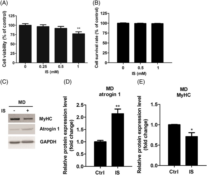
Methods: Mouse myoblast C2C12 cells are exposed to designated time and concentration of indoxyl sulfate (IS), a uremic toxin of chronic kidney disease. The proliferation, differentiation, and the expression of atrogin 1 are examined. The protein and mRNA expression of IS treated-C2C12 cells are inspected to distinguish the role of ER stress and oxidative stress underlying the sarcopenia.

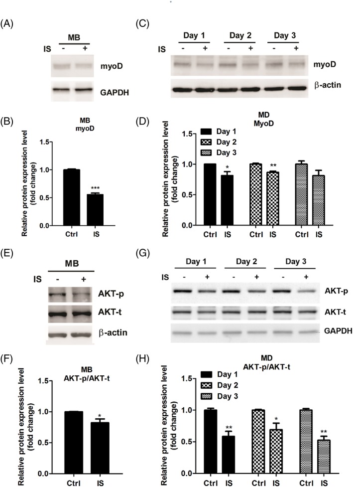
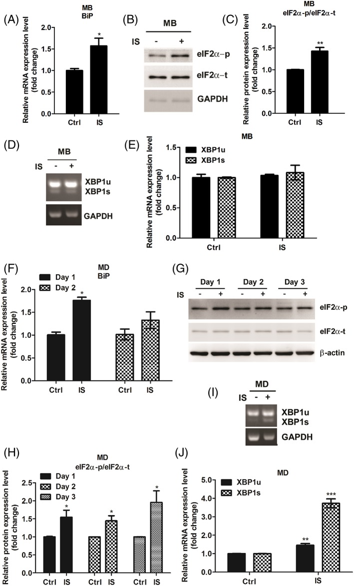
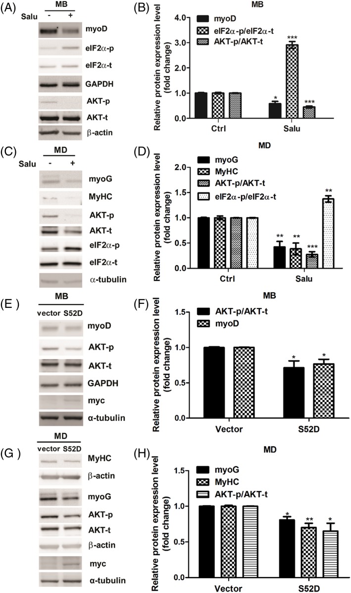
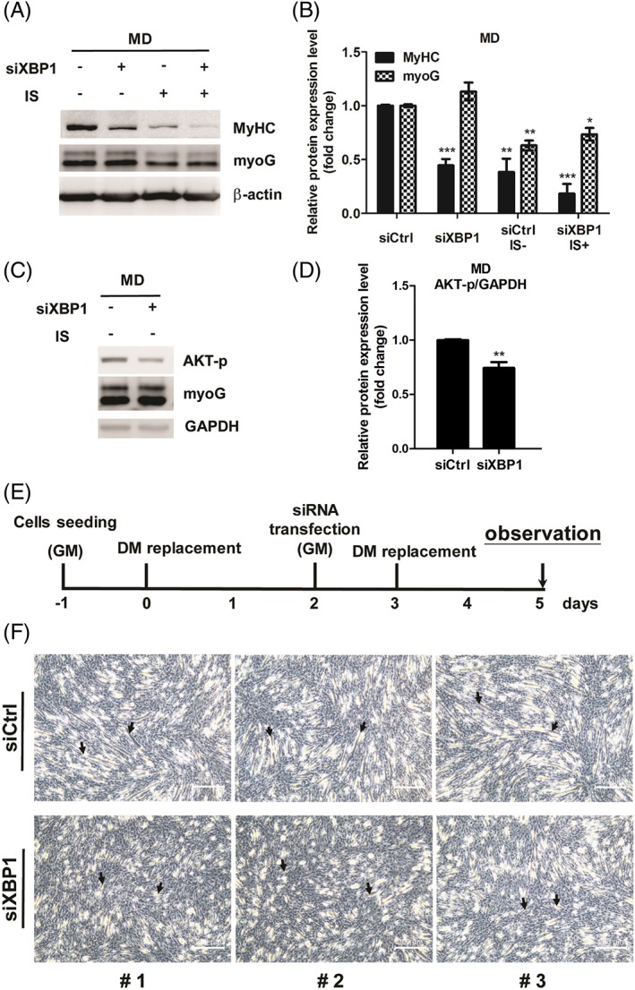
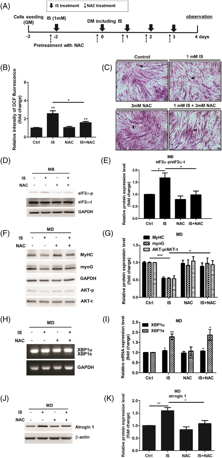
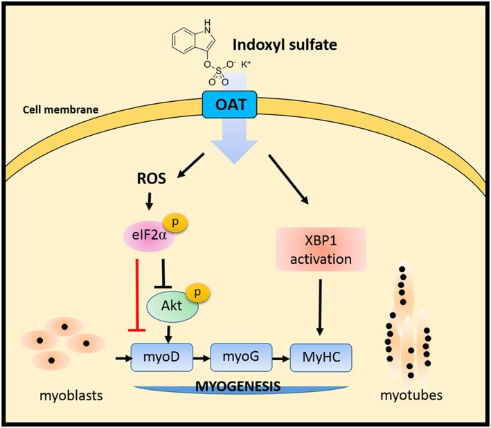
Results: Indoxyl sulfate inhibits myoblast differentiation. We demonstrate that as the number of multi-nuclei myotube decreased, the differentiation markers including myoD, myoG, and myosin heavy chain are also suppressed. Indoxyl sulfate inhibits myoblast proliferation and induces the myotubular atrophy marker atrogin-1 protein expression. Indoxyl sulfate stimulates eIF2α phosphorylation and XBP1 mRNA splicing in UPR. Interestingly, the oxidative stress is related to eIF2α phosphorylation but not XBP1 mRNA splicing. The eIF2α phosphorylation triggered by IS reduces myoD, myoG, and myosin heavy chain protein expression, which represents the anti-myogenic modulation on the early differentiation event. The XBP1 mRNA splicing induced by IS, however, is considered the adaptive response to restore the myogenic differentiation.

Conclusions: Our studies indicated that the ER stress and UPR modulation are critical in the chronic kidney disease uremic toxin-accumulated sarcopenia model. We believe that UPR-related signals showed great potential in clinical application.

Keywords: Chronic kidney disease; ER stress; Indoxyl sulfate; Myogenesis; Unfolded protein response.


Figures

Figure 1
Figure 1
IS impairs C2C12 myogenic differentiation (MD) in a dose‐dependent manner. (A) H&E staining of C2C12 myotubes under IS treatment, scale bars = 200 μm. The arrows indicate the fused myoblasts. (B) The number of nuclei per myotube at 4 days of differentiation were counted. The values were then classified in three categories and divided by the total number of myotubes in a field. The data were expressed as mean ± SED from three independent experiments. (C, E, and F) Protein expression of myogenin and MyHC expression in differentiated myoblasts treated with or without IS (1 mM) are examined by western blot and quantified. (D) The relative myogenin and MyHC mRNA expression levels are determined by real‐time PCR. Data represent means ± SEM for three independent experiments. *P < 0.05, ** P < 0.01, and *** P < 0.001, as compared with untreated control.
Figure 2
Figure 2
IS treatment inhibits myoblast proliferation and enhances atrogin 1 expression in differentiated myoblasts. (A) C2C12 myoblasts were incubated with growth medium containing indicated concentrations of IS for 48 h. Cell viability was determined via MTS assay. (B) Myoblast apoptotic potential are demonstrated by trypan blue exclusion assay. (C–E) Atrogin 1 and MyHC expression in differentiated myoblasts treated with or without IS (1 mM) are determined by western blot and quantified. Data represent means ± SEM for three independent experiments. *P < 0.05 and ** P < 0.01, as compared with untreated control.
Figure 3
Figure 3
IS treatment represses myoD expression and Akt phosphorylation during myoblast (MB) proliferation and myogenic differentiation (MD). (A and B) Protein expression of myoD in myoblsts treated with or without IS (1 mM) for 48 h is examined by western blot and quantified. (C and D) pPotein expression of myoD in differentiated myoblasts treated with or without IS (1 mM) at the indicated time points by western blot and quantified. (E–F) Akt expression and phosphorylation in myoblasts treated with or without IS (1 mM) for 48 h are determined by western blot and quantified. (G–H) Akt expression and phosphorylation during myoblasts differentiation treated with or without IS (1 mM) at the indicated time point are determined by western blot and quantified. Data represent means ± SEM for three independent experiments. *P < 0.05, ** P < 0.01, and *** P < 0.001, as compared with untreated control.
Figure 4
Figure 4
IS modulates myoblast and myotube UPR signalling pathways. (A–C) BiP mRNA expression level and phosphorylation of eIF2α are examined in C2C12 myoblasts (MB) after 2 days exposure of IS (1 mM). (D and E) The levels of both XBP1u or XBP1s mRNA were determined by semi‐quantitative PCR. (F–H) BiP mRNA expression level and phosphorylation of eIF2α increased are examined in IS‐treated well myogenic differentiation (MD) cells. (I and J) The level of XBP1u and XBP1s mRNA expression is determined in well MD cells with or without IS (1 mM) for 48 h. The data were expressed as mean ± SED from three independent experiments. *P < 0.05, ** P < 0.01, and *** P < 0.001, as compared with untreated control.
Figure 5
Figure 5
Salubrinal, an eIF2α dephosphorylation inhibitor, deregulates C2C12 myogenic differentiation (MD). (A and B) Myoblasts (MB) were treated with or without 20 μM salubrinal for 48 h. MyoD, phospho‐eIF2α, and phospho‐AKT expressions are examined with western blot and quantified. (C and D) Myoblasts under 48 h differentiation are simultaneously exposed with or without 20 μM salubrinal. MyoG, MyHC, phospho‐AKT, and phospho‐ eIF2α expression level are determined with western blot and quantified. (E and F) Myoblasts are overexpressed with or without mouse S52D phosphomimetic eIF2α mutant for 48 h. The expression of eIF2α S52D was detected with anti‐myc antibody. MyoD and phospho‐AKT expressions are examined with western blot and quantified. (G and H) myoblasts under 48 h differentiation are simultaneously transfected with or without S52D mutant. The expression of eIF2α S52D was detected with anti‐myc antibody. MyoG, MyHC, and phospho‐AKT expression level are determined with western blot and quantified.
Figure 6
Figure 6
Promyogenic role of XBP1 in myoblast differentiation (MD). (A–D) The expression of MyHC, myoG, and phospho‐AKT in XBP1‐dificent cells is detected by western bot and quantified. (E) Diagram illustrates timeline of experiment. (F) Phase contrast photomicroscopy revealed differentiated morphology in XBP1‐deficient myoblasts compared with ordinary myoblasts. The arrows indicate the fusion of myoblasts. Random views of three independent experiments at a magnification of ×100 were shown, scale bars = 200 μm.
Figure 7
Figure 7
NAC treatment reverses IS‐induced defect in myogenic differentiation (MD). (A) Diagram illustrates timeline of experiment. (B) Flow cytometric quantitation of DCF fluorescence. (C) H&E staining of C2C12 myotubes under NAC (3 mM) or IS (1 mM) treatment, scale bars = 100 μm. The arrows indicate the fused myotubes. (D and E) myoblasts (MB) were treated with NAC (3 mM) or IS (1 mM) for 48 h. Phospho‐eIF2α expression was examined with western blot and quantified. (f‐k) Myoblasts under 48 h differentiation are simultaneously exposed with NAC (3 mM) or IS (1 mM). MyHC, myoG, and phospho‐AKT expression were determined with western blot and quantified (F and G). XBP1u and XBP1s mRNA expression are examined by agarose gel electrophoresis (H and I). Atrogin 1 expression are measured by western blot and quantified (J and K). The data were expressed as mean ± SED from three independent experiments. *P < 0.05, ** P < 0.01, and *** P < 0.001, as compared with untreated control.
Figure 8
Figure 8
Model of IS‐induced defect in myoblast differentiation. The pro‐ and anti‐myogenesis roles of XBP1 and phospho‐eIF2α in different stages of myogenesis are pointed out. Notably, the ROS‐eIF2α axis is important for the antimyogenesis activity of IS. The red line indicates translational attenuation caused by phosphorylation of eIF2α probably results in decreased myoD expression. Overall, our research demonstrates the pivotal role of UPR signalling in age‐related muscle loss and sarcopenia.

References

    1. Metter EJ, Conwit R, Tobin J, Fozard JL. Age‐associated loss of power and strength in the upper extremities in women and men. J Gerontol A Biol Sci Med Sci 1997;52:B267–B276. - PubMed
    1. Maggio M, Lauretani F, Ceda GP. Sex hormones and sarcopenia in older persons. Curr Opin Clin Nutr Metab Care 2013;16:3–13. - PubMed
    1. Messier V, Rabasa‐Lhoret R, Barbat‐Artigas S, Elisha B, Karelis AD, Aubertin‐Leheudre M. Menopause and sarcopenia: a potential role for sex hormones. Maturitas 2011;68:331–336. - PubMed
    1. Frost RA, Lang CH. Protein kinase B/Akt: a nexus of growth factor and cytokine signaling in determining muscle mass. J Appl Physiol 2007;103:378–387. - PubMed
    1. Rygiel KA, Picard M, Turnbull DM. The ageing neuromuscular system and sarcopenia: a mitochondrial perspective. J Physiol 2016;594:4499–4512. - PMC - PubMed

Publication types