. 2017 Dec 28;10(1):19.
doi: 10.3390/toxins10010019.

Impact of the Oral Adsorbent AST-120 on Organ-Specific Accumulation of Uremic Toxins: LC-MS/MS and MS Imaging Techniques

Affiliations

Impact of the Oral Adsorbent AST-120 on Organ-Specific Accumulation of Uremic Toxins: LC-MS/MS and MS Imaging Techniques

Emiko Sato et al. Toxins (Basel). .

Abstract

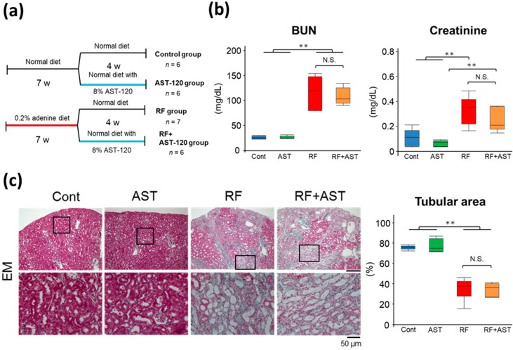
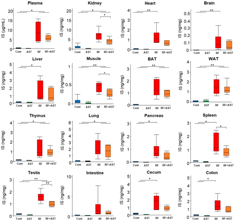
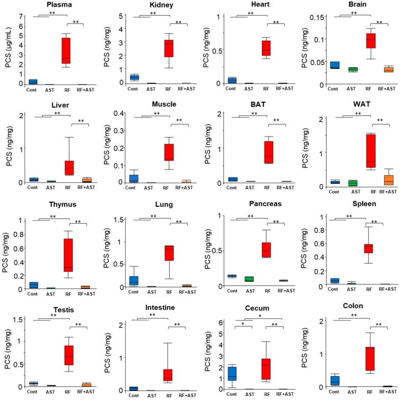
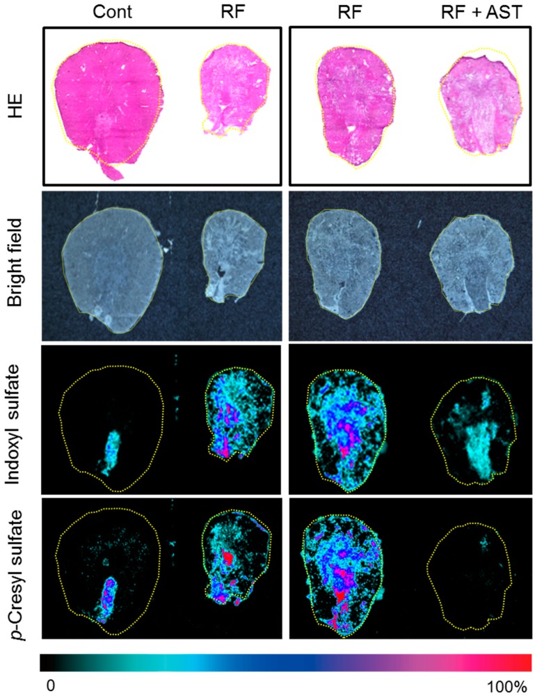
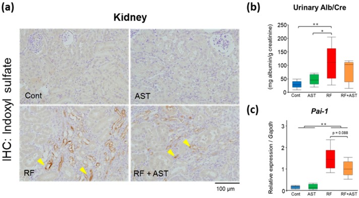
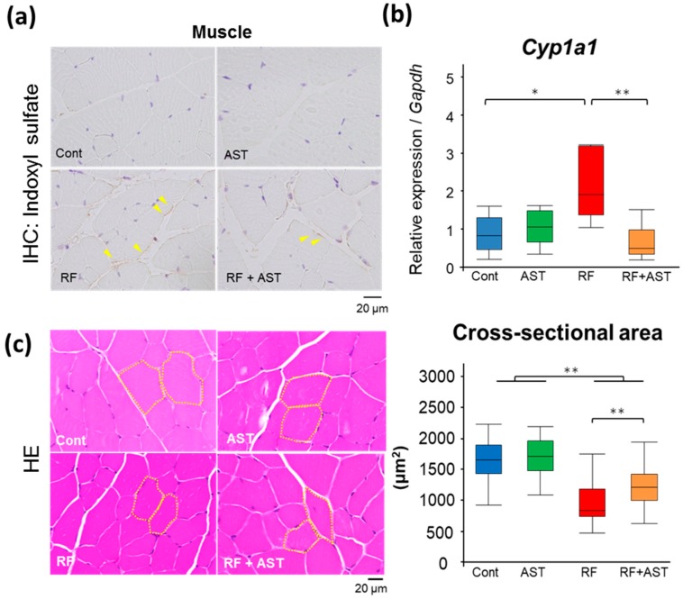
Elevated circulating uremic toxins are associated with a variety of symptoms and organ dysfunction observed in patients with chronic kidney disease (CKD). Indoxyl sulfate (IS) and p-cresyl sulfate (PCS) are representative uremic toxins that exert various harmful effects. We recently showed that IS induces metabolic alteration in skeletal muscle and causes sarcopenia in mice. However, whether organ-specific accumulation of IS and PCS is associated with tissue dysfunction is still unclear. We investigated the accumulation of IS and PCS using liquid chromatography/tandem mass spectrometry in various tissues from mice with adenine-induced CKD. IS and PCS accumulated in all 15 organs analyzed, including kidney, skeletal muscle, and brain. We also visualized the tissue accumulation of IS and PCS with immunohistochemistry and mass spectrometry imaging techniques. The oral adsorbent AST-120 prevented some tissue accumulation of IS and PCS. In skeletal muscle, reduced accumulation following AST-120 treatment resulted in the amelioration of renal failure-associated muscle atrophy. We conclude that uremic toxins can accumulate in various organs and that AST-120 may be useful in treating or preventing organ dysfunction in CKD, possibly by reducing tissue accumulation of uremic toxins.

Keywords: chronic kidney disease; indoxyl sulfate; mass spectrometry; p-cresyl sulfate; uremic toxin.


Conflict of interest statement

The authors declare no competing financial interests.

Figures

Figure 1
Figure 1
Renal histology and function in adenine-induced renal failure mice. (a) Experimental schedule. (b) Blood urea nitrogen (BUN) and plasma creatinine concentrations. n = 5–7 per group. (c) Representative histologic images of Elastica-Masson (EM) staining of kidney sections. Morphometric analysis of the percentage of the remaining cortical tubular area. The cortical tubular area was calculated from the EM staining images. Data are expressed as box plots. Tukey-Kramer test: ** p < 0.01. Cont, control; AST, AST-120; RF, renal failure; RF + AST, RF mice treated with AST-120; N.S., not significant.
Figure 2
Figure 2
Indoxyl sulfate (IS) accumulation in plasma and tissues. Comparison of plasma and tissue IS levels. Data are expressed as box plots. n = 6–7 per group. Tukey-Kramer test: * p < 0.05, ** p < 0.01. Cont, control; AST, AST-120; RF, renal failure; RF + AST, RF mice treated with AST-120; BAT, brown adipose tissue; WAT, white adipose tissue.
Figure 3
Figure 3
p-Cresyl sulfate (PCS) accumulation in plasma and tissues. Comparison of plasma and tissue PCS levels. Data are expressed as box plots. n = 6–7 for each group. Tukey-Kramer test: * p < 0.05, ** p < 0.01. Cont, control; AST, AST-120; RF, renal failure; RF + AST, RF mice treated with AST-120; BAT, brown adipose tissue; WAT, white adipose tissue.
Figure 4
Figure 4
Spatial distribution of indoxyl sulfate (IS) and p-cresyl sulfate (PCS) in kidney sections by mass spectrometry imaging. Hematoxylin and eosin (HE) and bright-field images of kidney sections. The second and third RF samples are same sample, but the slide section is different. Mass spectrometry imaging distribution of IS ([M − H], m/z 212) and p-cresyl sulfate ([M − H], m/z 187) in kidney. Cont, control; RF, renal failure; RF + AST, RF mice treated with AST-120.
Figure 5
Figure 5
Indoxyl sulfate (IS) immunostaining and expression of an inflammatory gene in kidney. (a) Renal immunostaining of IS using an anti-IS antibody. Arrowheads point to immunostaining-positive signals. (b) Urinary albumin excretion (creatinine ratio, Alb/Cre) in the four mouse groups. n = 5–7 per group. (c) Relative mRNA expression levels of plasminogen activator inhibitor 1 (Pai-1). Expression levels were normalized to those of Gapdh. n = 5–7 per group. Data are shown as box plots. Tukey-Kramer test: * p < 0.05, ** p < 0.01. For Pai-1 expression, Welch’s t-test was performed between the RF and RF + AST groups. Cont, control; AST, AST-120; RF, renal failure; RF + AST, RF mice treated with AST-120; IHC, immunohistochemistry.
Figure 6
Figure 6
Renal failure-induced skeletal muscle atrophy was ameliorated by AST-120 treatment. (a) Immunostaining of indoxyl sulfate (IS) using an anti-IS antibody on the gastrocnemius muscle. Arrowheads point to immunostaining-positive signals. (b) Relative mRNA expression levels of cytochrome P450 family 1 subfamily A member 1 (Cyp1a1) in skeletal muscle. Expression levels were normalized to those of Gapdh. n = 5–6 per group. Data are shown as box plots. (c) Representative images of hematoxylin and eosin (HE)-stained skeletal muscle sections and cross-sectional area size of the gastrocnemius muscle. Thirty cross-sections were randomly selected from each group for evaluation of cross-sectional area. Data are shown as box plots. Tukey-Kramer test: * p < 0.05, ** p < 0.01. Cont, control; AST, AST-120; RF, renal failure; RF + AST, RF mice treated with AST-120; IHC, immunohistochemistry.

References

    1. Barreto F.C., Barreto D.V., Liabeuf S., Drueke T.B., Massy Z.A. Effects of uremic toxins on vascular and bone remodeling. Semin. Dial. 2009;22:433–437. doi: 10.1111/j.1525-139X.2009.00595.x. - DOI - PubMed
    1. Ellis R.J., Small D.M., Vesey D.A., Johnson D.W., Francis R., Vitetta L., Gobe G.C., Morais C. Indoxyl sulphate and kidney disease: Causes, consequences and interventions. Nephrology. 2016;21:170–177. doi: 10.1111/nep.12580. - DOI - PubMed
    1. Kazama J.J., Iwasaki Y., Fukagawa M. Uremic osteoporosis. Kidney Int. Suppl. 2013;3:446–450. doi: 10.1038/kisup.2013.93. - DOI - PMC - PubMed
    1. Liabeuf S., Drueke T.B., Massy Z.A. Protein-bound uremic toxins: New insight from clinical studies. Toxins (Basel) 2011;3:911–919. doi: 10.3390/toxins3070911. - DOI - PMC - PubMed
    1. Saito S., Yisireyili M., Shimizu H., Ng H.Y., Niwa T. Indoxyl sulfate upregulates prorenin expression via nuclear factor-kappaB p65, signal transducer and activator of transcription 3, and reactive oxygen species in proximal tubular cells. J. Ren. Nutr. 2015;25:145–148. doi: 10.1053/j.jrn.2014.10.008. - DOI - PubMed

Publication types

LinkOut - more resources