. 2018 Jan-Mar;49(1):128-137.
doi: 10.1016/j.bjm.2017.03.014. Epub 2017 Aug 26.

Yersinia pestis detection by loop-mediated isothermal amplification combined with magnetic bead capture of DNA

Affiliations

Yersinia pestis detection by loop-mediated isothermal amplification combined with magnetic bead capture of DNA

Na Feng et al. Braz J Microbiol. 2018 Jan-Mar.

Abstract

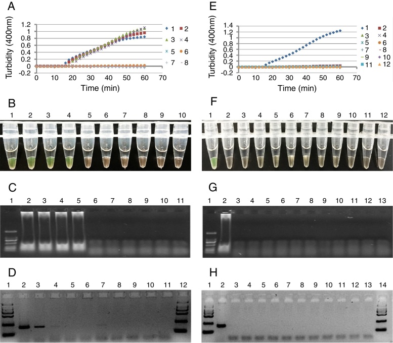
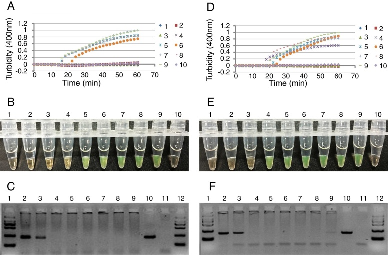
We developed a loop-mediated isothermal amplification (LAMP) assay for the detection of Y. pestis by targeting the 3a sequence on chromosome. All 11 species of the genus Yersinia were used to evaluate the specificity of LAMP and PCR, demonstrating that the primers had a high level of specificity. The sensitivity of LAMP or PCR was 2.3 or 23CFU for pure culture, whereas 2.3×10⁴ or 2.3×10⁶CFU for simulated spleen and lung samples. For simulated liver samples, the sensitivity of LAMP was 2.3×10⁶CFU, but PCR was negative at the level of 2.3×10⁷CFU. After simulated spleen and lung samples were treated with magnetic beads, the sensitivity of LAMP or PCR was 2.3×10³ or 2.3×10⁶CFU, whereas 2.3×10⁵ or 2.3×10⁷CFU for magnetic bead-treated liver samples. These results indicated that some components in the tissues could inhibit LAMP and PCR, and liver tissue samples had a stronger inhibition to LAMP and PCR than spleen and lung tissue samples. LAMP has a higher sensitivity than PCR, and magnetic bead capture of DNAs could remarkably increase the sensitivity of LAMP. LAMP is a simple, rapid and sensitive assay suitable for application in the field or poverty areas.

Keywords: Loop-mediated isothermal amplification; Magnetic beads; Plague; Yersinia pestis.


Figures

Fig. 1
Fig. 1
The sensitivity (A–D) of the LAMP or PCR for the detection of Y. pestis using serial dilutions of extracted DNA, and the specificity (E–H) of the LAMP or PCR for the detection of Y. pestis using 20 ng DNA from each bacterium of the genus Yersinia. (A) Detection of LAMP products with a real-time turbidity meter. 1–8: 20 ng–0.002 pg; 10: negative control. (B) Visualization of LAMP products stained with calcein and inspected under natural light. Tube 1–9: 20 ng–0.0002 pg; 10: negative control. (C) Detection of LAMP products with agarose gel electrophoresis. Lane 1: marker; 2–10: 20 ng–0.0002 pg; 11: negative control. (D) Electrophoretic analysis of PCR products with agarose gel. Lane 1: marker; 2–10: 20 ng–0.0002 pg; 11: negative control; 12: marker. (E) Detection of the LAMP products with a real-time turbidity meter. 1: Y. pestis; 2: Y. pseudotuberculosis; 3: Y. enterocolitica; 4: Y. frederiksenii; 5: Y. intermedia; 6: Y. kristensenii; 7: Y. bercovieri; 8: Y. mollaretii; 9: Y. rohdei; 10: Y. ruckeri; 11: Y. aldovae; 12: negative control. (F) Visualization of LAMP products stained with calcein and inspected under natural light. Tube 1: Y. pestis; 2: Y. pseudotuberculosis; 3: Y. enterocolitica; 4: Y. frederiksenii; 5: Y. intermedia; 6: Y. kristensenii; 7: Y. bercovieri; 8: Y. mollaretii; 9: Y. rohdei; 10: Y. ruckeri; 11: Y. aldovae; 12: negative control. (G) Detection of LAMP products with agarose gel electrophoresis. Lane 1: marker; 2: Y. pestis; 3: Y. pseudotuberculosis; 4: Y. enterocolitica; 5: Y. frederiksenii; 6: Y. intermedia; 7: Y. kristensenii; 8: Y. bercovieri; 9: Y. mollaretii; 10: Y. rohdei; 11: Y. ruckeri; 12: Y. aldovae; 13: negative control. (H) Electrophoretic analysis of PCR products with 1.5% agarose gel. Lane 1: marker; 2: Y. pestis; 3: Y. pseudotuberculosis; 4: Y. enterocolitica; 5: Y. frederiksenii; 6: Y. intermedia; 7: Y. kristensenii; 8: Y. bercovieri; 9: Y. mollaretii; 10: Y. rohdei; 11: Y. ruckeri; 12: Y. aldovae; 13: negative control; 14: marker.
Fig. 2
Fig. 2
The sensitivity (A–C) of the LAMP and PCR for the detection of Y. pestis by using the DNA extracted crudely from serial dilutions of bacterial solutions, and the specificity (D–F) of the LAMP and PCR for the detection of Y. pestis using crudely extracted DNA from each bacterium (2.3 × 107 CFU) of the genus Yersinia. (A) Detection of the LAMP products with a real-time turbidity meter. 1–9: 2.3 × 107–2.3 × 10−1 CFU of Y. pestis; 10: negative control. (B) Visualization of LAMP products stained with calcein and inspected under natural light. Tube 1–9: 2.3 × 107–2.3 × 10−1 CFU of Y. pestis; 10: negative control. (C) Electrophoretic analysis of PCR products with agarose gel. Lane 1: marker; Lane 2–10: 2.3 × 107–2.3 × 10−1 CFU of Y. pestis; 11: negative control; 12: marker. (D) Detection of LAMP products with a real-time turbidity meter. 1: Y. pestis; 2: Y. pseudotuberculosis; 3: Y. enterocolitica; 4: Y. frederiksenii; 5: Y. intermedia; 6: Y. kristensenii; 7: Y. bercovieri; 8: Y. mollaretii; 9: Y. rohdei; 10: Y. ruckeri; 11: Y. aldovae; 12: negative control. (E) Visualization of LAMP products stained with calcein and inspected under natural light. Tube 1: Y. pestis; 2: Y. pseudotuberculosis; 3: Y. enterocolitica; 4: Y. frederiksenii; 5: Y. intermedia; 6: Y. kristensenii; 7: Y. bercovieri; 8: Y. mollaretii; 9: Y. rohdei; 10: Y. ruckeri; 11: Y. aldovae; 12: negative control. (F) Electrophoretic analysis of PCR products with agarose gel. Lane 1: marker; 2: Y. pestis; 3: Y. pseudotuberculosis; 4: Y. enterocolitica; 5: Y. frederiksenii; 6: Y. intermedia; 7: Y. kristensenii; 8: Y. bercovieri; 9: Y. mollaretii; 10: Y. rohdei; 11: Y. ruckeri; 12: Y. aldovae; 13: negative control; 14: marker.
Fig. 3
Fig. 3
Detection of Y. pestis in simulated spleen samples by LAMP and PCR. The sensitivity of LAMP or PCR was determined by using the DNA extracted by boiling simulated spleen sample for 10 min (A–C) or by using the DNA captured by magnetic beads (D–F). (A) Detection of LAMP products with a real-time turbidity meter. 1–8: 2.3 × 100–2.3 × 107 CFU of Y. pestis; 9: positive control; 10: negative control. (B) Visualization of LAMP products stained with calcein and inspected under natural light. Tube 1–8: 2.3 × 100–2.3 × 107 CFU of Y. pestis; 9: positive control; 10: negative control. (C) Electrophoretic analysis of PCR products with agarose gel. Lane 1: marker; Lane 2–9: 2.3 × 107–2.3 × 100 CFU of Y. pestis; 10: positive control; 11: negative control; 12: marker. (D) Detection of LAMP products with a real-time turbidity meter. 1–8: 2.3 × 100–2.3 × 107 CFU of Y. pestis; 9: positive control; 10: negative control. (E) Visualization of LAMP products stained with calcein and inspected under natural light. Tube 1–8: 2.3 × 100–2.3 × 107 CFU of Y. pestis; 9: positive control; 10: negative control. (F) Electrophoretic analysis of PCR products with agarose gel. Lane 1: marker; Lane 2–9: 2.3 × 107–2.3 × 100 CFU of Y. pestis; 10: positive control (20 ng DNA); 11: negative control; 12: marker.
Fig. 4
Fig. 4
Detection of Y. pestis in simulated liver samples by LAMP and PCR. The sensitivity of LAMP or PCR was determined by using the DNA extracted by boiling simulated liver sample for 10 min (A-C) or by using the DNA captured by magnetic beads (D–F). (A) Detection of LAMP products with a real-time turbidity meter. 1–8: 2.3 × 100–2.3 × 107 CFU of Y. pestis; 9: positive control; 10: negative control. (B) Visualization of LAMP products stained with calcein and inspected under natural light. Tube 1–8: 2.3 × 100–2.3 × 107 CFU of Y. pestis; 9: positive control; 10: negative control. (C) Electrophoretic analysis of PCR products with agarose gel. Lane 1: marker; Lane 2–9: 2.3 × 107–2.3 × 100 CFU of Y. pestis; 10: positive control (20 ng DNA); 11: negative control; 12: marker. (D) Detection of LAMP products with a real-time turbidity meter. 1–8: 2.3 × 100–2.3 × 107 CFU of Y. pestis; 9: positive control; 10: negative control. (E) Visualization of LAMP products stained with calcein and inspected under natural light. Tube 1–8: 2.3 × 100–2.3 × 107 CFU of Y. pestis; 9: positive control; 10: negative control. (F) Electrophoretic analysis of PCR products with agarose gel. Lane 1: marker; Lane 2–9: 2.3 × 107–2.3 × 100 CFU of Y. pestis; 10: positive control (20 ng DNA); 11: negative control; 12: marker.
Fig. 5
Fig. 5
Detection of Y. pestis in simulated lung samples by LAMP and PCR. The sensitivity of LAMP or PCR was determined by using the DNA extracted by boiling simulated lung samples for 10 min (A–C) or by using the DNA captured by magnetic beads (D–F). (A) Detection of LAMP products with a real-time turbidity meter. 1–8: 2.3 × 100–2.3 × 107 CFU of Y. pestis; 9: positive control; 10: negative control. (B) Visualization of LAMP products stained with calcein and inspected under natural light. Tube 1–8: 2.3 × 100–2.3 × 107 CFU of Y. pestis; 9: positive control; 10: negative control. (C) Electrophoretic analysis of PCR products with agarose gel. Lane 1: marker; Lane 2–9: 2.3 × 107–2.3 × 100 CFU of Y. pestis; 10: positive control (20 ng DNA); 11: negative control; 12: marker. (D) Detection of LAMP products with a real-time turbidity meter. 1–8: 2.3 × 100–2.3 × 107 CFU of Y. pestis; 9: positive control; 10: negative control. (E) Visualization of LAMP products stained with calcein and inspected under natural light. Tube 1–8: 2.3 × 100–2.3 × 107 CFU of Y. pestis; 9: positive control; 10: negative control. (F) Electrophoretic analysis of PCR products with agarose gel. Lane 1: marker; Lane 2–9: 2.3 × 107–2.3 × 100 CFU of Y. pestis; 10: positive control (20 ng DNA); 11: negative control; 12: marker.

References

    1. Perry R.D., Fetherston J.D. Yersinia pestis-etiologic agent of plague. Clin Microbiol Rev. 1997;10:35–66. - PMC - PubMed
    1. Cui Y., Yu C., Yan Y. Historical variations in mutation rate in an epidemic pathogen, Yersinia pestis. Proc Natl Acad Sci U S A. 2013;110:577–582. - PMC - PubMed
    1. Achtman M., Zurth K., Morelli G., Torrea G., Guiyoule A., Carniel E. Yersinia pestis, the cause of plague, is a recently emerged clone of Yersinia pseudotuberculosis. Proc Natl Acad Sci U S A. 1999;96:14043–14048. - PMC - PubMed
    1. Greenfield R.A., Bronze M.S. Prevention and treatment of bacterial diseases caused by bacterial bioterrorism threat agents. Drug Discov Today. 2003;8:881–888. - PubMed
    1. Human plague. Review of regional morbidity and mortality 2004–2009. Wkly Epidemiol Rec. 2009;85:40–45. - PubMed

Publication types

MeSH terms

LinkOut - more resources