. 2016 Mar 16:7:340.
doi: 10.3389/fmicb.2016.00340. eCollection 2016.

Genomic Insights into a New Citrobacter koseri Strain Revealed Gene Exchanges with the Virulence-Associated Yersinia pestis pPCP1 Plasmid

Affiliations

Genomic Insights into a New Citrobacter koseri Strain Revealed Gene Exchanges with the Virulence-Associated Yersinia pestis pPCP1 Plasmid

Fabrice Armougom et al. Front Microbiol. .

Abstract

The history of infectious diseases raised the plague as one of the most devastating for human beings. Far too often considered an ancient disease, the frequent resurgence of the plague has led to consider it as a reemerging disease in Madagascar, Algeria, Libya, and Congo. The genetic factors associated with the pathogenicity of Yersinia pestis, the causative agent of the plague, involve the acquisition of the pPCP1 plasmid that promotes host invasion through the expression of the virulence factor Pla. The surveillance of plague foci after the 2003 outbreak in Algeria resulted in a positive detection of the specific pla gene of Y. pestis in rodents. However, the phenotypic characterization of the isolate identified a Citrobacter koseri. The comparative genomics of our sequenced C. koseri URMITE genome revealed a mosaic gene structure resulting from the lifestyle of our isolate and provided evidence for gene exchanges with different enteric bacteria. The most striking was the acquisition of a continuous 2 kb genomic fragment containing the virulence factor Pla of the Y. pestis pPCP1 plasmid; however, the subcutaneous injection of the CKU strain in mice did not produce any pathogenic effect. Our findings demonstrate that fast molecular detection of plague using solely the pla gene is unsuitable and should rather require Y. pestis gene marker combinations. We also suggest that the evolutionary force that might govern the expression of pathogenicity can occur through the acquisition of virulence genes but could also require the loss or the inactivation of resident genes such as antivirulence genes.

Keywords: Citrobacter koseri; bioinformatics; genomics and evolution; plague pathogenesis; virulence factors.


Figures

Figure 1
Figure 1
SNP-based phylogenetic tree of Citrobacter species. SNP-based phylogenetic tree using the SNP data collected from seven Citrobacter genomes. The branch supports were indicated as posterior probabilities.
Figure 2
Figure 2
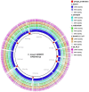
Genomic comparison of C. koseri URMITE with other Citrobacter species. High levels of sequence identity of the Citrobacter spp. genomes with the C. koseri URMITE genome are indicated as ring with colored tiles/blocks, whereas no or weak sequence identity is shown as white tiles/blocks. The C. koseri blue ring corresponds to the C. koseri ATCC BAA-895 genome. The C. koseri URMITE specific regions are mainly annotated as prophage (red tiles).
Figure 3
Figure 3
Phylogeny of strain-specific genes. The figure indicates the probable phylogenetic origin of the strain-specific genes at the genus level. The C. koseri URMITE and C. koseri ATCC BAA-895 specific genes are shown by square and star markers, respectively. The gene phylogeny with low branch supports or unresolved were not shown. The flow of gene exchange mainly involved Enterobacteriaceae spp.
Figure 4
Figure 4
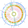
Overview and organization of the pCitro1 plasmid. From outside to inside circles. The light blue tiles are the pCitro1 ORFs. The violet tiles are Transposases/Resolvases; the ORFs related to the Y. pestis pPCP1 are in red; the tra operon is in dark blue and the blue ribbon indicates a 5 kb inverted repeat. The protein sequence identity with its best blast hit is plotted in the continuous gray (twilight zone <25%), orange (25–90%), and light green (>90%) circles. Below, ten circles showing the taxonomic origin of the ten best blast hits of each protein. Pink, orange, blue and red tiles belong to Escherichia, Citrobacter, Klebsiella and Yersinia genera, respectively. The yellow circle plot is the GC skew.
Figure 5
Figure 5
The taxonomic diversity of the plasmidome. Protein origins of the plasmidome using phylogenetic inference or best Blast hit. The pCitro1 proteins were related to multiple genera of the Enterobacteriaceae group including Klebsiella, Salmonella, Erwinia, Escherichia, Citrobacter and Yersinia. Four proteins of the pCitro1, including Pla, are exclusively shared with the virulence-associated 9.6 kb Y. pestis pPCP1 plasmid biovar Antiqua, Orientalis, Microtus, and Medievalis. The pCitro2 proteins were related to the Enterobacteriaceae group including the Yersinia genus. Several proteins of pCitro2 are identified as homologs to those from PY99, PY113, PY02, PY05, PY01 contigs of Peruvian Y. pestis species. We showed only five members of these Y. pestis strains but more than 50 Peruvian Y. pestis draft genomes exhibit a genomic fragment highly similar to the complete sequence of pCitro2.
Figure 6
Figure 6
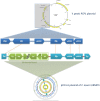
Comparison of the pla region of Y. pestis pPCP1 with the pCitro1. The 9.6 kb Y. pestis pPCP1 plasmid shows major similarity points with a pCitro1 region. The structural organization and the protein sequence identity (>99%) are highly conserved between pCitro1 and Y. pestis pPCP1 for Pla, PCP09 and PCP10 proteins. The PCP07 and the immunity Pim protein were not found in pCitro1. A fragment (54 amino acids) of the specific bacteriocin Pst protein is recovered downstream the 5 kb inverted repeat of pCitro1. Trp, Res and HP indicate transposase, resolvase, and hypothetical proteins, respectively.
Figure 7
Figure 7
Phylogeny inference of the main members of the omptin family including the predicted Pla protein of pCitro1. The phylogeny of the omptin family indicated that our predicted Pla protein is closely related to that of Y. pestis.

References

    1. Achtman M., Morelli G., Zhu P., Wirth T., Diehl I., Kusecek B., et al. (2004). Microevolution and history of the plague bacillus, Yersinia pestis. Proc. Natl. Acad. Sci. U.S.A. 101, 17837–17842. 10.1073/pnas.0408026101 - DOI - PMC - PubMed
    1. Adjemian J. Z., Adjemian M. K., Foley P., Chomel B. B., Kasten R. W., Foley J. E. (2008). Evidence of multiple zoonotic agents in a wild rodent community in the eastern Sierra Nevada. J. Wildl. Dis. 44, 737–742. 10.7589/0090-3558-44.3.737 - DOI - PubMed
    1. Aepfelbacher M., Trasak C., Ruckdeschel K. (2007). Effector functions of pathogenic Yersinia species. Thromb. Haemost. 98, 521–529. 10.1160/th07-03-0173 - DOI - PubMed
    1. Alikhan N. F., Petty N. K., Ben Zakour N. L., Beatson S. A. (2011). BLAST Ring Image Generator (BRIG): simple prokaryote genome comparisons. BMC Genomics 12:402. 10.1186/1471-2164-12-402 - DOI - PMC - PubMed
    1. Benson D. A., Cavanaugh M., Clark K., Karsch-Mizrachi I., Lipman D. J., Ostell J., et al. (2013). GenBank. Nucleic Acids Res. 41, D36–D42. 10.1093/nar/gks1195 - DOI - PMC - PubMed

LinkOut - more resources