Review
. 2015;37(2):209-22.
doi: 10.35946/arcr.v37.2.06.

The First Line of Defense: The Effects of Alcohol on Post-Burn Intestinal Barrier, Immune Cells, and Microbiome

Affiliations
Review

The First Line of Defense: The Effects of Alcohol on Post-Burn Intestinal Barrier, Immune Cells, and Microbiome

Adam M Hammer et al. Alcohol Res. 2015.

Abstract

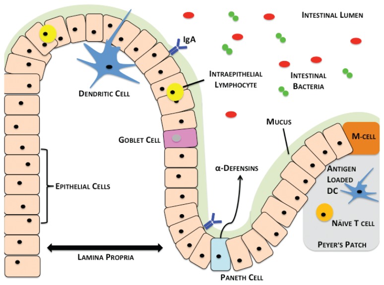
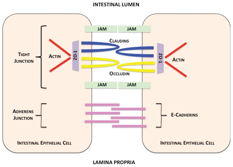
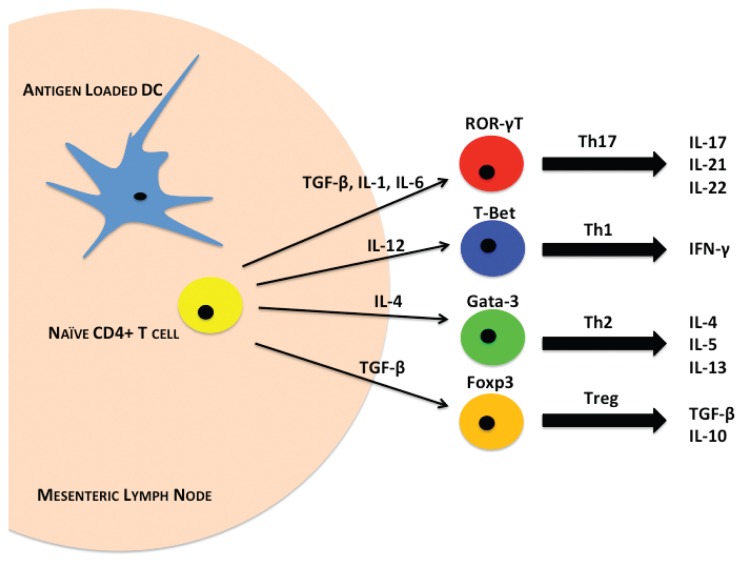
Alcohol (ethanol) is one of the most globally abused substances, and is one of the leading causes of premature death in the world. As a result of its complexity and direct contact with ingested alcohol, the intestine represents the primary source from which alcohol-associated pathologies stem. The gut is the largest reservoir of bacteria in the body, and under healthy conditions, it maintains a barrier preventing bacteria from translocating out of the intestinal lumen. The intestinal barrier is compromised following alcohol exposure, which can lead to life-threatening systemic complications including sepsis and multiple organ failure. Furthermore, alcohol is a major confounding factor in pathology associated with trauma. Experimental data from both human and animal studies suggest that alcohol perturbs the intestinal barrier and its function, which is exacerbated by a "second hit" from traumatic injury. This article highlights the role of alcohol-mediated alterations of the intestinal epithelia and its defense against bacteria within the gut, and the impact of alcohol on intestinal immunity, specifically on T cells and neutrophils. Finally, it discusses how the gut microbiome both contributes to and protects the intestines from dysbiosis after alcohol exposure and trauma.


Figures

Figure 1
Figure 1
Overview of the intestinal barrier, immune cells, and microbiome. Lumenal bacteria (red and green) are relegated to the lumen of the intestine by the intestinal barrier composed of the mucus (green), which contains IgA bound antibodies (blue) and epithelial cells. The epithelial-cell layer contains intraepithelial lymphocytes (yellow) and mucin-secreting goblet cells (pink). At the base of the intestinal crypts lie Paneth cells (light blue), which secrete alpha-defensins. Directly below the epithelial layer lies the lamina propria. Dendritic cells sample the lumenal bacterial contents and migrate to Peyer’s patches (gray) within the small intestine, where they interact with T cells (orange). M cells allow the passage of antigens into Peyer’s patches for uptake by resident antigen presenting cells.
Figure 2
Figure 2
Intestinal epithelial-cell junctions. Contents within the intestinal lumen are prevented from passing between epithelial cells by apical tight-junction complexes. Tight junctions are composed of claudin proteins (blue) and regulated by occludin proteins (yellow). Claudin and occludin proteins are transmembrane proteins attached to an adaptor molecule, zonula occludins protein 1 (ZO-1) (purple), which anchors tight-junction proteins to intracellular actin (red). Alcohol causes disruption of occludin and ZO proteins by an unknown mechanism. Junctional adhesion molecules (JAMs) (green) also support tight-junction interactions. Intestinal epithelial cells are further supported by adherens molecules, including E-cadherins (pink), which also contribute to cell–cell contact. These junctions allow selective separation of the intestinal lumen (top) and lamina propria (bottom).
Figure 3
Figure 3
Intestinal CD4+ T-cell differentiation. Antigen-loaded dendritic cells (DCs) interact with naïve CD4+ T cells (yellow) in mesenteric lymph nodes through MHC-II molecules. DCs secrete different cytokines depending on this interaction. Following alcohol and burn injury, antigen-presenting cells (APCs) such as DCs may have a significantly altered cytokine expression profile. The cytokine profiles present lead to the expression of different transcription factors that promote differentiation of T cells into either Th17 (red), Th1 (blue), Th2 (green), or Treg (orange) phenotypes. These T-cell subsets secrete different cytokines that lead to inflammatory or immunosuppressive immune responses. Combined alcohol and burn injury has been shown to suppress T-cell cytokines including interferon (IFN)-γ, interleukin (IL)-17, and IL-22 from T cells.

References

    1. Ahern PP, Schiering C, Buonocore S, et al. Interleukin-23 drives intestinal inflammation through direct activity on T cells. Immunity. 2010;33(2):279–288. - PMC - PubMed
    1. Akhtar S, Li X, Chaudry IH, Choudhry MA. Neutrophil chemokines and their role in IL-18-mediated increase in neutrophil O2-production and intestinal edema following alcohol intoxication and burn injury. American Journal of Physiology Gastrointestinal and Liver Physiology. 2009;297(2):G340–G347. - PMC - PubMed
    1. Akhtar S, Li X, Kovacs EJ, et al. Interleukin-18 delays neutrophil apoptosis following alcohol intoxication and burn injury. Molecular Medicine. 2011;17(1–2):88–94. - PMC - PubMed
    1. Amin PB, Diebel LN, Liberati DM. Ethanol effects proinflammatory state of neutrophils in shock. Journal of Surgical Research. 2007a;142(2):250–255. - PubMed
    1. Amin PB, Diebel LN, Liberati DM. The intestinal epithelial cell modulates the effect of alcohol on neutrophil inflammatory potential. Journal of Trauma. 2007b;63(6):1223–1229. - PubMed

Publication types