. 2016 Jan 12;113(2):380-5.
doi: 10.1073/pnas.1520947112. Epub 2015 Nov 30.

Human autoreactive T cells recognize CD1b and phospholipids

Affiliations

Human autoreactive T cells recognize CD1b and phospholipids

Ildiko Van Rhijn et al. Proc Natl Acad Sci U S A. .

Abstract

In contrast with the common detection of T cells that recognize MHC, CD1a, CD1c, or CD1d proteins, CD1b autoreactive T cells have been difficult to isolate in humans. Here we report the development of polyvalent complexes of CD1b proteins and carbohydrate backbones (dextramers) and their use in identifying CD1b autoreactive T cells from human donors. Activation is mediated by αβ T-cell receptors (TCRs) binding to CD1b-phospholipid complexes, which is sufficient to activate autoreactive responses to CD1b-expressing cells. Using mass spectrometry and T-cell responses to scan through the major classes of phospholipids, we identified phosphatidylglycerol (PG) as the immunodominant lipid antigen. T cells did not discriminate the chemical differences that distinguish mammalian PG from bacterial PG. Whereas most models of T-cell recognition emphasize TCR discrimination of differing self and foreign structures, CD1b autoreactive T cells recognize lipids with dual self and foreign origin. PG is rare in the cellular membranes that carry CD1b proteins. However, bacteria and mitochondria are rich in PG, so these data point to a more general mechanism of immune detection of infection- or stress-associated lipids.

Keywords: CD1b; T cell; dendritic cell; lipid antigen; self-antigen.


Conflict of interest statement

The authors declare no conflict of interest.

Figures
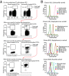
Fig. 1.
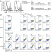
Fig. 1.
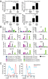
Generation of T-cell lines that recognize bacterial lipids presented by CD1b. (A) T cells from donor A25 that bind to CD1b dextramers loaded with S. Typhimurium lipids were enriched by multiple rounds of FACS sorting and expansion until they reached near-uniform binding of bacterial lipid extract-loaded CD1b dextramers. (B) Flow cytometric analysis of the cell line shown in A, as well as a S. aureus lipid-specific cell line from the same donor, using the indicated CD1 dextramers. Two additional T-cell lines were analyzed that were specific for B. melitensis and S. aureus lipids, derived from donor BC8. For each T-cell line, at least five flow cytometric experiments were performed with similar results.
Fig. 2.
Fig. 2.
Bacterial lipid-specific T cells are autoreactive to CD1b proteins on mammalian cells. Bacterial lipid-specific T-cell lines were stimulated with K562 cell lines stably transfected with the indicated CD1 isoform (a, CD1a; b, CD1b), with or without exogenously added S. Typhimurium (Sal.) or S. aureus (Staph.) lipids in an IFN-γ ELISPOT assay (A) or stimulated with mock-transfected or CD1b-transfected C1R lines in the absence of exogenous antigen, on which supernatants were harvested after 24 h and analyzed for cytokine content (B). Only those cytokines are shown that were produced upon stimulation with C1R.CD1b, but not on stimulation with mock-transfected C1R cells. In addition to the cytokines shown, 11 analytes were produced on stimulation with PMA and ionomycin, but not on stimulation with C1R.CD1b: IL-3, IL-6, IL-9, IL-10, IL-17, IL-22, IL-31, LT-α (TNF-β), chemokine (C-C motif) ligand 1 (CCL-1) (I-309), CXC chemokine ligand 1 (CXCL1) (IP-10), latency-associated peptide of TGF-β. Production of the following 15 analytes was tested but not detected in response to C1R.CD1b or PMA and ionomycin: IL-1 receptor antagonist (IL-1Ra), IL-5, IL-12, IL-21, IL-25, IFN-α, IFN-β, April, macrophage migration inhibitory factor, CCL17, CXCL, soluble programmed death 1 (sPD1), soluble Fas (sFas), sFas-L, and sTNF-RI. (C) The A25Salmonella T-cell line was stimulated with CD1a- or CD1b-transfected cells for 20 h before flow cytometric analysis of CD25 or CD3 expression. In A and C, one representative experiment of three is shown. For B, one experiment was performed on the pooled supernatants of triplicate wells. *P < 0.05; **P < 0.01; NS, not significant.
Fig. S1.
Fig. S1.
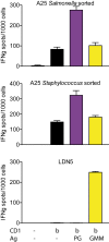
GMM-specificity and autoreactivity are distinct specificities. The GMM-specific T-cell line LDN5 and autoreactive T-cell lines were stimulated with wild-type or CD1b-transfected C1R cells, in the absence or presence of PG or glucose monomycolate in an IFN-γ ELISPOT assay.
Fig. S2.
Fig. S2.
CD1b expression by antigen-presenting cells. Flow cytometric analysis of CD1b-transfected K562 cells or human primary monocyte-derived DCs (GM-CSF and IL-4 treated monocytes), stained with the IgG1 isotype control antibody P3 or the anti CD1b antibody BCD1b.3, followed by goat-anti mouse Ig- phycoerythrin.
Fig. S3.
Fig. S3.
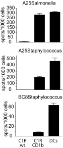
Untransfected antigen-presenting cells also stimulate autoreactive T cells. Bacterial lipid-specific T-cell lines were stimulated with wild-type or CD1b-transfected C1R cells, or with human monocyte-derived DCs, which are primary monocytes that had been treated with GM-CSF and IL-4 for 72 h, in an IFN-γ ELISPOT assay.
Fig. 3.
Fig. 3.
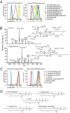
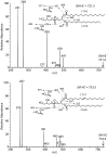
Phosphatidylglycerol is a major target of CD1b-autoreactive T cells. (A) CD1b dextramers were loaded with the major classes of phospholipids and used to stain bacterial lipid-specific T-cell lines. (B) Negative ions present in the methanol-eluted fractions from S. aureus and S. Typhimurium were analyzed by collisional mass spectrometry and generated patterns that are consistent with PG. (C) The same two cell lines that were shown in a were analyzed using CD1b dextramers loaded with PG from mammalian and bacterial sources, the structures of which are shown in D. A and C represent one of two independent experiments. Even though the branching pattern of fatty acids could not be determined directly, S. aureus fatty acids are drawn as anteiso forms because that is the most common form for C15 and C17 in this species (22). The C17 fatty acid in S. Typhimurium can be C17:1 or cyclopropyl C17 (38).
Fig. S4.
Fig. S4.
Collision-induced dissociation mass spectrometry. Negative ion mode collision-induced dissociation mass spectrometry of m/z 721.4 from S. aureus (Top) and m/z 733.4 in S. typhimurium (Bottom).
Fig. 4.
Fig. 4.
Single CD1b autoreactive TCRs recognize multiple phospholipids. Two TCRs that were cloned from the A25Salmonella T-cell line, named PG10 and PG90, were stably transduced into Jurkat cells. (A) The PG10 and PG90 TCR-expressing Jurkat cells were incubated overnight with wild-type C1R cells or CD1b-transfected C1R cells before they were stained with an antibody against CD69. PG10- and PG90-expressing Jurkat cells were stained with CD1b tetramers loaded with the indicated lipids (B). Results in B represent three or more independently performed experiments.

Comment in

References

    1. Dougan SK, Kaser A, Blumberg RS. CD1 expression on antigen-presenting cells. Curr Top Microbiol Immunol. 2007;314:113–141. - PubMed
    1. Jackman RM, et al. The tyrosine-containing cytoplasmic tail of CD1b is essential for its efficient presentation of bacterial lipid antigens. Immunity. 1998;8(3):341–351. - PubMed
    1. Porcelli S, Morita CT, Brenner MB. CD1b restricts the response of human CD4-8- T lymphocytes to a microbial antigen. Nature. 1992;360(6404):593–597. - PubMed
    1. Moody DB, et al. Structural requirements for glycolipid antigen recognition by CD1b-restricted T cells. Science. 1997;278(5336):283–286. - PubMed
    1. Gilleron M, et al. Diacylated sulfoglycolipids are novel mycobacterial antigens stimulating CD1-restricted T cells during infection with Mycobacterium tuberculosis. J Exp Med. 2004;199(5):649–659. - PMC - PubMed

Publication types