. 2016 Jan 1;310(1):G26-33.
doi: 10.1152/ajpgi.00293.2015. Epub 2015 Nov 5.

Glucagon-like peptide-1 receptor is present in pancreatic acinar cells and regulates amylase secretion through cAMP

Affiliations

Glucagon-like peptide-1 receptor is present in pancreatic acinar cells and regulates amylase secretion through cAMP

Yanan Hou et al. Am J Physiol Gastrointest Liver Physiol. .

Abstract

Glucagon-like peptide-1 (GLP-1) is a glucoincretin hormone that can act through its receptor (GLP-1R) on pancreatic β-cells and increase insulin secretion and production. GLP-1R agonists are used clinically to treat type 2 diabetes. GLP-1 may also regulate the exocrine pancreas at multiple levels, including inhibition through the central nervous system, stimulation indirectly through insulin, and stimulation directly on acinar cells. However, it has been unclear whether GLP-1R is present in pancreatic acini and what physiological functions these receptors regulate. In the current study we utilized GLP-1R knockout (KO) mice to study the role of GLP-1R in acinar cells. RNA expression of GLP-1R was detected in acutely isolated pancreatic acini. Acinar cell morphology and expression of digestive enzymes were not affected by loss of GLP-1R. GLP-1 induced amylase secretion in wild-type (WT) acini. In GLP-1R KO mice, this effect was abolished, whereas vasoactive intestinal peptide-induced amylase release in KO acini showed a pattern similar to that in WT acini. GLP-1 stimulated cAMP production and increased protein kinase A-mediated protein phosphorylation in WT acini, and these effects were absent in KO acini. These data show that GLP-1R is present in pancreatic acinar cells and that GLP-1 can regulate secretion through its receptor and cAMP signaling pathway.

Keywords: amylase secretion; cAMP; glucagon-like peptide-1 receptor; pancreatic acini.


Figures

Fig. 1.
Fig. 1.
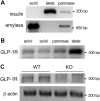
Glucagon-like peptide-1 (GLP-1) receptor (GLP-1R) is expressed in isolated pancreatic acinar cells. A: purity of hand-picked acini and islets was tested by RT-PCR using specific primers for insulin and amylase. B: expression of GLP-1R in 2 separate batches of hand-picked acini or representative islets and total pancreas was assessed by RT-PCR. C: expression of GLP-1R in isolated wild-type (WT) and GLP-1R knockout (KO) acini was examined by RT-PCR. Each lane represents samples from 1 mouse.
Fig. 2.
Fig. 2.
GLP-1R deficiency does not affect acinar cell morphology and expression of digestive enzymes. Pancreas tissues were obtained from WT (A and C) and GLP-1R KO (B and D) mice. A and B: hematoxylin-eosin-stained paraffin sections show a normal organization of pancreas in WT and GLP-1R KO mice, with zymogen granules (stained pink) in the apical region of acinar cells and basophilic (purple) staining in the acinar basal compartment. An islet in the section from the KO mouse (*) has normal morphology. C and D: fixed pancreas was cryosectioned at 6 μm and stained with Oregon Green 488-phalloidin to label actin (green), anti-amylase antibody (red), and 4′,6-diamidino-2-phenylindole (blue) to stain nuclei. Images were obtained using confocal microscopy; a projection of 4 or 5 optical slices (0.5 μm) is shown. Arrowheads in C and D denote subluminal actin. Distribution of apical zymogen granules and actin is similar in WT and KO mice.
Fig. 3.
Fig. 3.
GLP-1R deficiency does not affect expression of digestive enzymes. Total lysates were prepared from tissues obtained from WT and KO mice and then separated on a polyacrylamide gel. A: expression of major digestive enzymes in intact pancreas tissues was assessed by Western blotting using specific antibodies. Each lane represents 10-μg samples from 1 mouse. B: densitometry analysis of Western blot results on digestive enzymes. Values are means ± SE from 3 experiments, with 3–4 mice of each genotype in each experiment. CTL, control (WT). C: expression of major digestive enzymes in pancreas, liver, and kidney tissues was assessed by Western blotting using specific antibodies.
Fig. 4.
Fig. 4.
GLP-1 induces amylase release in acini from WT, but not GLP-1R KO, mice. Freshly isolated acini were incubated with 30 pM, 100 pM, 300 pM, 3 nM, and 30 nM GLP-1 (A and C) or 100 pM, 1 nM, and 10 nM vasoactive intestinal peptide (VIP, B and D) for 30 min. Amylase release is expressed as percentage of total acinar amylase content (A and B) or released amylase per milligram DNA (C and D). Values are means ± SE from 5–13 independent experiments. *P < 0.05, vs. WT control. #P < 0.05 vs. GLP-1R KO control.
Fig. 5.
Fig. 5.
GLP-1R deficiency caused a decrease in amylase release induced by GLP-1, but not most other secretagogues. Freshly isolated acini were incubated with 30 pM cholecystokinin (CCK), 1 μM carbachol (CCh), 3 nM GLP, 1 nM VIP, 2 μM A23187, or 100 μM 8-(4-chlorophenylthio)-2′-O-Me-cAMP (pCPT-cAMP) for 30 min. Amylase release is expressed as percentage of total acinar amylase content. Values are means ± SE from 5–9 independent experiments. *P < 0.05.
Fig. 6.
Fig. 6.
Loss of GLP-1R leads to GLP-1-induced decrease in cAMP formation. Freshly prepared acini were treated with IBMX for 3 min and then with GLP-1 or VIP for 12 min. cAMP was extracted, and levels were measured by enzyme immunoassay. Values are means ± SE from 6 independent experiments. *P < 0.05 vs. WT control. #P < 0.05 vs. GLP-1R KO control.
Fig. 7.
Fig. 7.
Deletion of GLP-1R causes attenuated response to cAMP activation. Freshly obtained acini were treated with GLP-1 or VIP for 5 min. A: anti-phosphorylated (Ser/Thr) PKA substrate blot was used to show cAMP activation. pPKA, phosphorylated PKA. B: quantification of p-PKA substrate signals. Values are means ± SE from 6 independent experiments. *P < 0.05 vs. WT control. #P < 0.05 vs. GLP-1R KO control.

References

    1. Ahmad SR, Swann J. Exenatide and rare adverse events. N Engl J Med 358: 1970–1972, 2008. - PubMed
    1. Baldescu R, Chirulescu Z. Study of interrelationship between Zn, Ca and Mg seric concentrations in healthy subjects comparatively with diverse forms of cancer. Rom J Intern Med 32: 203–208, 1994. - PubMed
    1. Ban K, Noyan-Ashraf MH, Hoefer J, Bolz SS, Drucker DJ, Husain M. Cardioprotective and vasodilatory actions of glucagon-like peptide 1 receptor are mediated through both glucagon-like peptide 1 receptor-dependent and -independent pathways. Circulation 117: 2340–2350, 2008. - PubMed
    1. Buteau J, Foisy S, Joly E, Prentki M. Glucagon-like peptide 1 induces pancreatic beta-cell proliferation via transactivation of the epidermal growth factor receptor. Diabetes 52: 124–132, 2003. - PubMed
    1. Buteau J, Foisy S, Rhodes CJ, Carpenter L, Biden TJ, Prentki M. Protein kinase Cξ activation mediates glucagon-like peptide-1-induced pancreatic beta-cell proliferation. Diabetes 50: 2237–2243, 2001. - PubMed

Publication types

MeSH terms

LinkOut - more resources