. 2015:2015:412946.
doi: 10.1155/2015/412946. Epub 2015 Oct 7.

Neuroprotective Effects of Clostridium butyricum against Vascular Dementia in Mice via Metabolic Butyrate

Affiliations

Neuroprotective Effects of Clostridium butyricum against Vascular Dementia in Mice via Metabolic Butyrate

Jiaming Liu et al. Biomed Res Int. 2015.

Abstract

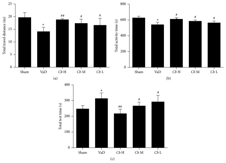
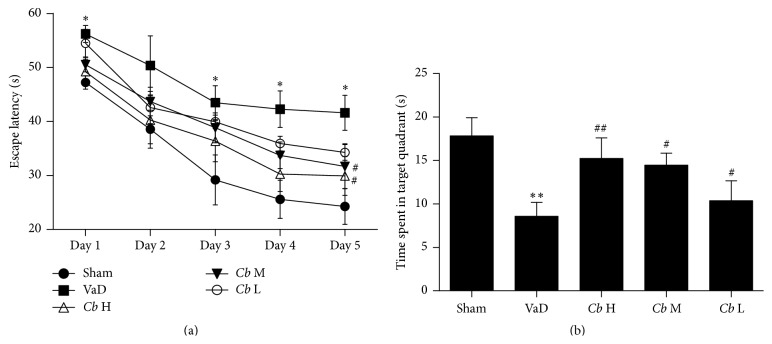
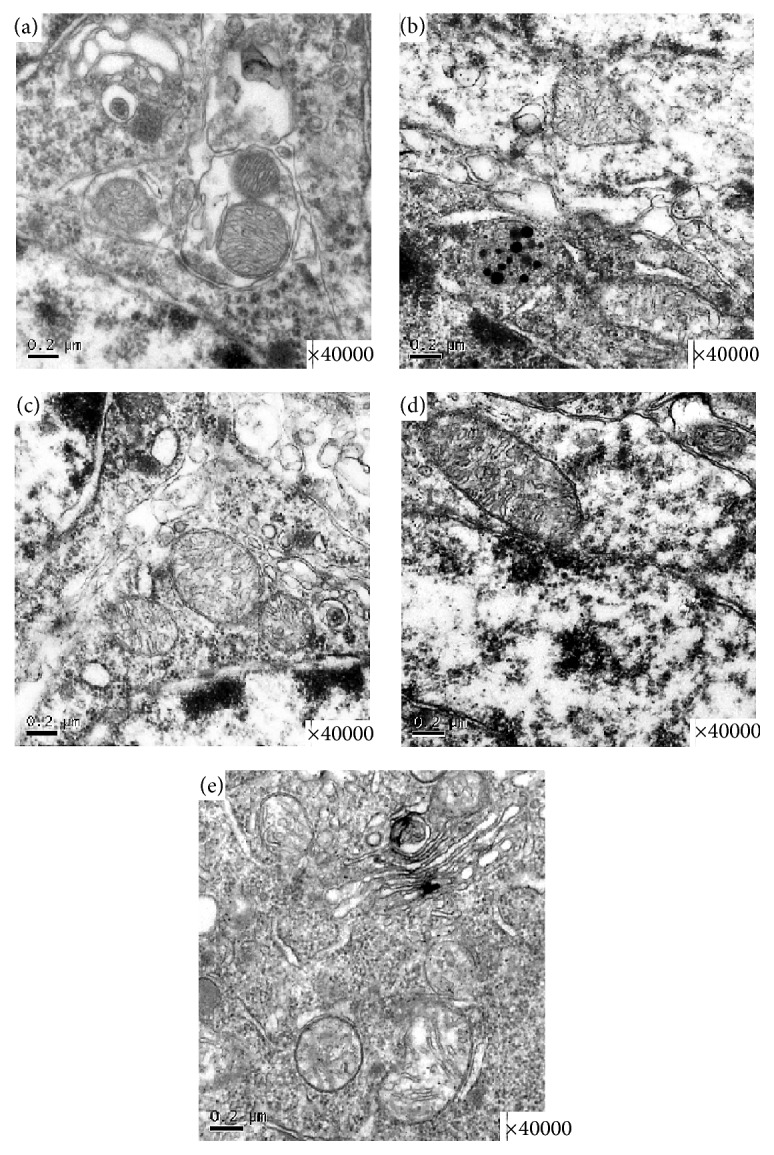
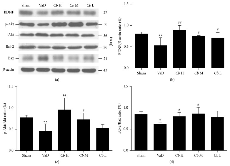
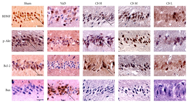
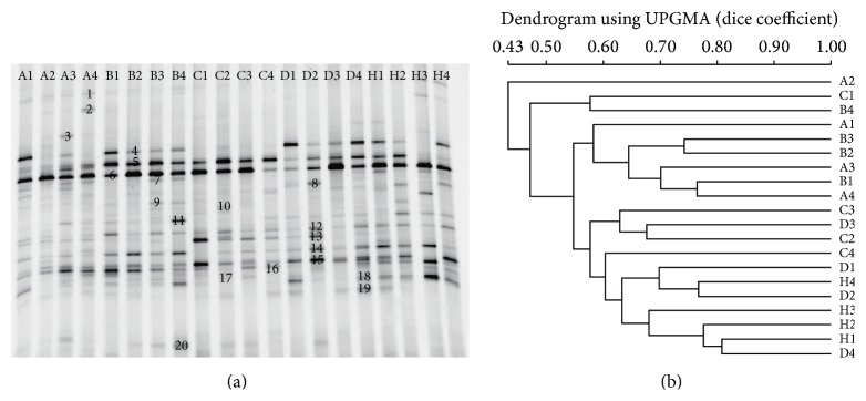
Probiotics actively participate in neuropsychiatric disorders. However, the role of gut microbiota in brain disorders and vascular dementia (VaD) remains unclear. We used a mouse model of VaD induced by a permanent right unilateral common carotid arteries occlusion (rUCCAO) to investigate the neuroprotective effects and possible underlying mechanisms of Clostridium butyricum. Following rUCCAO, C. butyricum was intragastrically administered for 6 successive weeks. Cognitive function was estimated. Morphological examination was performed by electron microscopy and hematoxylin-eosin (H&E) staining. The BDNF-PI3K/Akt pathway-related proteins were assessed by western blot and immunohistochemistry. The diversity of gut microbiota and the levels of butyrate in the feces and the brains were determined. The results showed that C. butyricum significantly attenuated the cognitive dysfunction and histopathological changes in VaD mice. C. butyricum not only increased the levels of BDNF and Bcl-2 and decreased level of Bax but also induced Akt phosphorylation (p-Akt) and ultimately reduced neuronal apoptosis. Moreover, C. butyricum could regulate the gut microbiota and restore the butyrate content in the feces and the brains. These results suggest that C. butyricum might be effective in the treatment of VaD by regulating the gut-brain axis and that it can be considered a new therapeutic strategy against VaD.


Figures

Figure 1
Figure 1
Open field test. (a) The total distance travelled over 1 h, (b) the time spent in the central area, and (c) the total rest time are presented for each group. Sham, sham-operated group; VaD, VaD model group; Cb H, C. butyricum- (1 × 10⁸ CFU-) treated group; Cb M, C. butyricum- (1 × 10⁷ CFU-) treated group; and Cb L, C. butyricum- (1 × 10⁶ CFU-) treated group. Error bars indicate SEM; P < 0.05 versus sham group; # P < 0.05 and ## P < 0.01 versus VaD group.
Figure 2
Figure 2
Morris water maze. (a) Escape latency (s). (b) The time spent in the target quadrant (s). Sham, sham-operated group; VaD, VaD model group; Cb H, C. butyricum- (1 × 10⁸ CFU-) treated group; Cb M, C. butyricum- (1 × 10⁷ CFU-) treated group; and Cb L, C. butyricum- (1 × 10⁶ CFU-) treated group. Error bars indicate SEM; P < 0.05 and ∗∗ P < 0.01 versus sham group; # P < 0.05 and ## P < 0.01 versus VaD group.
Figure 3
Figure 3
Representative photomicrographs of the ultrastructural changes observed in brain tissue. (a) A representative nucleolus in a hippocampal neuron of the sham-operated group. (b) A representative nucleolus of cerebral ischemia in the VaD group. (c) A representative nucleolus in the Cb L group, Cb M group (d), and Cb H group (e). Sham, sham-operated group; VaD, VaD model group; Cb H, C. butyricum- (1 × 10⁸ CFU-) treated group; Cb M, C. butyricum- (1 × 10⁷ CFU-) treated group; and Cb L, C. butyricum- (1 × 10⁶ CFU-) treated group.
Figure 4
Figure 4
Representative photomicrographs of the histopathological changes in area CA1 of the hippocampus in mice. (a) HE staining. (b) TUNEL staining. Cells with a brown-stained cytoplasm are considered positive. Sham, sham-operated group; VaD, VaD model group; Cb H, C. butyricum- (1 × 10⁸ CFU-) treated group; Cb M, C. butyricum- (1 × 10⁷ CFU-) treated group; and Cb L, C. butyricum- (1 × 10⁶ CFU-) treated group. Magnification: 400x. Scale bar = 20 μm.
Figure 5
Figure 5
Western blot analysis. (a) Western blot of the expression levels of BDNF, p-Akt, Akt, Bcl-2, and Bax in mouse hippocampus. (b) A quantitative analysis of the protein levels of BDNF from each group normalized to the loading control β-actin (c). (d) Bar graphs showing the protein level ratios of p-Akt/Akt and Bcl-2/Bax from each group; Sham, sham-operated group; VaD, VaD model group; Cb H, C. butyricum- (1 × 10⁸ CFU-) treated group; Cb M, C. butyricum- (1 × 10⁷ CFU-) treated group; and Cb L, C. butyricum- (1 × 10⁶ CFU-) treated group. Error bars indicate SD; n = 8 for each group. P < 0.01 and ∗∗ P < 0.01 versus sham group; # P < 0.05 and ## P < 0.01 versus VaD group.
Figure 6
Figure 6
Immunohistochemical staining. Representative images of the immunohistochemical staining of BDNF, p-Akt, Bcl-2, and Bax in area CA1 of the hippocampus. Cells with a brown-stained cytoplasm are considered positive. Sham, sham-operated group; VaD, VaD model group; Cb H, C. butyricum- (1 × 10⁸ CFU-) treated group; Cb M, C. butyricum- (1 × 10⁷ CFU-) treated group; and Cb L, C. butyricum- (1 × 10⁶ CFU-) treated group. Magnification: 400x. Scale bar = 20 μm.
Figure 7
Figure 7
PCR-DGGE analysis of the predominant fecal microbiota in mice. (a) PCR-DGGE fingerprints used to analyze the fecal microbiota of the samples from the VaD model group (A), sham-operated group (H), and C. butyricum- (B, 1 × 10⁶ CFU-; C, 1 × 10⁷ CFU-; and D, 1 × 10⁸ CFU-) treated groups. Each lane represents one subject that was randomly selected from each group. The bands marked in the DGGE gel were identified by cloning and sequencing to facilitate the interpretation of the figure. Bands: 1: Bacteroides sp.; 2: Hespellia sp.; 3: Clostridium XVIII sp.; 4: TM7 genera incertae sedis; 5: TM7 genera incertae sedis; 6: Anaerostipes sp.; 7: Barnesiella sp.; 8: Barnesiella sp.; 9: Roseburia sp.; 10: Acinetobacter sp.; 11: unclassified Helicobacteraceae sp.; 12: Streptobacillus sp.; 13: Barnesiella sp.; 14: Clostridium XIVa sp.; 15: Alistipes sp.; 16: unclassified Lachnospiraceae sp.; 17: Lachnospiraceae incertae sedis Alistipes sp.; 18: Anaerostipes sp.; 19: unclassified Porphyromonadaceae sp.; and 20: Butyricimonas sp. (b) Dendrogram of the DGGE profiles shown in (a).
Figure 8
Figure 8
Absolute concentration of butyrate (μmol g−1 wet weight). Sham, sham-operated group; VaD, VaD model group; Cb H, C. butyricum- (1 × 10⁸ CFU-) treated group; Cb M, C. butyricum- (1 × 10⁷ CFU-) treated group; and Cb L, C. butyricum- (1 × 10⁶ CFU-) treated group. Error bars indicate SD; n = 8 for each group. P < 0.05 versus sham group, ∗∗ P < 0.01 versus sham group, and # P < 0.05 and ## P < 0.01 versus VaD group.

References

    1. Sahathevan R., Brodtmann A., Donnan G. A. Dementia, stroke, and vascular risk factors; a review. International Journal of Stroke. 2012;7(1):61–73. doi: 10.1111/j.1747-4949.2011.00731.x. - DOI - PubMed
    1. Zhao Y., Gong C.-X. From chronic cerebral hypoperfusion to Alzheimer-like brain pathology and neurodegeneration. Cellular and Molecular Neurobiology. 2015;35(1):101–110. doi: 10.1007/s10571-014-0127-9. - DOI - PMC - PubMed
    1. Snyder H. M., Corriveau R. A., Craft S., et al. Vascular contributions to cognitive impairment and dementia including Alzheimer's disease. Alzheimer's & Dementia. 2015;11(6):710–717. doi: 10.1016/j.jalz.2014.10.008. - DOI - PMC - PubMed
    1. Zhang J., Guo W., Tian B., et al. Puerarin attenuates cognitive dysfunction and oxidative stress in vascular dementia rats induced by chronic ischemia. International Journal of Clinical and Experimental Pathology. 2015;8(5):4695–4704. - PMC - PubMed
    1. Sun Z.-K., Ma X.-R., Jia Y.-J., Liu Y.-R., Zhang J.-W., Zhang B.-A. Effects of resveratrol on apoptosis in a rat model of vascular dementia. Experimental and Therapeutic Medicine. 2014;7(4):843–848. doi: 10.3892/etm.2014.1542. - DOI - PMC - PubMed

Publication types

MeSH terms