. 2015 Jul;40(8):2001-14.
doi: 10.1038/npp.2015.50. Epub 2015 Feb 23.

Parabrachial Nucleus Contributions to Glucagon-Like Peptide-1 Receptor Agonist-Induced Hypophagia

Affiliations

Parabrachial Nucleus Contributions to Glucagon-Like Peptide-1 Receptor Agonist-Induced Hypophagia

Jennifer C Swick et al. Neuropsychopharmacology. 2015 Jul.

Abstract

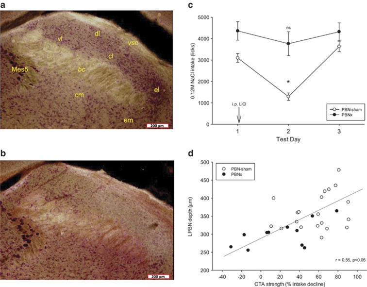
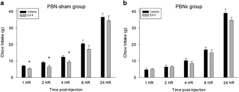
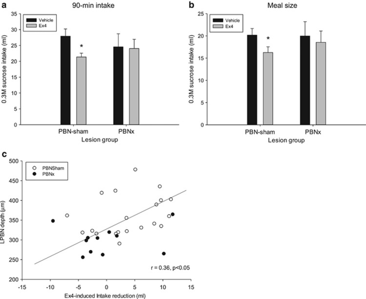
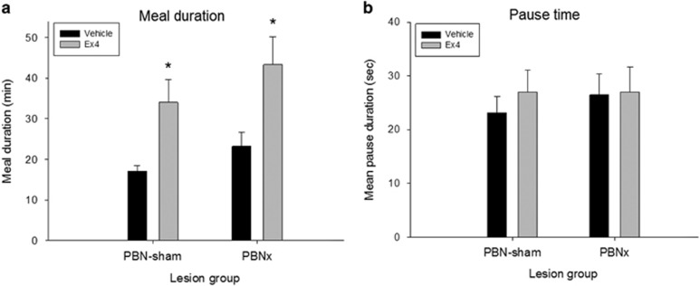
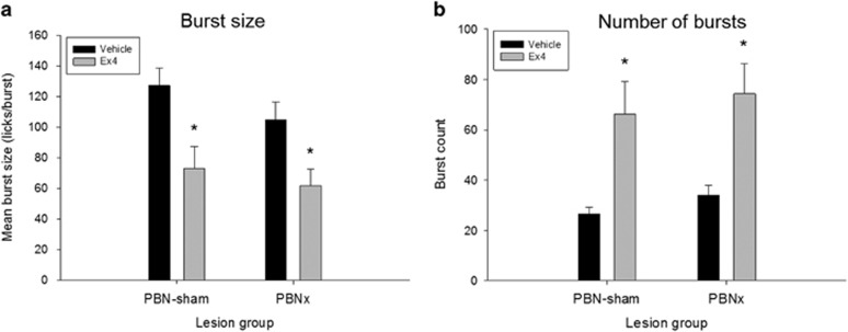
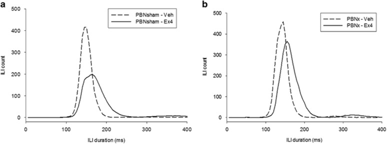
Exendin-4 (Ex4), a glucagon-like peptide-1 receptor (GLP-1R) agonist approved to treat type 2 diabetes mellitus, is well known to induce hypophagia in human and animal models. We evaluated the contributions of the hindbrain parabrachial nucleus (PBN) to systemic Ex4-induced hypophagia, as the PBN receives gustatory and visceral afferent relays and descending input from several brain nuclei associated with feeding. Rats with ibotenic-acid lesions targeted to the lateral PBN (PBNx) and sham controls received Ex4 (1 μg/kg) before 24 h home cage chow or 90 min 0.3 M sucrose access tests, and licking microstructure was analyzed to identify components of feeding behavior affected by Ex4. PBN lesion efficacy was confirmed using conditioned taste aversion (CTA) tests. As expected, sham control but not PBNx rats developed a CTA. In sham-lesioned rats, Ex4 reduced chow intake within 4 h of injection and sucrose intake within 90 min. PBNx rats did not show reduced chow or sucrose intake after Ex4 treatment, indicating that the PBN is necessary for Ex4 effects under the conditions tested. In sham-treated rats, Ex4 affected licking microstructure measures associated with hedonic taste evaluation, appetitive behavior, oromotor coordination, and inhibitory postingestive feedback. Licking microstructure responses in PBNx rats after Ex4 treatment were similar to sham-treated rats with the exception of inhibitory postingestive feedback measures. Together, the results suggest that the PBN critically contributes to the hypophagic effects of systemically delivered GLP-1R agonists by enhancing visceral feedback.


Figures

Figure 1
Figure 1
Left: Representative dark-field microphotographs (x40 magnification) of the lateral parabrachial nucleus (LPBN) in an intact sham-lesioned rat (a) and an ibotenic acid-lesioned rat (b). In (a), clusters of neutral red-stained somata in all regions dorsal to the brachium conjuctivum are evident, and these include the ventral lateral, dorsal lateral, central lateral, and external lateral subnuclei. In the ibotenic-acid-lesioned rat there is a clear paucity of stained somata in all of these subnuclei, although somata loss is weaker in the rostral ventral lateral subnucleus. Note also the distinct lateral border of the lesion where intact somata from the external medial subnucleus are visible. In several PBN-lesioned brains the depth of the region dorsal to the brachium was also significantly attenuated. (c) NaCl intake responses in PBN-sham and PBN-lesioned rats. Water-deprived rats in both groups initially avidly consumed a normally preferred NaCl solution as measured by mean lick count (±SEM) during a 30-min test. Both groups were injected with LiCl but only PBNsham-lesioned rats (open circles) exhibited intake reduction on the second test day. The CTA rapidly extinguished by the third day of testing. *P<0.05. (d) The extent of intake suppression induced by LiCl was positively and significantly correlated with the size of the LPBN. Intact sham-lesioned rats (open circles) with larger LPBN regions tended to exhibit fewer licks for NaCl after LiCl treatment. bc, brachium conjuctivum; cl, central lateral subnucleus; dl, dorsal lateral subnucleus; el, external lateral subnucleus; em, external medial subnucleus; Mes5, mesencephalic trigeminal motor nucleus; vl, ventral lateral subnucleus; vsc, ventral spinocerebellar tract.
Figure 2
Figure 2
Exendin-4 (1 μg/kg, i.p.) significantly reduced chow intake in intact (PBNsham) but not PBN-lesioned (PBNx) rats. (a) Cumulative chow intake (mean+SEM) measured at 1, 2, 4, 8, and 24 h time intervals in sham-lesioned rats (n=20), *p<0.05. (b) Cumulative chow intake (mean+SEM) measured at 1, 2, 4, 8, and 24 h time intervals in rats with bilateral PBN lesions (n=12).
Figure 3
Figure 3
(a) Mean 90 min 0.3 M sucrose intake (+SEM) after vehicle (saline) or exendin-4 (Ex4; 1 μg/kg, i.p.) injection in intact sham-lesioned rats (PBNsham) and rats with ibotenic-acid lesions of the PBN (PBNx). Ex4 significantly reduced 0.3 M sucrose solution intake in PBNsham rats but not PBNx rats. (b) The 0.3 M sucrose meal size (+SEM) after vehicle or Ex4 injection in PBNsham and PBNx rats. Ex4 significantly reduced 0.3 M sucrose solution intake PBNsham rats but not PBNx rats. *P<0.05. (c) The extent of intake suppression induced by Ex4 was positively and significantly correlated with the size of the lateral PBN (LPBN). Sham-lesioned rats (open circles); PBNx rats (filled circles). Rats with larger LPBN regions tended to show more sucrose intake reduction after Ex4 treatment (vehicle condition intake minus Ex4 condition intake (ml)).
Figure 4
Figure 4
Exendin-4 (Ex4; 1 μg/kg, i.p.) significantly prolonged meal duration in both groups but did not prolong pauses between bursts of licking. (a) Mean meal duration (+SEM) after vehicle (saline) or exendin-4 (Ex4) injection in intact sham-lesioned rats (PBNsham) and rats with ibotenic-acid lesions of the PBN (PBNx). Ex4 significantly prolonged the meal in both groups. (b) Mean pause duration (+SEM) after vehicle or Ex4 injection in PBNsham and PBNx rats. Ex4 did not affect the mean interval between bursts of licking in either lesion group. *P<0.05.
Figure 5
Figure 5
(a) Mean (+SEM) burst size per meal after vehicle (saline) or exendin-4 (Ex4; 1 μg/kg, i.p.) injection in intact PBN sham-lesioned rats (PBNsham) and rats with ibotenic-acid lesions of the PBN (PBNx). Ex4 significantly reduced the average burst size in both groups. (b) Mean (+SEM) number of bursts per meal after vehicle or Ex4 injection in PBNsham and PBNx rats. Ex4 significantly increased the number of meal bursts in both groups. *P<0.05.
Figure 6
Figure 6
Mean ingestion rate plotted by meal thirds (means±SEM) after vehicle (saline) or exendin-4 (Ex4; 1 μg/kg) injection in intact sham-lesioned rats (PBNsham) and rats with ibotenic-acid lesions of the PBN (PBNx). Meals were temporally divided into thirds and the average lick rate (licks/min) was determined for each meal third. (a) Mean lick rate by meal thirds after vehicle (open triangles) and Ex4 injection (filled triangles) in PBNsham rats. Ex4 significantly slowed the rate of ingestion in PBNsham rats relative to the vehicle condition for all three phases of the meal. (b) Mean lick rate by meal thirds after vehicle (open triangles) and Ex4 (filled triangles) injection in PBNx rats. PBNx rats showed a less robust reduction in ingestion rate after Ex4 injection, with no differences relative to vehicle in the last two thirds of the meal. *P<0.05.
Figure 7
Figure 7
Mean interlick interval (ILI) counts (5 ms bins) after vehicle (saline) or exendin-4 (Ex4; 1 μg/kg) injection in (a) intact sham-lesioned rats (PBNsham) and (b) rats with ibotenic-acid lesions of mostly the lateral PBN (PBNx). Ex4 significantly slowed the rate of licking within bursts in both lesion groups as indicated by a comparable right shift in the distribution of ILIs <250 ms. Note that PBNx rats exhibited more ILIs (and therefore, licks) after Ex4 injection relative to the PBNsham group, reflecting the lack of intake reduction after Ex4 in PBNx rats.

References

    1. Alhadeff AL, Baird J-P, Swick JC, Hayes MR, Grill HJ (2014). Glucagon-like peptide-1 receptor signaling in the lateral parabrachial nucleus contributes to the control of food intake and motivation to feed. Neuropsychopharmacology 39: 2233–2243. - PMC - PubMed
    1. Alhadeff AL, Grill HJ (2014). Hindbrain nucleus tractus solitarius glucagon-like peptide-1 receptor signaling reduces appetitive and motivational aspects of feeding. Am J Physiol Regul Integr Comp Physiol 307: R465–R470. - PMC - PubMed
    1. Antin J, Gibbs J, Holt J, Young RC, Smith GP (1975). Cholecystokinin elicits the complete behavioral sequence of satiety in rats. J Comp Physiol Psychol 89: 784–790. - PubMed
    1. Asarian L, Corp ES, Hrupka B, Geary N (1998). Intracerebroventricular glucagon-like peptide-1 (7-36) amide inhibits sham feeding in rats without eliciting satiety. Physiol Behav 64: 367–372. - PubMed
    1. Baird J-P, Choe A, Loveland JL, Beck J, Mahoney CE, Lord JS et al (2009). Orexin-A hyperphagia: hindbrain participation in consummatory feeding responses. Endocrinology 150: 1202–1216. - PMC - PubMed

Publication types

MeSH terms