. 2015 Jan 27;14(1):10.
doi: 10.1186/s12943-014-0274-0.

Activation of the NF-κB pathway as a mechanism of alcohol enhanced progression and metastasis of human hepatocellular carcinoma

Affiliations

Activation of the NF-κB pathway as a mechanism of alcohol enhanced progression and metastasis of human hepatocellular carcinoma

Fei Wang et al. Mol Cancer. .

Expression of concern in

Abstract

Background: Hepatocellular carcinoma (HCC), the most common form of primary liver cancer, is the third leading cause of cancer-related death in human. Alcohol is a known risk factor for HCC. However it is still unclear whether and how alcohol enhances the progression and metastasis of existing HCC.

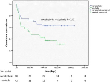
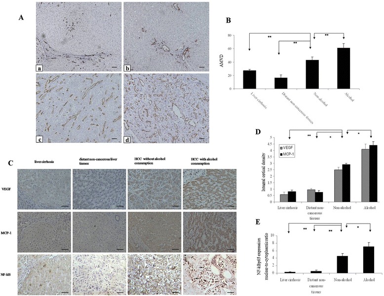
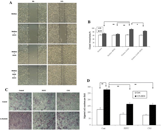
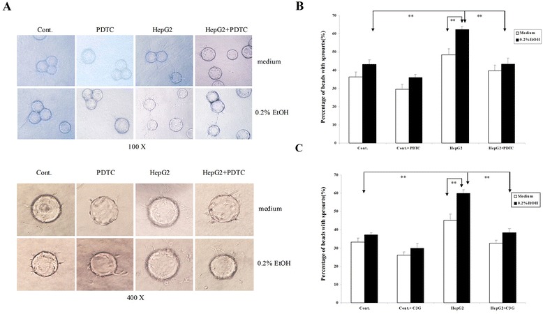
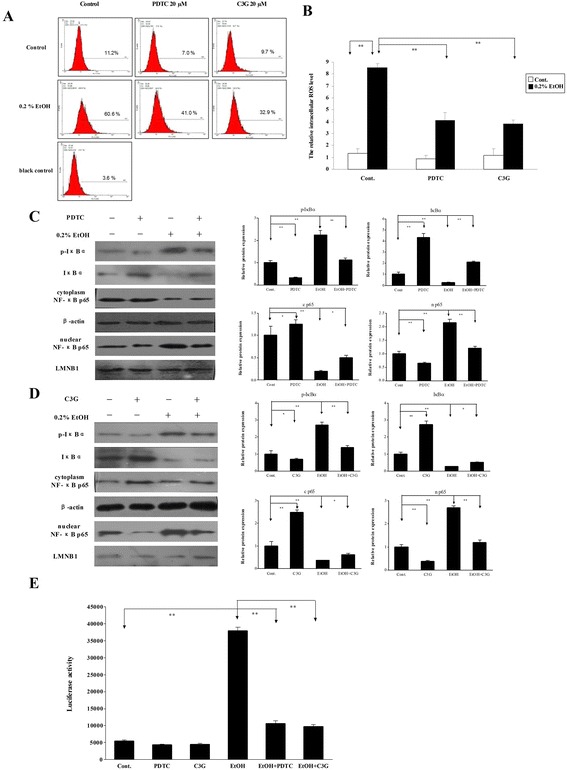
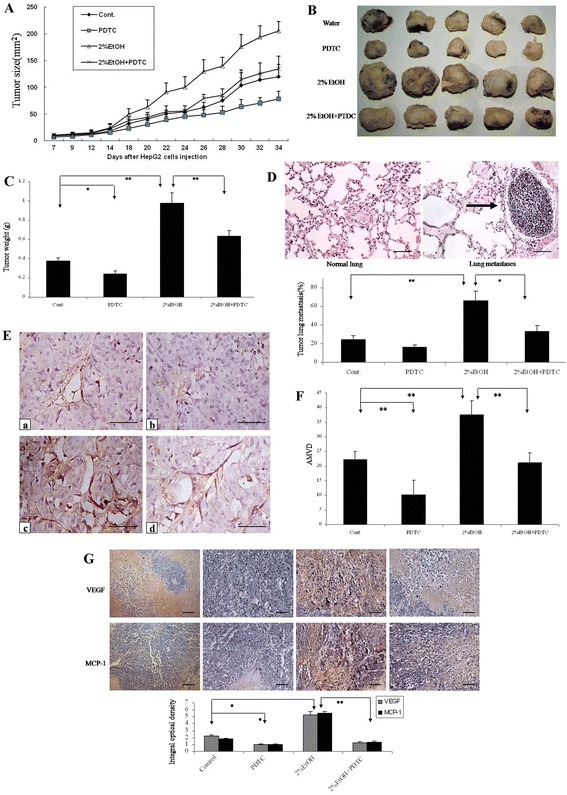
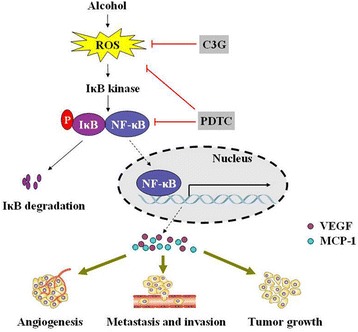
Methods and results: We first retrospectively investigated 52 HCC patients (24 alcohol-drinkers and 28 non-drinkers), and found a positive correlation between alcohol consumption and advanced Tumor-Node-Metastasis (TNM) stages, higher vessel invasion and poorer prognosis. In vitro and in vivo experiments further indicated that alcohol promoted the progression and migration/invasion of HCC. Specifically, in a 3-D tumor/endothelial co-culture system, we found that alcohol enhanced the migration/invasion of HepG2 cells and increased tumor angiogenesis. Consistently, higher expression of VEGF, MCP-1 and NF-κB was observed in HCC tissues of alcohol-drinkers. Alcohol induced the accumulation of intracellular reactive oxygen species (ROS) and the activation of NF-κB signaling in HepG2 cells. Conversely, blockage of alcohol-mediated ROS accumulation and NF-κB signaling inhibited alcohol-induced expression of VEGF and MCP-1, the tumor growth, angiogenesis and metastasis.

Conclusion: This study suggested that chronic moderate alcohol consumption may promote the progression and metastasis of HCC; the oncogenic effect may be at least partially mediated by the ROS accumulation and NF-ĸB-dependent VEGF and MCP-1 up-regulation.


Figures

Figure 1
Figure 1
Effects of alcohol consumption on the prognosis of patients with hepatocellular cancer. The overall survival time in 82 HCC patients (40 none-drinkers and 42 alcohol drinkers) during the period of 2006–2009 was analyzed and presented with Kaplan-Meier curves. Mean survival time was 27.4 months for non-drinkers and 47.8 months for drinkers, respectively. Alcohol consumption significantly shorted life span of patients (P = 0.021). The number of patients at risk was listed below the survival curves.
Figure 2
Figure 2
Effects of chronic alcohol consumption on the expression of CD31, VEGF, MCP-1 and NF-kB in HCC patients. (A) Immunohistochemical analysis of new blood vessels with anti-CD31 staining in different liver tissues. a: Liver cirrhosis; b: Distant non-cancerous liver tissues in HCC patients; c: HCC tissues of no-alcohol drinking patients; d: HCC tissues of alcohol-drinking patients; a-d: (Bar: 200 μm). The brown staining indicates the presence of microvessels. (B) Quantitative analysis of average microvessel density (AMVD, the number of microvessels per mm2 area) indicated that alcohol consumption significantly increased the number of new blood vessels (61.0 ± 6.78 in alcohol drinking patient vs 42.6 ± 4.82 in no-alcohol drinking patients, ** P < 0.01). (C) Immunohistochemical analysis of VEGF, MCP-1 and NF-κBp65 in liver tissues and HCC. (Bar: 100 μm). Arrows indicate NF-kBp65+ brown stain, which mainly found in the nuclei of tumor cells. (D) Quantitative analysis of VEGF, MCP-1 was determined by integral optical density. (E) Quantitative analysis of NF-κBp65 immunohistochemistry, as given by the nuclear-to-cytoplasmic ratio of NF-κBp65 positivity, n = 3.
Figure 3
Figure 3
Effects of ethanol on the migration/invasion of HepG2 cells in vitro. (A) The migration of HepG2 cells was analyzed by the wound healing assay as described in the Materials and methods. HepG2 cells were exposed to ethanol (0.2%) with/without PDTC (20 μM) for 24 hours. Representative images of wound healing at 0 and 24 hours are shown. (B) Quantification of the migration of HepG2 cells. (C) The invasion of HepG2 cells was analyzed by a matrigel invasion assay. HepG2 cells were plated into the upper compartments of the matrigel invasion chambers and exposed to ethanol (0.2%) with/without PDTC (20 μM) or C3G (20 μM) for 48 hours. Images of cells migrating through the chambers were shown. (D) The cells migrated through the chamber were quantified. *P <0.05, **P <0.01, n = 3.
Figure 4
Figure 4
Effects of ethanol on tumor angiogenesis in a 3-D model in vitro. Tumor angiogenesis was analyzed using a 3-D co-culture system as described in Materials and methods. (A) Representative images of sprouts from HUVEC cells with bead under various culture conditions are shown. (B) HUVEC and HepG2 co-cultures were treated with ethanol (0.2%) with/without PDTC (20 μM). The angiogenesis which was indicated by the growth of endothelial sprouts was measured. (C) HUVEC and HepG2 co-cultures were treated with ethanol (0.2%) with/without C3G (20 μM). The angiogenesis which was indicated by the growth of endothelial sprouts was measured. **P < 0.01, n = 3.
Figure 5
Figure 5
Effects of ethanol on intracellular ROS and the activation of NF-kB. (A) HepG2 cells were treated with ethanol (0.2%) with/without PTDC or C3G for 0.5 hours. Intracellular ROS was detected by flow cytometry as described in Materials and methods. (B) The relative levels of intracellular ROS were quantified. **P < 0.01. (C-D) HepG2 cells were treated with ethanol (0.2%) with/without PTDC or C3G for 2 hours. Expression of IkBα, p-IkBα and NF-kB p65 in cytoplasm and nuclei was analyzed by immunoblotting (left panel). The relative expression levels were quantified with a densitometry (right panel). *P < 0.05, **P < 0.01. (E) NF-kB activity was detected by the luciferase reporter gene assay. **P < 0.01, n = 3.
Figure 6
Figure 6
Effects of ethanol on tumor development in a xenograft model. (A) HepG2 cells were implanted subcutaneously in nude mice. The mice were exposed to ethanol in drinking water (0 or 2% ethanol). Mice were injected with PDTC (0 or 100 mg/kg). The size of tumor was measured every other day. (B) At the end of experiments, tumors were removed for subsequent analyses. A representative image shows tumors from control and ethanol-exposed mice with/without PDTC treatment. (C) Tumor weight was determined and presented as the means ± SD (n = 12). (D) Tumor metastases in the lungs were analyzed. A representative microphotograph shows metastatic carcinoma nodes in the lungs of ethanol-exposed mice (arrow, top panel; Bar: 100 μm). The percentage of mice which were positive for lung metastasis was quantified (bottom panel). (E) Microvessels in tumor tissues were detected by CD31 immunohistochemistry (Bar: 50 μm). a: water control, b: water plus PDTC, c: 2% ethanol, d: 2% ethanol plus PDTC. (F) AVMD was determined as described in Figure 2. (G) VEGF and MCP-1 in tumor tissues were detected by IHC (Bar: 100 μm). *P < 0.05, **P < 0.01, n = 3.
Figure 7
Figure 7
Proposed working model for the role of the NF-kB signaling pathway in the regulation of ethanol-enhanced the progression of HCC. (1) ROS induced by alcohol mediated activation of the NF-kB pathway. This effect can be abolished by C3G or PDTC. (2) Nuclear translocation of NF-kB via phosphorylation by the IkB kinase, which then binds target DNA that regulates VEGF and MCP-1 gene expression. Ultimately, the changes stimulated by alcohol enhance angiogenesis, tumor growth and metastasis.

References

    1. Mittal S, El-Serag HB. Epidemiology of hepatocellular carcinoma: consider the population. J Clin Gastroenterol. 2013;47(Suppl):S2–6. doi: 10.1097/MCG.0b013e3182872f29. - DOI - PMC - PubMed
    1. Gluer AM, Cocco N, Laurence JM, Johnston ES, Hollands MJ, Pleass HC, et al. Systematic review of actual 10-year survival following resection for hepatocellular carcinoma. HPB (Oxford) 2012;14(5):285–90. doi: 10.1111/j.1477-2574.2012.00446.x. - DOI - PMC - PubMed
    1. Venook AP, Papandreou C, Furuse J, de Guevara LL. The incidence and epidemiology of hepatocellular carcinoma: a global and regional perspective. Oncologist. 2010;15(Suppl 4):5–13. doi: 10.1634/theoncologist.2010-S4-05. - DOI - PubMed
    1. McKillop IH, Schrum LW. Role of alcohol in liver carcinogenesis. Semin Liver Dis. 2009;29(2):222–32. doi: 10.1055/s-0029-1214377. - DOI - PubMed
    1. Severi T, van Malenstein H, Verslype C, van Pelt JF. Tumor initiation and progression in hepatocellular carcinoma: risk factors, classification, and therapeutic targets. Acta Pharmacol Sin. 2010;31(11):1409–20. doi: 10.1038/aps.2010.142. - DOI - PMC - PubMed

Publication types

MeSH terms

LinkOut - more resources