Review
. 2014:209:191-205.
doi: 10.1016/B978-0-444-63274-6.00010-2.

Cardiorespiratory coupling: common rhythms in cardiac, sympathetic, and respiratory activities

Affiliations
Review

Cardiorespiratory coupling: common rhythms in cardiac, sympathetic, and respiratory activities

Thomas E Dick et al. Prog Brain Res. 2014.

Abstract

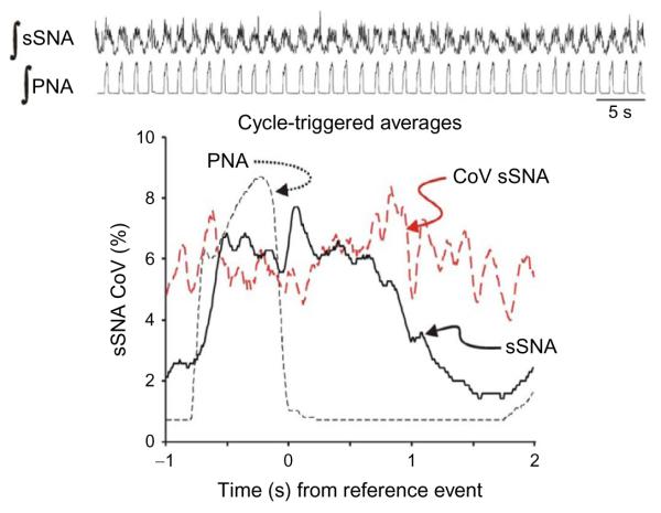
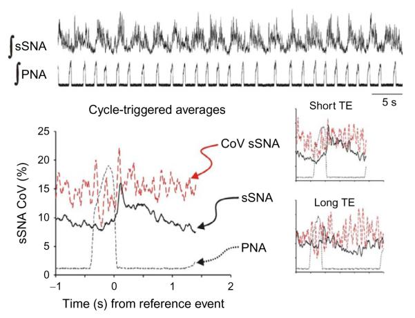
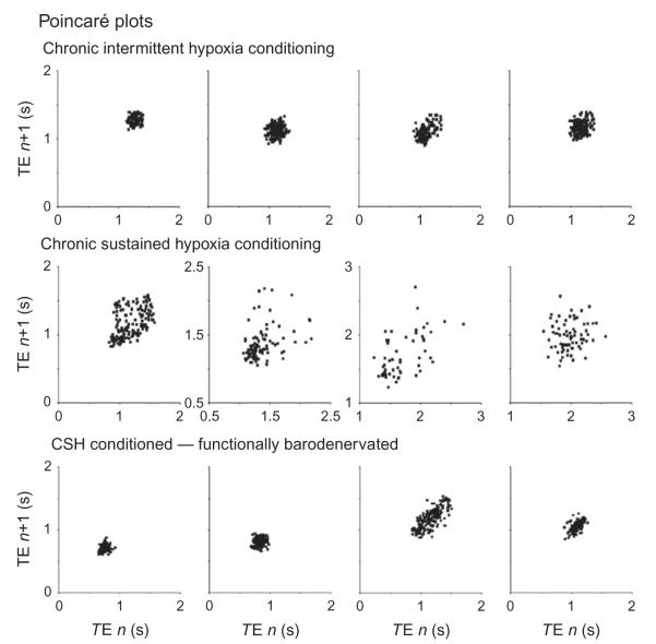
Cardiorespiratory coupling is an encompassing term describing more than the well-recognized influences of respiration on heart rate and blood pressure. Our data indicate that cardiorespiratory coupling reflects a reciprocal interaction between autonomic and respiratory control systems, and the cardiovascular system modulates the ventilatory pattern as well. For example, cardioventilatory coupling refers to the influence of heart beats and arterial pulse pressure on respiration and is the tendency for the next inspiration to start at a preferred latency after the last heart beat in expiration. Multiple complementary, well-described mechanisms mediate respiration's influence on cardiovascular function, whereas mechanisms mediating the cardiovascular system's influence on respiration may only be through the baroreceptors but are just being identified. Our review will describe a differential effect of conditioning rats with either chronic intermittent or sustained hypoxia on sympathetic nerve activity but also on ventilatory pattern variability. Both intermittent and sustained hypoxia increase sympathetic nerve activity after 2 weeks but affect sympatho-respiratory coupling differentially. Intermittent hypoxia enhances sympatho-respiratory coupling, which is associated with low variability in the ventilatory pattern. In contrast, after constant hypobaric hypoxia, 1-to-1 coupling between bursts of sympathetic and phrenic nerve activity is replaced by 2-to-3 coupling. This change in coupling pattern is associated with increased variability of the ventilatory pattern. After baro-denervating hypobaric hypoxic-conditioned rats, splanchnic sympathetic nerve activity becomes tonic (distinct bursts are absent) with decreases during phrenic nerve bursts and ventilatory pattern becomes regular. Thus, conditioning rats to either intermittent or sustained hypoxia accentuates the reciprocal nature of cardiorespiratory coupling. Finally, identifying a compelling physiologic purpose for cardiorespiratory coupling is the biggest barrier for recognizing its significance. Cardiorespiratory coupling has only a small effect on the efficiency of gas exchange; rather, we propose that cardiorespiratory control system may act as weakly coupled oscillator to maintain rhythms within a bounded variability.

Keywords: neural control of heart rate; neural control of respiration; neural control of sympathetic nerve activity; weakly coupled oscillators.


Figures

FIGURE 1
FIGURE 1
Schematic of cardiorespiratory coupling. The bidirectional arrows between each limit cycle (blue (left), respiratory and red (right), cardiac) represent the reciprocal coupling scheme between respiration and autonomic, cardiac rhythms, which are depicted as harmonics (here the cardiac: respiratory rhythm is 4:1 entrainment). Multiple mechanisms mediate the respiratory influence on the cardiac cycle, whereas a single mechanism mediates the influence of the cardiac/sympathetic activity onthe respiratory cycle. The proposed mechanism is through baror eceptors and their beat-to-beat increase in activity.
FIGURE 2
FIGURE 2
Sympatho-respiratory coupling is high following chronic intermittent hypoxia. The bursts of integrated splanchnic sympathetic nerve activity (∫ sSNA) and integrated phrenic nerve activity (∫ PNA) are highly correlated; this is reflected in the cycle-triggered average of ∫ sSNA in which the nadir is close to 0 and the coefficient of variation (CoV) is low across the respiratory cycle. Traces for this figure and Fig. 3: top, ∫ sSNA and ∫ PNA. Graph: Cycle-triggered averages of ∫ sSNA (black continuous line), ∫ PNA (black dashed line), CoV of ∫ sSNA (red dashed line). Note: The y-axis is scaled for the CoV of ∫ sSNA. Even though the scale of the averages of ∫ sSNA and ∫ PNA is arbitrary because it depends on the amplification of each nerve recording, it does range from zero to a normalized maximum, so the signal-to-ratio is depicted by the averages for that component of the signal that is correlated temporally to the reference event, the offset of inspiration.
FIGURE 3
FIGURE 3
Sympatho-respiratory coupling is low following chronic-sustained hypoxia. The bursts of ∫ sSNA do not appear time-locked to ∫ PNA. Instead of a 1:1 coupling pattern, 2 bursts of ∫ sSNA are coupled to three bursts of ∫ PNA. Nevertheless, the cycle-triggered average of ∫ sSNA has a “postinspiratory” burst of activity or a peak of activity associated with the inspiratory–expiratory phase transition; the nadir of ∫ sSNA is not close to 0 with the lowest values of ∫ sSNA occurring after the start of inspiration; and the coefficient of variation (CoV) is two-to-three times that of the chronic intermittent hypoxic-conditioned rat across the respiratory cycle. Insets: Cycle-triggered averages; even selecting the short and long respiratory cycles (duration of expiration, TE) does not improve the signal-to-noise ratio of the ∫ sSNA. Traces as in Fig. 2.
FIGURE 4
FIGURE 4
Poincaré plots. The duration of expiration (TE) of the next cycle is plotted against the TE of the current cycle to display cycle-to-cycle variability. In four chronic intermittent hypoxic-conditioned rats, the variability of TE is small and forms a tight cluster points. Compare this to four chronic-sustained hypoxic-conditioned rats in which the points are distributed widely. However, the distribution forms a tight cluster after both the aortic depressor and carotid sinus nerve are transected.

References

    1. Heart rate variability. Standards of measurement, physiological interpretation, and clinical use. Task Force of the European Society of Cardiology and the North American Society of Pacing and Electrophysiology. Eur. Heart. J. 1996;17:354–381. - PubMed
    1. Abdala AP, Rybak IA, Smith JC, Paton JF. Abdominal expiratory activity in the rat brainstem-spinal cord in situ: patterns, origins and implications for respiratory rhythm generation. J. Physiol. 2009;587:3539–3559. - PMC - PubMed
    1. Baekey DM, Dick TE, Paton JF. Pontomedullary transection attenuates central respiratory modulation of sympathetic discharge, heart rate and the baroreceptor reflex in the in situ rat preparation. Exp. Physiol. 2008;93:803–816. - PubMed
    1. Baekey DM, Molkov YI, Paton JF, Rybak IA, Dick TE. Effect of baroreceptor stimulation on the respiratory pattern: insights into respiratory-sympathetic interactions. Respir. Physiol. Neurobiol. 2010;174:135–145. - PMC - PubMed
    1. Ben-Tal A, Shamailov SS, Paton JF. Evaluating the physiological significance of respiratory sinus arrhythmia: looking beyond ventilation-perfusion efficiency. J. Physiol. 2012;590:1989–2008. - PMC - PubMed

Publication types