. 2014 Mar 12;15(3):374-81.
doi: 10.1016/j.chom.2014.02.006.

Gut microbiota promote hematopoiesis to control bacterial infection

Affiliations

Gut microbiota promote hematopoiesis to control bacterial infection

Arya Khosravi et al. Cell Host Microbe. .

Abstract

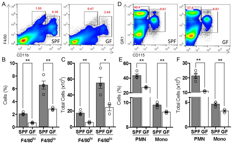
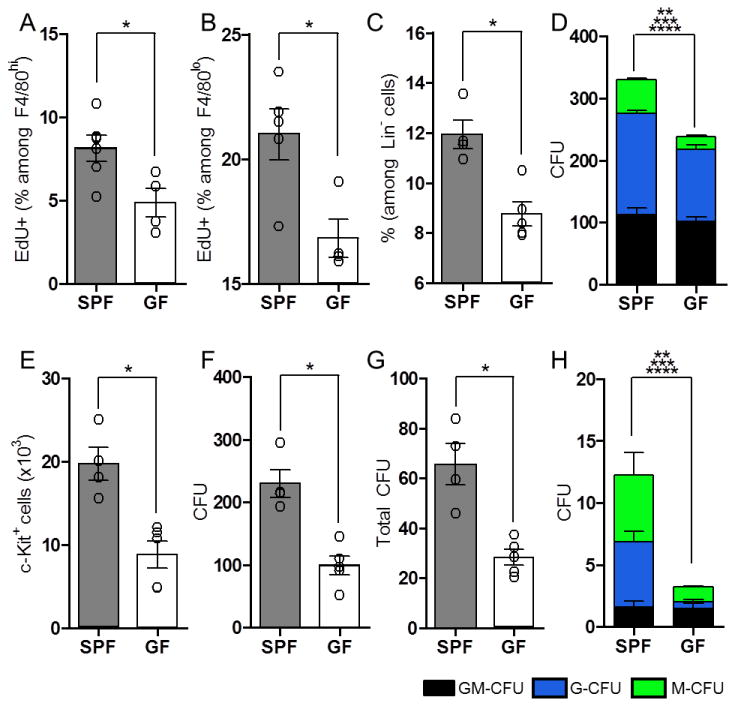
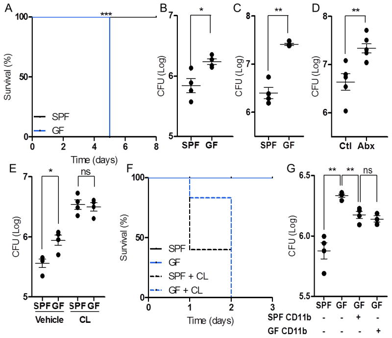
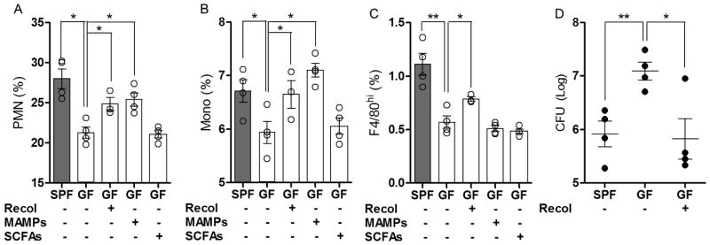
The commensal microbiota impacts specific immune cell populations and their functions at peripheral sites, such as gut mucosal tissues. However, it remains unknown whether gut microbiota control immunity through regulation of hematopoiesis at primary immune sites. We reveal that germ-free mice display reduced proportions and differentiation potential of specific myeloid cell progenitors of both yolk sac and bone marrow origin. Homeostatic innate immune defects may lead to impaired early responses to pathogens. Indeed, following systemic infection with Listeria monocytogenes, germ-free and oral-antibiotic-treated mice display increased pathogen burden and acute death. Recolonization of germ-free mice with a complex microbiota restores defects in myelopoiesis and resistance to Listeria. These findings reveal that gut bacteria direct innate immune cell development via promoting hematopoiesis, contributing to our appreciation of the deep evolutionary connection between mammals and their microbiota.


Conflict of interest statement

The authors declare no conflicts of interest related to this work.

Figures

Figure 1
Figure 1. GF Mice Are Deficient in Resident Myeloid Cell Populations in the Spleen and Bone Marrow
(A-C) Splenic phagocyte profile among SPF and GF mice. Representative flow cytometry plots (A), cell proportions (B), and total cell number (C) of CD11blo F4/80hi and CD11bhi F4/80lo splenic cells in SPF and GF mice. (D-F) Bone marrow populations of neutrophils (Gr1hi CD115neg) and monocytes (Gr1hi CD115hi) among SPF and GF mice. Representative flow cytometry plots (D), cell proportions (E) and total cell number (F) within the bone marrow of SPF and GF mice. For all panels, data are representative of at least 3 independent trials with n≥ 4 mice / group. Each symbol represents data from a single animal. Error bars represent standard error of mean (SEM). *p<0.05, **p<0.01. PMN: polymorphonuclear cells; Mono: monocytes. See also Figure S1.
Figure 2
Figure 2. The Microbiota Directs Myelopoiesis
The percentage of F4/80hi CD11b+ (A) and F4/80lo CD11b+ (B) splenocytes with incorporated EdU, following single dose administration. (C) The frequency of LKS- CD34+ Fc γ Rhi granulocyte and/or monocyte progenitors (GMPs) among lineage negative (Lin-) progenitors from bone marrow of SPF and GF mice, as assessed by flow cytometry. (D) Distribution of cell types following purified LKS- CD34+ Fc γ Rhi cell culture in methylcellulose medium. Colonies were identified and counted to assess the proportion of granulocyte-monocytes (GM-CFU; black), granulocytes (G-CFU; blue) and monocytes (M-CFU; green). (E) Total numbers of c-Kit+ CD11b- progenitors from methylcellulose cultures of LKS- CD34+ Fc γ Rhi progenitors, as assessed by flow cytometry. (F) Cells harvested from methylcellulose cultures of LKS- CD34+ Fc γ Rhi progenitors were re-plated at equal numbers in fresh methylcellulose, and cultured to assess their colony forming capacity. (G and H) Splenic cells isolated from SPF and GF mice were cultured in methylcellulose to assess the colony forming capacity of progenitors from SPF and GF mice. Total CFUs (G), and GM-CFUs, G-CFUs and M-CFUs (H) are shown. For each panel, data are representative of at least 2-3 independent trials with n≥ 4/ group. Each symbol represents data from a single animal. Error bars represent SEM. *p<0.05 for all panels. **p<0.05 (comparing total CFU between SPF and GF for (D) and (H)), ***p<0.05 (comparing G-CFU between SPF and GF for (D) and (H)), ****p<0.05 (comparing M-CFU between SPF and GF for (D) and (H)). CFU: colony forming units. See also Figure S2.
Figure 3
Figure 3. The Microbiota Promotes Early Resistance to Systemic Infection by L. monocytogenes via Tissue-Resident Cells
(A-C) SPF and GF mice infected with L. monocytogenes and assessed for survival (A) and splenic bacterial burden at 24 (B) and 72 (C) hours post- infection (hpi). SPF mice treated with antibiotics (Abx) and untreated controls (Ctl) were infected with L. monocytogenes and splenic bacterial burden was measured 72 hpi (D). SPF and GF mice depleted of tissue-resident cells prior to infection with L. monocytogenes and assessed for splenic bacterial burden 24 hpi (E) and survival (F). Splenic bacterial burden, 24 hpi, following transfer of splenic CD11b+ cells from SPF or GF donors (G). For all panels, data are representative of at least 2-3 independent trials with n≥ 4/ group. Each symbol represents data from a single animal. Error bars represent SEM. *p<0.05, **p<0.01, *** p<0.05 log-rank test used for survival curves in (A). CL: clodronate-loaded liposomes. See also Figure S3.
Figure 4
Figure 4. Re-colonization of GF Mice Restores Immune Integrity Against Systemic Listeriosis
(A) Neutrophil (GR1hi CD115-) and (B) monocyte (GR1hi CD115+) bone marrow profiles from SPF, GF, re-colonized GF mice and MAMP or SCFA-treated GF mice. (C) F4/80hi splenic macrophage profile among SPF, GF, re-colonized GF mice and GF mice treated with MAMPs or SCFAs. (D) Splenic bacterial burden 72 hpi among SPF, GF and re-colonized GF mice infected with L. monocytogenes. For all panels, data are representative of at least 2 independent trials with n≥4/ group. Each symbol represents data from a single animal. Error bars represent standard error of mean (SEM). *p<0.05, **p<0.01. Recol: re-colonized; MAMPs: molecular associated molecular patterns; SCFAs: short chain fatty acids. See also Figure S4.

References

    1. Aichele P, Zinke J, Grode L, Schwendener RA, Kaufmann SH, Seiler P. Macrophages of the splenic marginal zone are essential for trapping of blood-borne particulate antigen but dispensable for induction of specific T cell responses. J Immunol. 2003;171:1148–1155. - PubMed
    1. Akashi K, Traver D, Miyamoto T, Weissman IL. A clonogenic common myeloid progenitor that gives rise to all myeloid lineages. Nature. 2000;404:193–197. - PubMed
    1. Atarashi K, Tanoue T, Oshima K, Suda W, Nagano Y, Nishikawa H, Fukuda S, Saito T, Narushima S, Hase K, et al. Treg induction by a rationally selected mixture of Clostridia strains from the human microbiota. Nature. 2013;500:232–236. - PubMed
    1. Belkaid Y, Naik S. Compartmentalized and systemic control of tissue immunity by commensals. Nat Immunol. 2013;14:646–653. - PMC - PubMed
    1. Bugl S, Wirths S, Radsak MP, Schild H, Stein P, André MC, Müller MR, Malenke E, Wiesner T, Märklin M, et al. Steady-state neutrophil homeostasis is dependent on TLR4/TRIF signaling. Blood. 2013;121:723–733. - PubMed

Publication types