Review
. 2013 Dec;23(6):1034-40.
doi: 10.1016/j.conb.2013.09.012. Epub 2013 Oct 22.

Phagocytic glial cells: sculpting synaptic circuits in the developing nervous system

Affiliations
Review

Phagocytic glial cells: sculpting synaptic circuits in the developing nervous system

Dorothy P Schafer et al. Curr Opin Neurobiol. 2013 Dec.

Abstract

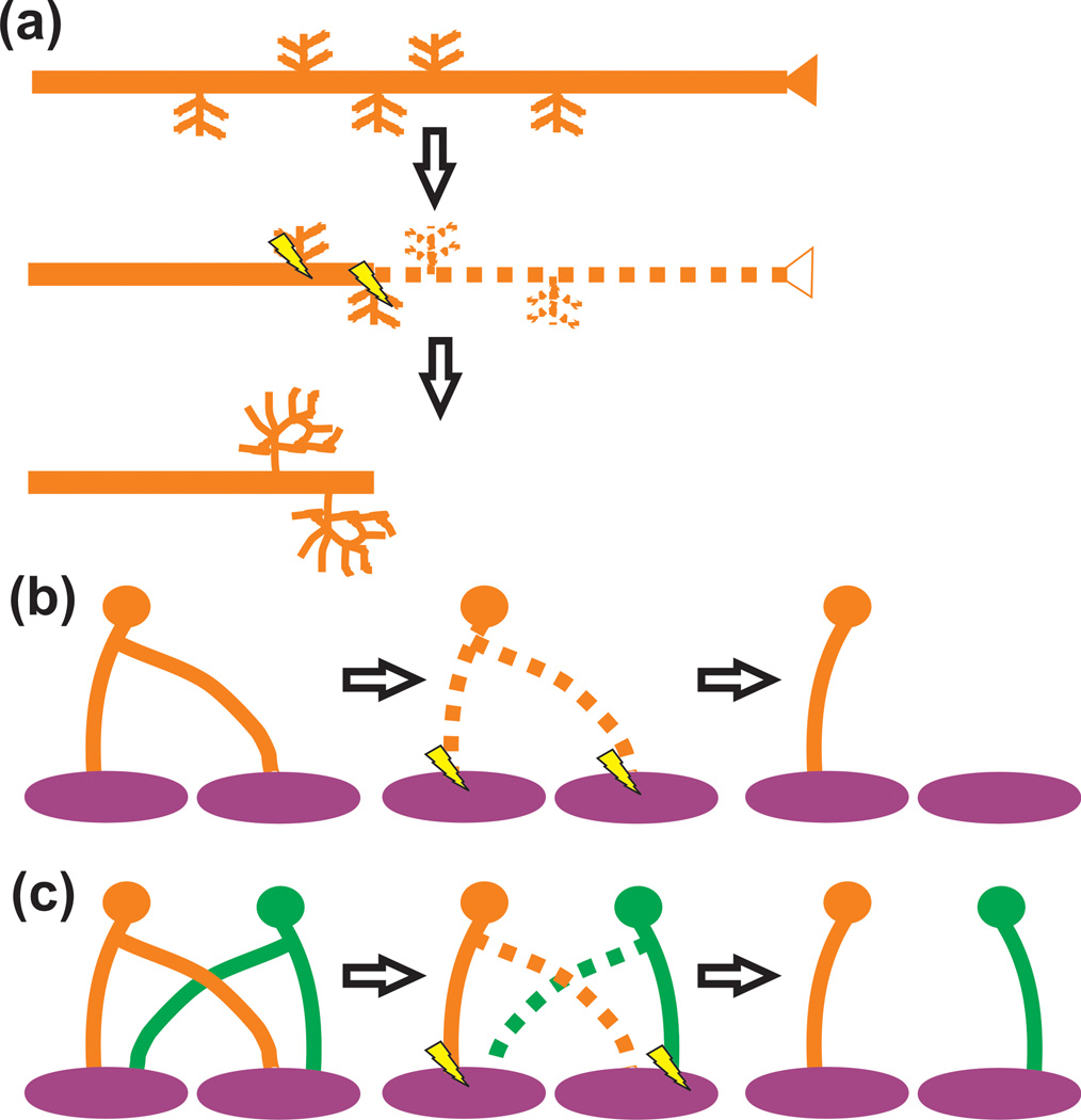
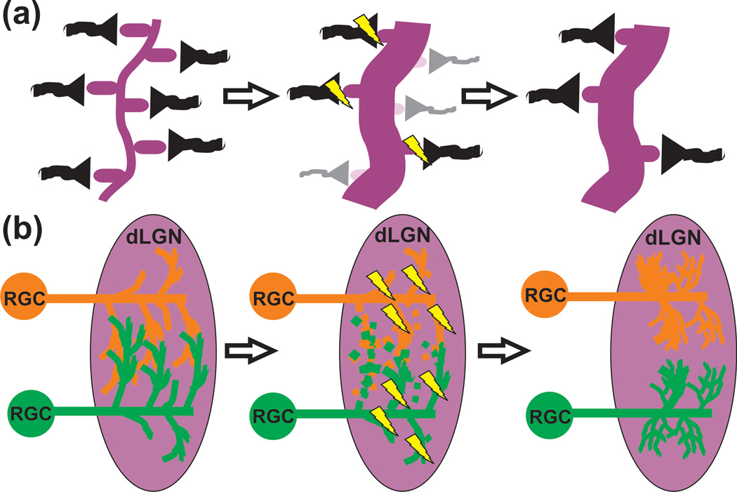
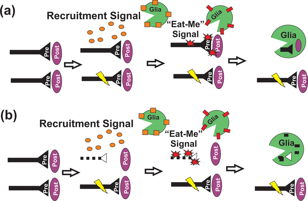
In the developing nervous system, synaptic connections are formed in excess and must remodel to achieve the precise synaptic connectivity characteristic of the mature organism. Synaptic pruning is a developmental process in which subsets of synapses are eliminated while the remaining synapses are preserved and strengthened. Recent findings have demonstrated unexpected roles for glial cells in this developmental process. These data demonstrate that phagocytic glia engulf synaptic and/or axonal elements in the developing nervous system and disruptions in this process result in sustained deficits in synaptic connectivity. These new findings highlight the importance of glia for nervous system development and function and may shed new light on mechanisms underlying nervous system disease.


Figures

Figure 1
Figure 1. Axonal pruning in the nervous system
(a) A presynaptic input (orange) with interstitial branches overshoots its target or is inappropriately targeted. Subsequently this input and small branches are pruned and remaining interstitial branches are elaborated (e.g., callosal projections). (b) A presynaptic input (orange) forms synapses on a postsynaptic target during early development (purple). These synaptic connections are subsequently eliminated and reform to form circuitry necessary for processing in the mature animal (e.g., Drosophila mushroom body). (c) Axon pruning involving the elimination of an axon collateral (orange and green dotted lines) from the postsynaptic target (purple) (e.g., NMJ). In all cases, axon pruning is driven by neural activity. Those synapses that are more active (lightning bolts) are maintained and strengthened.
Figure 2
Figure 2. Pruning of local synaptic circuits in the CNS
(a) Small scale pruning (e.g., visual cortex) can involve the local elimination of dendritic spines (light purple) and axon terminals (gray). Those presynaptic inputs (black) that are more active (lightning bolts) will be maintained and strengthened. (b) In the case of the retinogeniculate system, RGCs form transient, weak presynaptic inputs that innervate primarily the soma of postsynaptic dLGN relay neurons. Inputs from both eyes (red and green) are intermingled. During developmental pruning, smaller branches from both eyes that are less active (dotted lines) are removed. The remaining inputs that are more active (lightning bolts) are maintained, elaborated, and strengthened to form eye-specific territories within the dLGN.
Figure 3
Figure 3. Models of glia-mediated synaptic pruning
In both cases, glial cells may be attracted to sites of activity-dependent pruning by a local, soluble recruitment signal (orange circles; e.g., Cx3cl1) which binds receptors (orange squares; e.g., Cx3cr1) on the glial cell surface. Subsequently, glia engulf synaptic and/or axonal material through a local “eat-me” signal (red starbursts; e.g., C3) localized to the synapse and/or axon. This engulfment is mediated via a corresponding receptor (red rectangle; e.g., CR3, Draper, etc.) localized to the glial cell surface. The remaining, more active synapse (lightning bolt) is maintained and strengthened. This process could occur through local elimination of (a) intact, pre and postsynaptic components and/or (b) following intrinsic, degeneration (dotted line) of presynaptic components.

References

    1. Sanes JR, Lichtman JW. Development of the vertebrate neuromuscular junction. Annual Review of Neuroscience. 1999;22:389–442. - PubMed
    1. Hua JY, Smith SJ. Neural activity and the dynamics of central nervous system development. Nat Neurosci. 2004;7:327–332. - PubMed
    1. Katz L, Shatz C. Synaptic activity and the constuction of cortical circuits. Science. 1996;274:1133–1138. - PubMed
    1. Luo L, O'Leary DD. Axon retraction and degeneration in development and disease. Annu Rev Neurosci. 2005;28:127–156. - PubMed
    1. Kano M, Hashimoto K. Synapse elimination in the central nervous system. Curr Opin Neurobiol. 2009;19:154–161. - PubMed

Publication types

LinkOut - more resources