. 2013 Jun 18;8(6):e67102.
doi: 10.1371/journal.pone.0067102. Print 2013.

Disruption of the Circadian Clock in Mice Increases Intestinal Permeability and Promotes Alcohol-Induced Hepatic Pathology and Inflammation

Affiliations

Disruption of the Circadian Clock in Mice Increases Intestinal Permeability and Promotes Alcohol-Induced Hepatic Pathology and Inflammation

Keith C Summa et al. PLoS One. .

Abstract

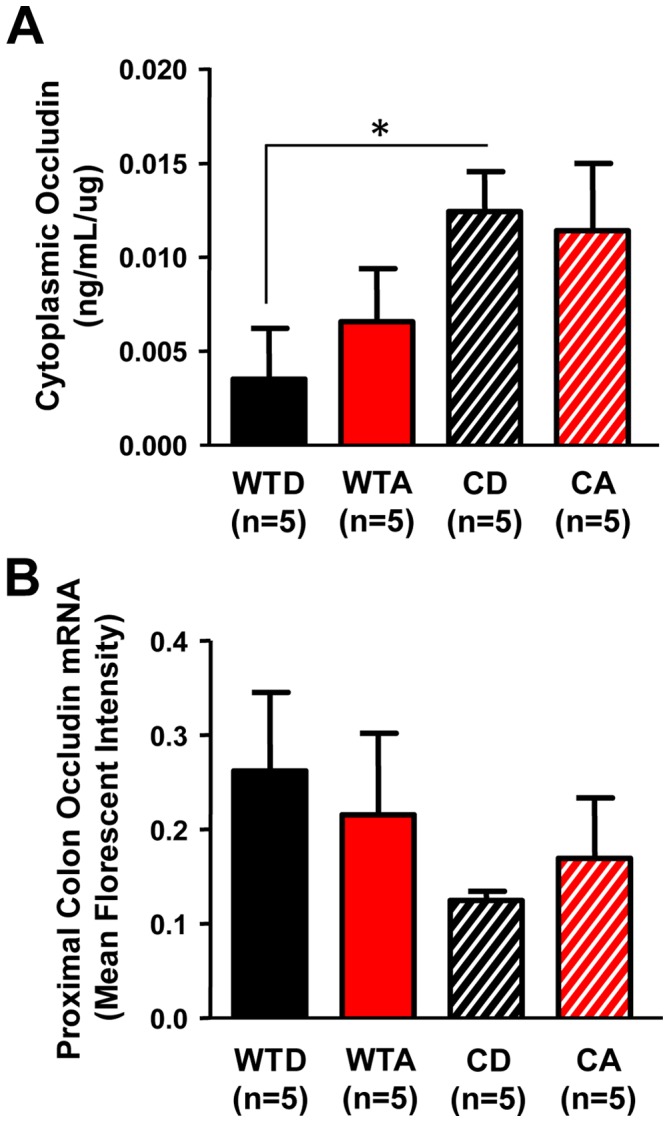
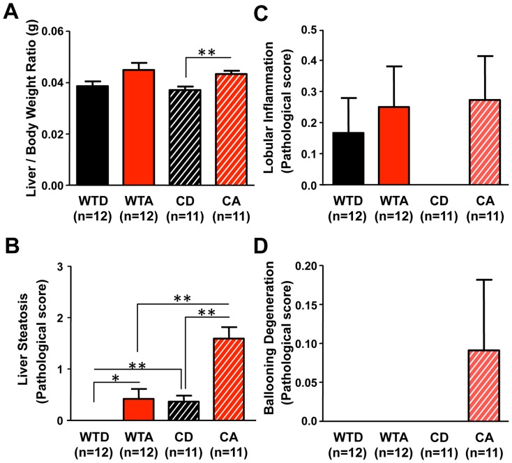
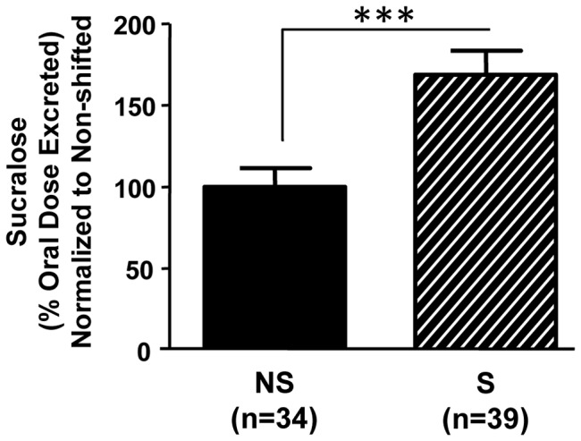
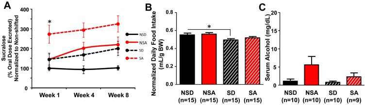
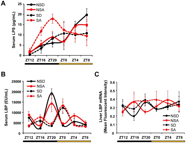
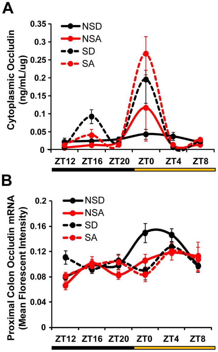
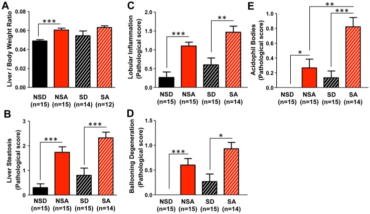
The circadian clock orchestrates temporal patterns of physiology and behavior relative to the environmental light:dark cycle by generating and organizing transcriptional and biochemical rhythms in cells and tissues throughout the body. Circadian clock genes have been shown to regulate the physiology and function of the gastrointestinal tract. Disruption of the intestinal epithelial barrier enables the translocation of proinflammatory bacterial products, such as endotoxin, across the intestinal wall and into systemic circulation; a process that has been linked to pathologic inflammatory states associated with metabolic, hepatic, cardiovascular and neurodegenerative diseases - many of which are commonly reported in shift workers. Here we report, for the first time, that circadian disorganization, using independent genetic and environmental strategies, increases permeability of the intestinal epithelial barrier (i.e., gut leakiness) in mice. Utilizing chronic alcohol consumption as a well-established model of induced intestinal hyperpermeability, we also found that both genetic and environmental circadian disruption promote alcohol-induced gut leakiness, endotoxemia and steatohepatitis, possibly through a mechanism involving the tight junction protein occludin. Circadian organization thus appears critical for the maintenance of intestinal barrier integrity, especially in the context of injurious agents, such as alcohol. Circadian disruption may therefore represent a previously unrecognized risk factor underlying the susceptibility to or development of alcoholic liver disease, as well as other conditions associated with intestinal hyperpermeability and an endotoxin-triggered inflammatory state.


Conflict of interest statement

Competing Interests: The authors have declared that no competing interests exist.

Figures

Figure 1
Figure 1. Models of circadian disruption and chronic alcohol consumption experimental protocols.
(A) Young adult (7–9 week) male ClockΔ19/Δ19 mutant mice and wild-type littermates (C57BL/6J coisogenic) were individually housed and maintained on a constant 12∶12 LD cycle for the duration of the experiment. Mice of each genotype were randomly assigned to receive either the alcohol-containing or isocaloric control diet, resulting in four experimental groups: wild-type dextrose control (WTD), wild-type alcohol (WTA), ClockΔ19/Δ19 mutant dextrose control (CD) and ClockΔ19/Δ19 mutant alcohol (CA). There was a gradual two week increase in alcohol concentration (open arrow; weeks −2–0; 0–29% total calories from alcohol) followed by eight weeks on the full alcohol diet (weeks 0–8; 29% total calories from alcohol; 4.5% v/v). Intestinal permeability was measured at weeks 1, 4 and 8 (closed arrows). At the end of week 8, mice were euthanized at ZT6 and tissues were collected for analyses. (B) Young adult (7–9 week) male C57BL/6J mice were individually housed. Mice were randomized into one of two light schedules: non-shifted mice were maintained on a constant 12∶12 LD cycle and shifted mice were subjected to a weekly 12 hour phase shift of the LD cycle for 12 weeks. Shifted and non-shifted mice were randomized into one of two diet treatments: alcohol or control, resulting in four experimental groups: non-shifted control diet (NSD), non-shifted alcohol (NSA), shifted control diet (SD) and shifted alcohol (SA). There was a gradual two week increase in alcohol as described in (A). Intestinal permeability was measured at weeks 1, 4 and 8 (closed arrows). At the end of week 8, mice were euthanized in groups every four hours across the diurnal cycle (at ZT0, ZT4, ZT8, ZT12, ZT16 and ZT20).
Figure 2
Figure 2. Genetic disruption of circadian organization increases intestinal permeability.
Young adult (7–9 week) male ClockΔ19/Δ19 mutant mice (C, n = 10) exhibited increased intestinal permeability, as measured by the urinary excretion of orally administered sucralose, a non-metabolized, non-absorbed sugar, compared to wild-type littermates (WT, n = 10). ***p<0.001, Student's t-test.
Figure 3
Figure 3. Genetic disruption of circadian organization promotes alcohol-induced intestinal hyperpermeability.
(A) Urinary sucralose excretion was measured as an index of colon permeability. Alcohol-fed ClockΔ19/Δ19 mutant mice (CA, n = 8–14/week) exhibited increased intestinal permeability compared to alcohol-fed wild-type mice (WTA, n = 8–11/week), control-fed ClockΔ19/Δ19 mutants (CD, n = 8–10/week) and control-fed wild-type mice (WTD, n = 8–9/week). Overall, significant effects of diet (p<0.01) and genotype (p<0.01) were observed. *p<0.05, three-way ANOVA followed by post-hoc Tukey-Kramer Multiple-Comparison test. (B) Average daily diet intake (normalized to body weight) over the duration of the experiment. Alcohol-fed ClockΔ19/Δ19 mutant mice (CA) did not consume more alcohol than wild-type littermates (WTA). *p<0.05, two-way ANOVA followed by post-hoc Tukey-Kramer Multiple-Comparison test. (C) Serum collected at ZT6 was assessed for alcohol levels using gas chromatography. Alcohol-fed ClockΔ19/Δ19 mutant mice (CA) exhibited significantly increased serum alcohol compared to alcohol-fed wild-type mice (WTA) and control diet-fed ClockΔ19/Δ19 mutants (CD). *p<0.05, **p<0.01; two-way ANOVA followed by post-hoc Tukey-Kramer Multiple-Comparison test.
Figure 4
Figure 4. Serum lipopolysaccharide (LPS) levels are altered and affected by alcohol in ClockΔ19/Δ19 mutant mice.
(A) Compared to wild-type littermates (WTD), ClockΔ19/Δ19 mutant mice on the control diet (CD) exhibited significantly reduced serum LPS levels. ClockΔ19/Δ19 mutant mice on alcohol (CA) had significantly elevated serum LPS compared to mutants on the control diet (CD), with an overall significant genotype × diet interaction observed (p<0.01). *p<0.05, two-way ANOVA followed by post-hoc Tukey-Kramer Multiple-Comparison. There were no significant differences in serum LPS binding protein (LBP) levels (B) or liver LBP mRNA expression (C).
Figure 5
Figure 5. Cytoplasmic tight junction protein occludin levels in the proximal colon are significantly elevated in ClockΔ19/Δ19 mutant mice.
(A) ClockΔ19/Δ19 mutant mice on the control diet (CD) had significantly elevated levels of cytoplasmic occludin in the proximal colon compared to wild-type littermates on the control diet (WTD), with an overall significant effect of genotype (p<0.05). *p<0.05, two-way ANOVA followed by post-hoc Fisher's LSD Multiple-Comparison test. (B) Occludin mRNA was not significantly altered.
Figure 6
Figure 6. Genetic disruption of circadian organization promotes alcohol-induced hepatic steatosis.
(A) Alcohol-fed ClockΔ19/Δ19 mutant mice (CA) had a significantly greater liver/body weight ratio than control-fed mutants (CD), with an overall significant effect of diet (p<0.01). **p<0.01, two-way ANOVA followed by post-hoc Tukey-Kramer Multiple-Comparison test. (B) Alcohol-fed ClockΔ19/Δ19 mutant mice (CA) exhibited significant steatosis compared to all other experimental groups, with a significant overall effect of genotype (p<0.001), diet (p<0.001), as well as a significant interaction (p<0.05). *p<0.05, **p<0.01; two-way ANOVA followed by post-hoc Tukey-Kramer Multiple-Comparison test. No between group differences were observed for histological assessment of lobular inflammation (C) or ballooning degeneration (D). Histological assessments were performed by a blinded gastrointestinal pathologist. Steatosis score was based on % hepatocyte involvement: 0 = <5%, 1 = 5–33%, 2 = 34–66%, 3 = >67%. Lobular inflammation score was based on the number of foci/200× field: 0 = none, 1 = 1, 2 = 2–4, 3 = >4. Ballooning degeneration score was based on the presence and frequency of ballooned cells: 0 = none, 1 = few, 2 = prominent/many.
Figure 7
Figure 7. Environmental disruption of circadian organization increases intestinal permeability.
Young adult (7–9 week) male C57BL/6J mice underwent three months of weekly 12 hour phase shifts of the LD cycle (S) or maintained on a constant 12∶12 LD cycle (NS). Shifted animals had significantly greater intestinal permeability. ***, p<0.001, Student's t-test.
Figure 8
Figure 8. Environmental disruption of circadian organization augments alcohol-induced intestinal hyperpermeability.
(A) Alcohol-fed shifted mice (SA, n = 13–15/week) exhibited significantly increased intestinal permeability compared to alcohol-fed non-shifted mice (NSA, n = 13–15/week), control-fed shifted mice (SD, n = 11–12/week) and control-fed non-shifted mice (NSD, n = 8–13/week). Overall significant effects of diet (p<0.01) and schedule (shifted vs. non-shifted, p<0.05) were observed at each time point. *, p<0.05, Tukey-Kramer Multiple-Comparison test. (B) Average daily diet intake (normalized to body weight) for the duration of the experiment in all groups. Shifted control-fed mice (SD) consumed less than non-shifted mice (NSD). Shifted mice (SA) did not consume more alcohol than non-shifted mice (NSA). *p<0.05, two-way ANOVA followed by post-hoc Tukey-Kramer Multiple-Comparison test. (C) There was a significant effect of diet on serum alcohol levels (F(1,39) = 4.986, p<0.05). Two-way ANOVA.
Figure 9
Figure 9. Environmental disruption of circadian organization impacts serum LPS and LBP levels.
(A) Serum LPS levels exhibited a significant time-of-day effect (p<0.001), with a significant schedule (i.e., shifted vs. non-shifted) × time-of-day interaction (p<0.01). Three-way ANOVA. (B) There was an apparent delayed peak in serum LBP of shifted mice, independent of diet, that failed to reach statistical significance. (C) LBP mRNA expression in liver was not significantly affected by schedule or alcohol.
Figure 10
Figure 10. Altered regulation of the tight junction protein occludin in the proximal colon by alcohol and chronic circadian disruption.
(A) Cytoplasmic occludin protein levels in the proximal colon were elevated at ZT0 in shifted animals (SA and SD) on both the control and alcohol diet, with an overall significant effect of schedule (i.e., shifted vs. non-shifted; p<0.01) and time-of-day (i.e., ZT; p<0.001). There was also a significant schedule × time-of-day interaction (p<0.001). Three-way ANOVA. (B) There were significant time-of-day (p<0.001) and diet (p<0.05) effects on proximal colon occludin mRNA expression, with a significant schedule × time-of-day interaction (p<0.01). Three-way ANOVA.
Figure 11
Figure 11. Environmental disruption of circadian organization promotes alcohol-induced liver pathology.
(A) There was an overall effect of diet for the liver/body weight ratio (p<0.001). The ratio was significantly elevated in non-shifted alcohol-fed mice (NSA) compared to non-shifted control-fed mice (NSD). There was a trend for increased liver/body weight ratio in alcohol-fed shifted mice (SA) compared to control-fed shifted mice (SD), although the difference failed to reach statistical significance. ***p<0.001, two-way ANOVA followed by post-hoc Tukey-Kramer Multiple-Comparison test. Histological assessment of liver steatosis (p<0.001) (B), lobular inflammation (p<0.001) (C), ballooning degeneration (p<0.001) (D) and the presence of acidophil bodies (p<0.001) (E) all revealed significant effects of diet, with ballooning degeneration (p<0.05) and acidophil bodies (p<0.05) also demonstrating significant effects of schedule (i.e, shifted vs. non-shifted). *p<0.05, **p<0.01, ***p<0.001; two-way ANOVA followed by post-hoc Tukey-Kramer Multiple-Comparison test. Histological assessment of liver samples was performed by a blinded gastrointestinal pathologist. Steatosis score was based on % hepatocyte involvement: 0 = <5%, 1 = 5–33%, 2 = 34–66%, 3 = >67%. Lobular inflammation score was based on the number of foci/200× field: 0 = none, 1 = 1, 2 = 2–4, 3 = >4. Ballooning degeneration score was based on the presence and frequency of ballooned cells: 0 = none, 1 = few, 2 = prominent/many. Acidophil body score was based on the presence of acidophil bodies: 0 = absent, 1 = focal apoptosis (few), 2 = many, 3 = confluent necrosis.

References

    1. Lowrey PL, Takahashi JS (2011) Genetics of circadian rhythms in Mammalian model organisms. Adv Genet 74: 175–230. - PMC - PubMed
    1. Hoogerwerf WA, Hellmich HL, Cornelissen G, Halberg F, Shahinian VB, et al. (2007) Clock gene expression in the murine gastrointestinal tract: endogenous rhythmicity and effects of a feeding regimen. Gastroenterology 133: 1250–1260. - PubMed
    1. Douris N, Kojima S, Pan X, Lerch-Gaggl AF, Duong SQ, et al. (2011) Nocturnin regulates circadian trafficking of dietary lipid in intestinal enterocytes. Curr Biol 21: 1347–1355. - PMC - PubMed
    1. Hoogerwerf WA, Shahinian VB, Cornelissen G, Halberg F, Bostwick J, et al. (2010) Rhythmic changes in colonic motility are regulated by period genes. Am J Physiol Gastrointest Liver Physiol 298: G143–150. - PMC - PubMed
    1. Marra G, Anti M, Percesepe A, Armelao F, Ficarelli R, et al. (1994) Circadian variations of epithelial cell proliferation in human rectal crypts. Gastroenterology 106: 982–987. - PubMed

Publication types

LinkOut - more resources