. 2013 Jul;94(1):171-82.
doi: 10.1189/jlb.1212659. Epub 2013 Apr 26.

Alcohol-induced IL-1β in the brain is mediated by NLRP3/ASC inflammasome activation that amplifies neuroinflammation

Affiliations

Alcohol-induced IL-1β in the brain is mediated by NLRP3/ASC inflammasome activation that amplifies neuroinflammation

Dora Lippai et al. J Leukoc Biol. 2013 Jul.

Abstract

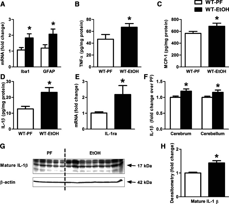
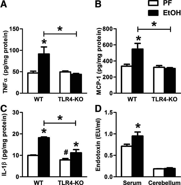
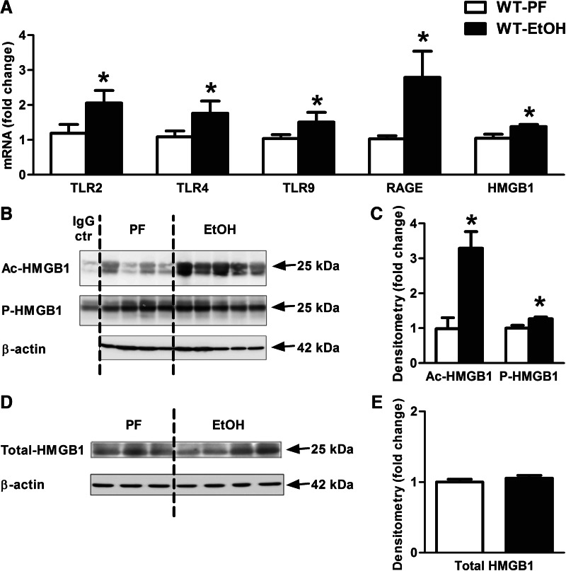
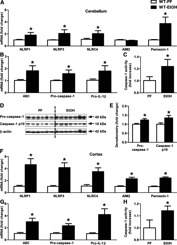
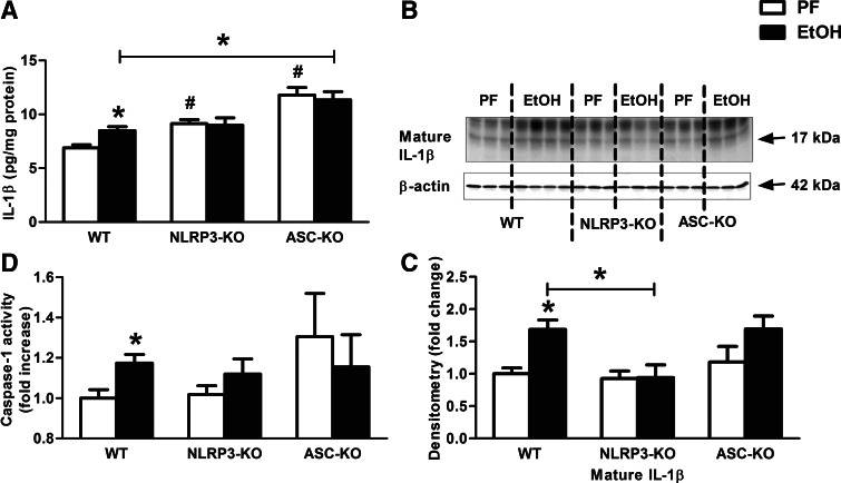
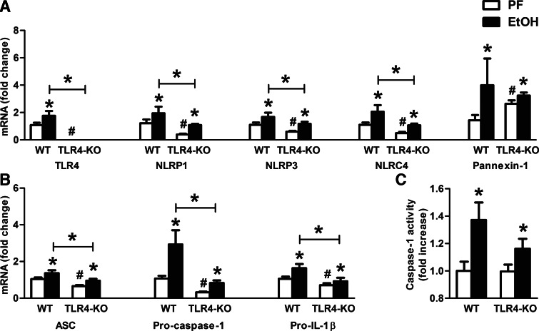
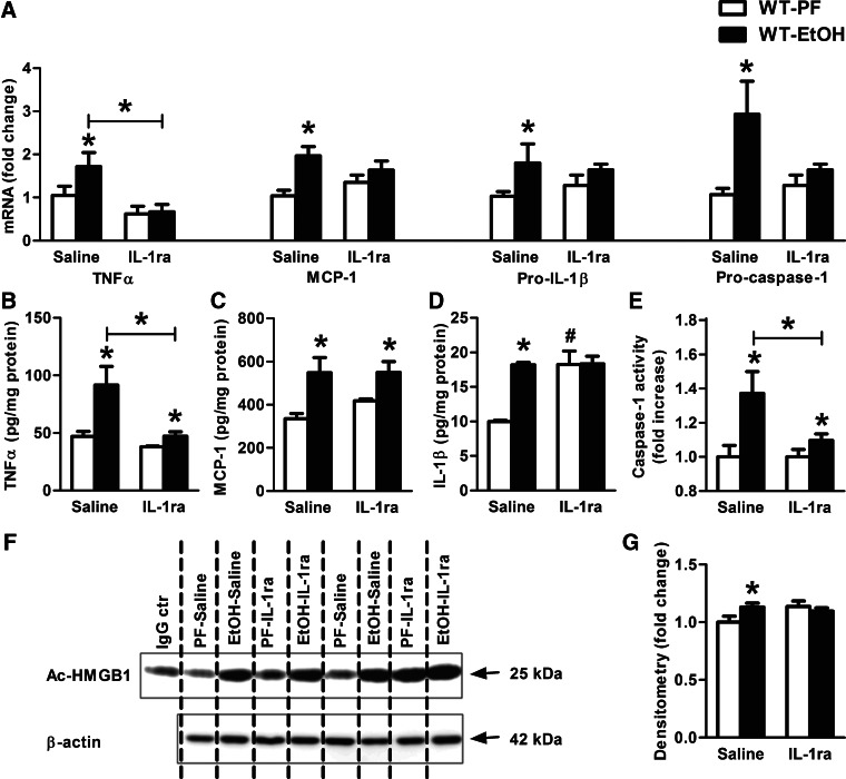
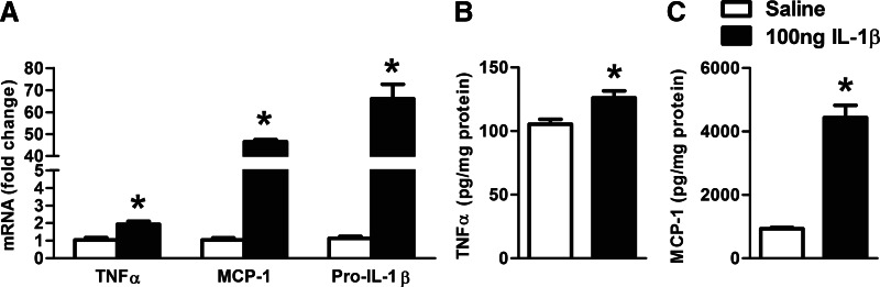
Alcohol-induced neuroinflammation is mediated by proinflammatory cytokines, including IL-1β. IL-1β production requires caspase-1 activation by inflammasomes-multiprotein complexes that are assembled in response to danger signals. We hypothesized that alcohol-induced inflammasome activation contributes to increased IL-1β in the brain. WT and TLR4-, NLRP3-, and ASC-deficient (KO) mice received an ethanol-containing or isocaloric control diet for 5 weeks, and some received the rIL-1ra, anakinra, or saline treatment. Inflammasome activation, proinflammatory cytokines, endotoxin, and HMGB1 were measured in the cerebellum. Expression of inflammasome components (NLRP1, NLRP3, ASC) and proinflammatory cytokines (TNF-α, MCP-1) was increased in brains of alcohol-fed compared with control mice. Increased caspase-1 activity and IL-1β protein in ethanol-fed mice indicated inflammasome activation. TLR4 deficiency protected from TNF-α, MCP-1, and attenuated alcohol-induced IL-1β increases. The TLR4 ligand, LPS, was not increased in the cerebellum. However, we found up-regulation of acetylated and phosphorylated HMGB1 and increased expression of the HMGB1 receptors (TLR2, TLR4, TLR9, RAGE) in alcohol-fed mice. NLRP3- or ASC-deficient mice were protected from caspase-1 activation and alcohol-induced IL-1β increase in the brain. Furthermore, in vivo treatment with rIL-1ra prevented alcohol-induced inflammasome activation and IL-1β, TNF-α, and acetylated HMGB1 increases in the cerebellum. Conversely, intracranial IL-1β administration induced TNF-α and MCP-1 in the cerebellum. In conclusion, alcohol up-regulates and activates the NLRP3/ASC inflammasome, leading to caspase-1 activation and IL-1β increase in the cerebellum. IL-1β amplifies neuroinflammation, and disruption of IL-1/IL-1R signaling prevents alcohol-induced inflammasome activation and neuroinflammation. Increased levels of acetylated and phosphorylated HMGB1 may contribute to alcoholic neuroinflammation.

Keywords: CNS; HMGB1; MCP-1; TNF-α; cerebellum.


Figures

Figure 1.
Figure 1.. Proinflammatory cytokines are increased in alcohol-induced brain injury.
WT mice were fed with an EtOH (n=6) or a control (PF; n=8) diet for 5 weeks. Astrocyte (GFAP) and microglia (Iba1) markers (A), as well as IL-1ra (E) were assessed by real-time PCR from whole cerebellar RNA extract, normalized to 18S. Inflammatory cytokines, TNF-α (B) and MCP-1 (C) of whole cerebellar lysates, and IL-1β (D and F) of cerebellar and cerebral lysates were measured by specific ELISAs. Mature IL-1β of whole cerebellar lysates were assessed by Western blot using β-actin as a loading control (G) and quantified further by densitometry (H). Bars represent mean ± sem (*P<0.05, relative to appropriate PF controls by the Kruskal-Wallis nonparametric test).
Figure 2.
Figure 2.. Cerebella of TLR4-deficient, alcohol-fed mice are partially protected against inflammatory cytokine production.
WT (n=8 or 7) and TLR4-KO (n=8 or 13) mice were fed with a control (PF) or EtOH diet for 5 weeks, respectively. Inflammatory cytokines, TNF-α (A), MCP-1 (B), and IL-1β (C) of whole cerebellar lysates, were measured by specific ELISAs. Endotoxin measurement was executed in serum and whole cerebellar lysates of WT mice by LAL assay (D). Bars represent mean ± sem (*P<0.05, relative to appropriate PF or WT-EtOH controls; #P<0.05, relative to appropriate WT-PF controls by the Kruskal-Wallis nonparametric test).
Figure 3.
Figure 3.. Active endogenous danger molecule, HMGB1, and its receptors are elevated in alcohol-fed mice in the cerebellum.
WT mice were fed with an EtOH (n=6) or a control (PF; n=8) diet for 5 weeks. TLR2, TLR4, TLR9, and RAGE receptors and HMGB1 (A) were assessed by real-time PCR from whole cerebellar RNA extracts, normalized to 18S. Acetyl (Ac)- and phospho (P)-HMGB1 of whole cerebellar lysates were analyzed by IP (B) and assessed further by densitometry, using β-actin Western blot for loading control on a separate gel (C). Cerebellar lysate of one representative ethanol-fed mouse was applied for IgG control (ctr), using the same amount of protein. Total HMGB1 of whole cerebellar lysates was analyzed by Western blot using β-actin as a loading control (D) and quantified further by densitometry (E). Bars represent mean ± sem (*P<0.05, relative to appropriate PF controls by the Kruskal-Wallis nonparametric test).
Figure 4.
Figure 4.. Inflammasome complex is up-regulated and activated in alcohol-fed mice in the cerebellum.
WT mice were fed with an EtOH (n=6) or a control (PF; n=8) diet for 5 weeks. Various inflammasome sensors (NLRP1, NLRP3, NLRC4, AIM2) and Pannexin-1 (A) and the inflammasome adaptor (ASC), the inflammasome effector (procaspase-1), and pro-IL-1β (B) were assessed by real-time PCR from whole cerebellar RNA extracts, normalized to 18S. Inflammasome activity was measured by a caspase-1 colorimetric assay (C) from whole cerebellar lysates. The caspase-1 p10 level (D) was visualized on a Western blot, using β-actin as a loading control, and quantified by densitometry (E). NLRP1, NLRP3, NLRC4, AIM2, and Pannexin-1 (F) and ASC, procaspase-1, and pro-IL-1β (G) were assessed by real-time PCR from whole cortical RNA extracts. Inflammasome activity was measured by a caspase-1 colorimetric assay (H) from whole cortical lysates. Bars represent mean ± sem (*P<0.05, relative to appropriate PF controls by the Kruskal-Wallis nonparametric test).
Figure 5.
Figure 5.. NLRP3 or ASC deficiency prevents caspase-1 activity and IL-1β production in alcohol-fed mice in the cerebellum.
WT (n=7 or 14), NLRP3 (n=8 or 10), and ASC-KO (n=9 or 12) mice were fed with a control (PF) or an EtOH diet for 5 weeks, respectively. IL-1β protein level of whole cerebellar lysates was measured by specific ELISA (A). Mature IL-1β of whole cerebellar lysates was assessed by Western blot using β-actin as a loading control (B) and quantified further by densitometry (C). Inflammasome activity was evaluated by a caspase-1 colorimetric assay from whole cerebellar lysates (D). Bars represent mean ± sem (*P<0.05, relative to appropriate PF or WT-EtOH controls; #P<0.05, relative to appropriate WT-PF controls by the Kruskal-Wallis nonparametric test).
Figure 6.
Figure 6.. TLR4 deficiency does not decrease caspase-1 activity in alcohol-fed mice in the cerebellum.
WT (n=8 or 7) and TLR4-KO (n=8 or 13) mice were fed with a control (PF) or an EtOH diet for 5 weeks, respectively. TLR4, various inflammasome receptors (NLRP1, NLRP3, NLRC4), and Pannexin-1 (A) and the inflammasome adaptor ASC, the inflammasome effector procaspase-1, and pro-IL-1β (B) were assessed by real-time PCR from whole cerebellar RNA extracts, normalized to 18S. Inflammasome activity was evaluated by the caspase-1 colorimetric assay from whole cerebellar lysates (C). Bars represent mean ± sem (*P<0.05, relative to appropriate PF or WT-EtOH controls; #P<0.05, relative to appropriate WT-PF controls by the Kruskal-Wallis nonparametric test).
Figure 7.
Figure 7.. IL-1ra, anakinra, treatment attenuates the effect of alcohol-feeding on caspase-1 activity and IL-1β production in murine cerebella.
WT mice were fed with a PF or an EtOH diet for 5 weeks and received daily i.p. IL-1ra (anakinra: 25 mg/kg) or an equal amount of saline injections. Inflammatory cytokines (TNF-α, MCP-1, and pro-IL-1β) and procaspase-1 (A) were assessed by real-time PCR from whole cerebellar RNA extract, normalized to 18S. Proinflammatory cytokines, TNF-α (B), MCP-1 (C), and IL-1β (D) of whole cerebellar lysates were measured by specific ELISAs. Inflammasome activity was measured by the caspase-1 colorimetric assay from whole cerebellar lysates (E). Acetyl-HMGB1 of whole cerebellar lysates were analyzed by IP (F) and assessed by densitometry, using β-actin Western blot for loading control on a separate gel (G). The cerebellar lysate of one representative ethanol-fed, IL-1ra-treated mouse was applied for IgG control, using the same amount of protein. Bars represent mean ± sem (*P<0.05, relative to appropriate PF or saline treated-EtOH controls; #P<0.05, relative to appropriate saline-treated PF controls by the Kruskal-Wallis nonparametric test. PF-saline, n = 8; PF-IL-1ra, n = 5; EtOH-saline, n = 7; EtOH-IL-1ra, n=9).
Figure 8.
Figure 8.. IL-1β induces TNF-α and MCP-1 in the cerebellum.
WT mice received 100 ng (30 μl) intracranial mouse rIL-1β or an equal amount of saline solution (n=7 or 8). Inflammatory cytokines (TNF-α, MCP-1, and pro-IL-1β) were assessed by real-time PCR from whole cerebellar RNA extract, normalized to 18S (A). Proinflammatory cytokines, TNF-α (B) and MCP-1 (C), of whole cerebellar lysates were measured by specific ELISAs. Bars represent mean ± sem (*P<0.05, relative to appropriate saline-treated controls by the Kruskal-Wallis nonparametric test).

References

    1. Holley M. M., Kielian T. (2012) Th1 and Th17 cells regulate innate immune responses and bacterial clearance during central nervous system infection. J. Immunol. 188, 1360–1370 - PMC - PubMed
    1. Glass C. K., Saijo K., Winner B., Marchetto M. C., Gage F. H. (2010) Mechanisms underlying inflammation in neurodegeneration. Cell 140, 918–934 - PMC - PubMed
    1. Tracey K. J. (2010) Understanding immunity requires more than immunology. Nat. Immunol. 11, 561–564 - PMC - PubMed
    1. Alfonso-Loeches S., Pascual-Lucas M., Blanco A. M., Sanchez-Vera I., Guerri C. (2010) Pivotal role of TLR4 receptors in alcohol-induced neuroinflammation and brain damage. J. Neurosci. 30, 8285–8295 - PMC - PubMed
    1. Szabo G., Mandrekar P., Petrasek J., Catalano D. (2011) The unfolding web of innate immune dysregulation in alcoholic liver injury. Alcohol Clin. Exp. Res. 35, 782–786 - PMC - PubMed

Publication types

MeSH terms

Substances