. 2013 Jan;43(1):219-27.
doi: 10.1002/eji.201242468. Epub 2012 Nov 7.

Dexamethasone promotes tolerance in vivo by enriching CD11clo CD40lo tolerogenic macrophages

Affiliations

Dexamethasone promotes tolerance in vivo by enriching CD11clo CD40lo tolerogenic macrophages

Guoxing Zheng et al. Eur J Immunol. 2013 Jan.

Abstract

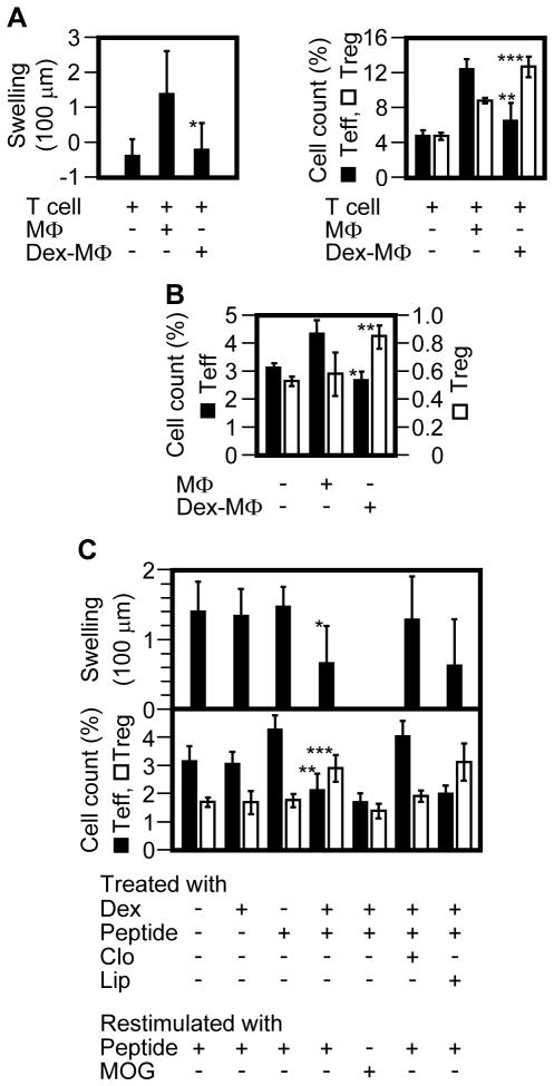
We previously showed that antigen immunization in the presence of the immunosuppressant dexamethasone (a strategy we termed "suppressed immunization") could tolerize established recall responses of T cells. However, the mechanism by which dexamethasone acts as a tolerogenic adjuvant has remained unclear. In the present study, we show that dexamethasone enriches CD11c(lo) CD40(lo) macrophages in a dose-dependent manner in the spleen and peripheral lymph nodes of mice by depleting all other CD11c(+) CD40(+) cells including dendritic cells. The enriched macrophages display a distinct MHC class II (MHC II)(lo) CD86(hi) phenotype. Upon activation by antigen in vivo, CD11c(lo) CD40(lo) macrophages upregulate IL-10, a classic marker for tolerogenic antigen-presenting cells, and elicit a serum IL-10 response. When presenting antigen in vivo, these cells do not elicit recall responses from memory T cells, but rather stimulate the expansion of antigen-specific regulatory T cells. Moreover, the depletion of CD11c(lo) CD40(lo) macrophages during suppressed immunization diminishes the tolerogenic efficacy of the treatment. These results indicate that dexamethasone acts as a tolerogenic adjuvant partly by enriching the CD11c(lo) CD40(lo) tolerogenic macrophages.


Conflict of interest statement

Conflicts of interest: The authors declare no financial or commercial conflict of interest.

Figures

Figure 1
Figure 1
Dex enriches CD11clo cells in the spleen and LNs by depleting CD11chi cells. Balb/c or C57BL/6 mice (n = 3) were injected (s.c.) with PBS (“NT”) or 4.5 mg/kg of Dex; 1 day later, splenocytes, pooled LN cells, and blood leukocytes were analyzed by flow cytometry. T cells (CD11cCD40) and B cells (CD11cCD40hi) were gated out, except for an arbitrary small portion used as internal controls for CD11c and CD40 staining. Shown is 1 of 3 experiments with similar results. Major CD11c+CD40+ populations are circled. Total CD11c+CD40+ cells are gated by R2, of which the CD11cloCD40lo population is gated by R3. Numbers in parentheses indicate percentages of R1 (live)-gated cells. a, dermis-derived DCs; b, Langerhans cells; c, CD11b+ DCs.
Figure 2
Figure 2
Dex-enriched CD11clo cells are monocyte-derived macrophages. Balb/c mice (n = 3) were injected with PBS (“NT”) or Dex (4.5 mg/kg); 1day later, pooled LN cells and splenocytes were analyzed by flow cytometry. Solid line, specific staining for the indicated marker; dotted line, isotype control. (A) Expression of various markers in the LN and spleen cells. CD11clo cells and CD11chi cells were gated by R3 and R2 –R3 (depicted in Fig. 1), respectively. (B) Depletion by Dex of spleen pDCs from the CD11clo population. CD11clo cells and CD11chi cells were separately gated. (C) Depletion of spleen cDCs by Dex. Total CD11c+ cells were gated. Shown in each panel is 1 of 3 or more experiments with similar results.
Figure 3
Figure 3
Dex-MΦs display the phenotype of tolerogenic APCs. (A) Balb/c mice (n ≥ 3 per dose point) were injected with various doses of Dex; 1 day later, splenocytes were analyzed by flow cytometry for each mouse. CD11clo cells were gated. Regression curves were plotted with the Sigmaplot software. Shown is 1 of 3 experiments with similar results. Open circle, CD86; filled circle, MHC II. (B) Four groups of OVA-sensitized Balb/c mice (n = 3) were each injected with Dex alone (4.5mg/kg, s.c., on day 1), OVA323-339 peptide alone (50 μg/mouse, i.v., on day 2), both Dex (on day 1) and the peptide (on day 2), or neither. On day 3, splenocytes were analyzed by flow cytometry. CD11cloF4/80hi cells were gated. Shown is 1 of 2 experiments with similar results. Thick line, specific staining for the indicated marker; thin line, isotype control. (C) Splenocytes from the same set of mice in (B) were analyzed for intracellular IL-10 by flow cytometry. CD11cloF4/80hi cells were gated. Shown is 1 of 3 experiments with similar results. Thin line, NT; dotted line, Dex; dashed line, peptide; thick line, Dex + peptide. (D –F) OVA-sensitized Balb/c mice were divided into groups (n = 3); each group was injected with the indicated agents, as described in panel B. Clodronate liposomes (Clo) were given intravenously at 100 μl/mouse on day 1. Blood samples were taken from all groups on day 3. Serum IL-10 and IL-12 (p70 heterodimer) levels were analyzed by ELISA (R&D Systems). The results were expressed as pg/ml of serum. Shown in each panel are the combined results of 2 or more separate experiments (mean ± SD). The difference between the Dex + peptide combination group and any of the other groups is statistically significant (p < 0.001 by Student’s t test). Filled bar, IL-10; open bar, IL-12.
Figure 4
Figure 4
Dex-MΦs function as tolerogenic APCs. (A) Donor T cells (eFluor 670-labeled CD4+ T cells from OVA-sensitized Balb/c Foxp3-eGFP mice) and donor APCs (Ag-primed Dex-MΦs or Ag-primed control MΦs) were co-injected (s.c.) into a footpad of naïve Balb/c mice. At 24 h, DTH was measured by footpad swelling (left panel). On day 4, the draining LN cells were analyzed by flow cytometry (right panel). Donor memory T cells (CD3+CD44+eFluor 670+) were gated, of which the percentages of proliferating Teff (eFluor670loGFP) vs. Treg (eFluor670loGFP+) were determined. Shown are the combined results of 2 separate experiments involving a total of 6 mice per group (mean ± SD). Cell count (%) indicates the relative number of proliferating Teff or Treg within donor memory T cells. Filled bar, Teff; open bar, Treg. *, p = 0.04; **, p = 0.006; ***, p = 0.005 (all between the Dex-MΦ group and the MΦ group; Student’s t test). (B) Ag-primed MΦs or Ag-primed Dex-MΦs were injected (i.v.) into OVA-sensitized Foxp3-eGFP mice (n = 3) on day 1. BrdU was injected (i.p.) on day 3; 12 hrs later, splenic CD4+ T cells were isolated from the mice and analyzed by flow cytometry. Memory T cells (CD3+CD44+) were gated, of which the percentages of proliferating Teff (BrdU+GFP) vs. Treg (BrdU+GFP+) were determined. Shown is 1 of 2 experiments with similar results (mean ± SD). Cell count (%) indicates the relative number of proliferating Teff or Treg within memory CD4+ T cells. Filled bar, Teff; open bar, Treg. *, p = 0.007; **, p = 0.04 (all between the Dex-MΦ group and the MΦ group; Student’s t test). (C) OVA-sensitized Balb/c mice (n = 5) were treated as indicated, and DTH was rechecked 2 month later (upper panel). One month after that, the mice were restimulated twice (i.v.) with 50 μg of OVA323-339 or a control peptide, MOG, on days 1 and 3. Of note, group 4 (from the left) of the top panel was divided into 2 subgroups (the lower panel, groups 4 and 5 from the left) during restimulation. BrdU were injected (i.p.) on day 3; 12 hrs later, splenic CD4+ T cells were isolated, immunostained for CD3, CD44, Foxp3, and BrdU, and analyzed by flow cytometry. Memory T cells (CD3+CD44+) were gated, of which the percentages of proliferating Teff (BrdU+Foxp3) vs. Treg (BrdU+ Foxp3+) were determined (lower panel). Shown is 1 of 2 experiments (mean ± SD). Cell count (%) indicates the relative number of proliferating Teff or Treg within memory CD4+ T cells. Filled bar, Teff; open bar, Treg; Clo, clodronate liposomes; Lip, control liposomes; *, p = 0.002; **, p = 0.04; ***, p = 0.01 [all between column 1 (nontreated) and column 4 (suppressed immunization); Student’s t test].
Figure 5
Figure 5
Effect of clodronate liposomes on suppressed immunization may involve bone marrow, but not liver, macrophages. (A) Balb/c mice were injected with the indicated agents. At 16 h, the liver and bone marrow cells were isolated, immunostained for MHC II and F4/80, and the MHC II+F4/80+ cells (macrophages) were counted by flow cytometry as a percentage of total live-gated liver or bone marrow cells (mean ± SD). Cell count (%) indicates the relative number of MHC II+F4/80+ cells in total liver or bone marrow cells. The difference between the Dex group and the control group (left panel) and that between the Clo group and any of the other groups (right panel) are statistically significant (p < 0.0001 by Student’s t test). (B) Bone marrow cells from Dex-treated mice were immunostained for MHC II, F4/80, CD11c, and CD40 and analyzed by flow cytometry. MHC II+F4/80+ cells were gated (R2) and analyzed in comparison with the cells outside of R2 (i.e. “- R2”). CD11cloCD40lo cells are indicated. Shown in each panel is 1 of 3 or more experiments with similar results.

Comment in

  • DEXterity of tolerogenic APCs.
    Stagliano KE, Oppenheim JJ. Stagliano KE, et al. Eur J Immunol. 2013 Jan;43(1):38-41. doi: 10.1002/eji.201243184. Eur J Immunol. 2013. PMID: 23322692

References

    1. Coffman RL, Sher A, Seder RA. Vaccine adjuvants: putting innate immunity to work. Immunity. 2010;33:492–503. - PMC - PubMed
    1. Chen X, Oppenheim JJ, Winkler-Pickett RT, Ortaldo JR, Howard OM. Glucocorticoid amplifies IL-2-dependent expansion of functional FoxP3(+)CD4(+)CD25(+) T regulatory cells in vivo and enhances their capacity to suppress EAE. Eur J Immunol. 2006;36:2139–2149. - PubMed
    1. Chen X, Murakami T, Oppenheim JJ, Howard OM. Differential response of murine CD4+CD25+ and CD4+CD25− T cells to dexamethasone-induced cell death. Eur J Immunol. 2004;34:859–869. - PubMed
    1. Hackstein H, Thomson AW. Dendritic cells: emerging pharmacological targets of immunosuppressive drugs. Nat Rev Immunol. 2004;4:24–34. - PubMed
    1. Kang Y, Xu L, Wang B, Chen A, Zheng G. Cutting edge: immunosuppressant as adjuvant for tolerogenic immunization. J Immunol. 2008;180:5172–5176. - PMC - PubMed

Publication types

MeSH terms