. 2012 Jul 6;287(28):23582-93.
doi: 10.1074/jbc.M112.375378. Epub 2012 May 17.

Structure of the cytoplasmic region of PelD, a degenerate diguanylate cyclase receptor that regulates exopolysaccharide production in Pseudomonas aeruginosa

Affiliations

Structure of the cytoplasmic region of PelD, a degenerate diguanylate cyclase receptor that regulates exopolysaccharide production in Pseudomonas aeruginosa

John C Whitney et al. J Biol Chem. .

Abstract

High cellular concentrations of bis-(3',5')-cyclic dimeric guanosine mono-phosphate (c-di-GMP) regulate a diverse range of phenotypes in bacteria including biofilm development. The opportunistic pathogen Pseudomonas aeruginosa produces the PEL polysaccharide to form a biofilm at the air-liquid interface of standing cultures. Among the proteins required for PEL polysaccharide production, PelD has been identified as a membrane-bound c-di-GMP-specific receptor. In this work, we present the x-ray crystal structure of a soluble cytoplasmic region of PelD in its apo and c-di-GMP complexed forms. The structure of PelD reveals an N-terminal GAF domain and a C-terminal degenerate GGDEF domain, the latter of which binds dimeric c-di-GMP at an RXXD motif that normally serves as an allosteric inhibition site for active diguanylate cyclases. Using isothermal titration calorimetry, we demonstrate that PelD binds c-di-GMP with low micromolar affinity and that mutation of residues involved in binding not only decreases the affinity of this interaction but also abrogates PEL-specific phenotypes in vivo. Bioinformatics analysis of the juxtamembrane region of PelD suggests that it contains an α-helical stalk region that connects the soluble region to the transmembrane domains and that similarly to other GAF domain containing proteins, this region likely forms a coiled-coil motif that mediates dimerization. PelD with Alg44 and BcsA of the alginate and cellulose secretion systems, respectively, collectively constitute a group of c-di-GMP receptors that appear to regulate exopolysaccharide assembly at the protein level through activation of their associated glycosyl transferases.


Figures
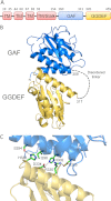
FIGURE 1.
FIGURE 1.
Overall structure of PelD156–455. A, domain organization of full-length PelD. Predicted transmembrane domains are abbreviated as TM. The approximate boundaries of each domain are indicated on the diagram. The relative size of each domain is not proportional to its molecular weight because the diagram is only intended to illustrate the linear arrangement of the domains. B, overall structure of PelD156–455 displayed in cartoon representation. The GAF and GGDEF domains are shown in blue and yellow, respectively. The locations of the N and C termini are indicated with N and C, respectively. The disordered linker region (residues 309–317) is indicated by the black dashed line. C, stick representation of the residues involved in interactions between the GAF and GGDEF domains. Carbon, nitrogen, and oxygen atoms are colored green, blue, and red, respectively (see also supplemental Figs. S1 and S2).
FIGURE 2.
FIGURE 2.
PelD contains a degenerate GGDEF domain with a conserved I-site. A, multiple sequence alignment of the GGDEF and I-site residues (or lack thereof) found in the GGDEF domains of C. crescentus PleD (Protein Data Bank code 1W25), P. aeruginosa WspR (Protein Data Bank code 3BRE), M. aquaeolei Maqu_2607 (Protein Data Bank code 3IGN), P. aeruginosa FimX (Protein Data Bank code 3HVA), and P. fluorescens LapD (Protein Data Bank code 3PJX). The alignments are based on three-dimensional superposition using the secondary structure matching algorithm in COOT. The catalytic and I-site residues are indicated in green and red, respectively. B, structural comparison of the GGDEF domains of PelD and the active DGC PleD as shown from two opposing views. The structures are shown as a cartoon representation with PleD displayed in gray and PelD displayed in yellow. In the left panel, secondary structure elements in PelD that are not present (α2 and β7) or are displaced significantly (α4/α5) compared with those found in canonical GGDEF domains, such as in PleD, are labeled. In the right panel, secondary structure elements that contain (α3/α4) or are adjacent to (α2/α3) the conserved I-site residues are indicated. In both structures, the catalytic and I-site residues are colored green and red, respectively. In the instances where a secondary structure element has two labels, the secondary structure of PelD is indicated first. C, comparison of the active site of PleD in complex with guanosine 5′-[α-thio]triphosphate (GTPαS) and PelD. Single amino acid labels indicate nucleotide or magnesium binding residues in PleD for which there are no structural equivalents in PelD. Double amino acid labels indicate conserved residues between the two proteins (with the PelD residue number indicated first). GTPαS is shown as a stick representation with the carbon, nitrogen, oxygen, phosphorous, and sulfur atoms colored green, blue, red, orange, and yellow, respectively. The magnesium ions in the PleD structure are shown as magenta spheres (see also supplemental Fig. S3).
FIGURE 3.
FIGURE 3.
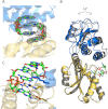
Comparison of the apo and holo forms of PelD156–455. A, (|Fo| − |Fc|) electron density map of (c-di-GMP)2 after molecular replacement contoured at 3 σ and shown as a gray mesh. Cyclic di-GMP is shown as a stick representation with carbon, nitrogen, oxygen, and phosphorous atoms colored green, blue, red, and orange, respectively. B, Cα superposition of the GGDEF domains of the apo and c-di-GMP bound structures of PelD156–455. The ligand free and ligand bound structures of PelD156–455 are displayed as a cartoon representation and colored in gray and yellow/blue, respectively. C, close-up of I-site/c-di-GMP interactions. Cyclic di-GMP binding residues Arg161, Arg367, Asp370, and Arg402 are shown as sticks and are colored in the same manner as their associated domains (see also supplemental Fig. S4).
FIGURE 4.
FIGURE 4.
PelD156–455 binds c-di-GMP with micromolar affinity. Isothermal titration calorimetry of c-di-GMP with wild-type PelD156–455 and the indicated site-directed mutants. In each experiment, the top panel displays the heats of injection, whereas the bottom panel shows the normalized integration data as a function of the molar syringe and cell concentrations. Where binding was observed (top panels), the red line represents the fit of the integrated data to a single-site binding model. The calculated dissociation constants (KD) are also indicated for each experiment where binding was observed.
FIGURE 5.
FIGURE 5.
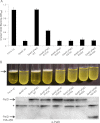
Effect of I-site mutations on biofilm formation. Microtiter dish biofilm assay (A) and pellicle assay (B) of P. aeruginosa PA14, PA14ΔpelD, PA14ΔpelD pPelD WT, or the indicated point mutants and PA14ΔpelD pPelD156–455 strains. The amount of crystal violet staining for each sample is the mean of three independent replicates. The error bars represent the standard errors. The pellicles form at the air-liquid interface of standing cultures as indicated by the black arrow. The α-PelD Western blot of each of the indicated strains shows expression of the various PelD mutants. The asterisk indicates a nonspecific band that was used as a loading control.
FIGURE 6.
FIGURE 6.
Full-length PelD likely forms a homodimer that self-associates via its α-helical stalk region. Cartoon representation of P. aeruginosa PelD, P. aeruginosa PA5279, and H. sapiens PDE2A. For each protein, the GAF domain(s) is displayed in blue with its corresponding dimerization helix highlighted in red. Because the proposed dimerization helix lies upstream of the crystallized fragment of PelD, a red cartoon cylinder is used to indicate this helix. The GGDEF domain of PelD, the catalytic phosphodiesterase domain of PDE2A, and the noncrystallographic symmetry copies of PA5279 and PDE2A are colored in yellow, green, and gray, respectively (see also Supplemental Fig. S5).
FIGURE 7.
FIGURE 7.
Inner membrane c-di-GMP receptors activate exopolysaccharide polymerization in the PEL, alginate, and cellulose secretion systems. Cartoon schematic of the proteins required for the polymerization of the PEL, alginate, and cellulose polysaccharides. The putative PEL synthase PelF and its associated c-di-GMP receptor PelD are colored light blue and orange, respectively. The P. aeruginosa alginate synthase protein Alg8 and its associated c-di-GMP receptor Alg44 are colored blue and red, respectively. The E. coli cellulose synthase protein BcsA is colored green. GT-2 and GT-4 indicate the family of glycosyl transferase to which each of the polysaccharide synthases is predicted to belong. The periplasmic domain of Alg44 is predicted to form a membrane fusion protein domain (MFP) as indicated. The chains of hexagons containing ?, M, and G represent the unknown composition of the PEL polysaccharide, 1,4-linked β-d-mannuronic acid (alginate), and 1,4-linked β-d-glucose (cellulose), respectively.

References

    1. Merighi M., Lee V. T., Hyodo M., Hayakawa Y., Lory S. (2007) The second messenger bis-(3′-5′)-cyclic-GMP and its PilZ domain-containing receptor Alg44 are required for alginate biosynthesis in Pseudomonas aeruginosa. Mol. Microbiol. 65, 876–895 - PubMed
    1. Lee V. T., Matewish J. M., Kessler J. L., Hyodo M., Hayakawa Y., Lory S. (2007) A cyclic-di-GMP receptor required for bacterial exopolysaccharide production. Mol. Microbiol. 65, 1474–1484 - PMC - PubMed
    1. Tischler A. D., Camilli A. (2004) Cyclic diguanylate (c-di-GMP) regulates Vibrio cholerae biofilm formation. Mol. Microbiol. 53, 857–869 - PMC - PubMed
    1. Tischler A. D., Camilli A. (2005) Cyclic diguanylate regulates Vibrio cholerae virulence gene expression. Infect. Immun. 73, 5873–5882 - PMC - PubMed
    1. Duerig A., Abel S., Folcher M., Nicollier M., Schwede T., Amiot N., Giese B., Jenal U. (2009) Second messenger-mediated spatiotemporal control of protein degradation regulates bacterial cell cycle progression. Genes Dev. 23, 93–104 - PMC - PubMed

Publication types

MeSH terms

LinkOut - more resources