. 2012 Jul;26(5):766-77.
doi: 10.1016/j.bbi.2011.10.003. Epub 2011 Oct 17.

Lipopolysaccharide-induced interleukin (IL)-4 receptor-α expression and corresponding sensitivity to the M2 promoting effects of IL-4 are impaired in microglia of aged mice

Affiliations

Lipopolysaccharide-induced interleukin (IL)-4 receptor-α expression and corresponding sensitivity to the M2 promoting effects of IL-4 are impaired in microglia of aged mice

Ashley M Fenn et al. Brain Behav Immun. 2012 Jul.

Abstract

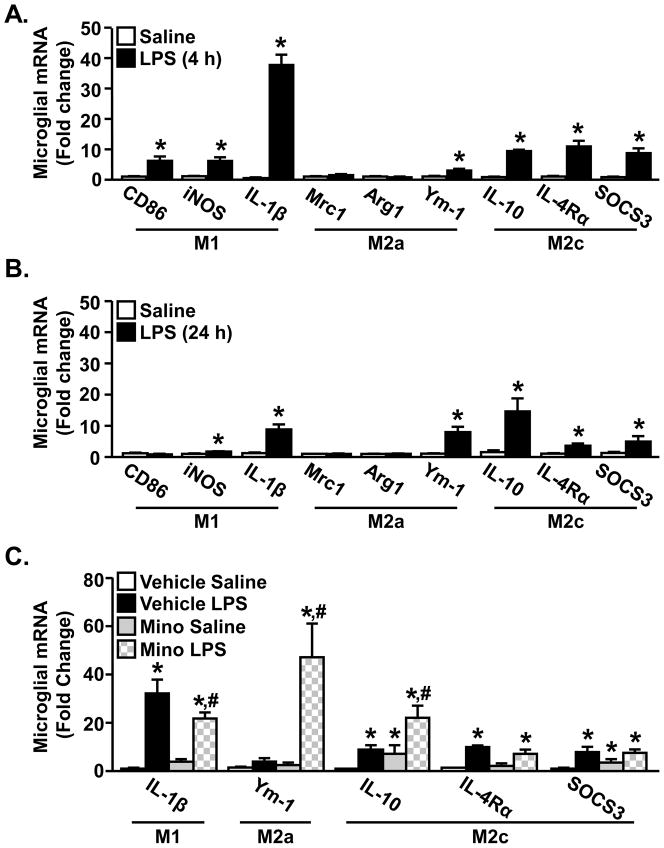
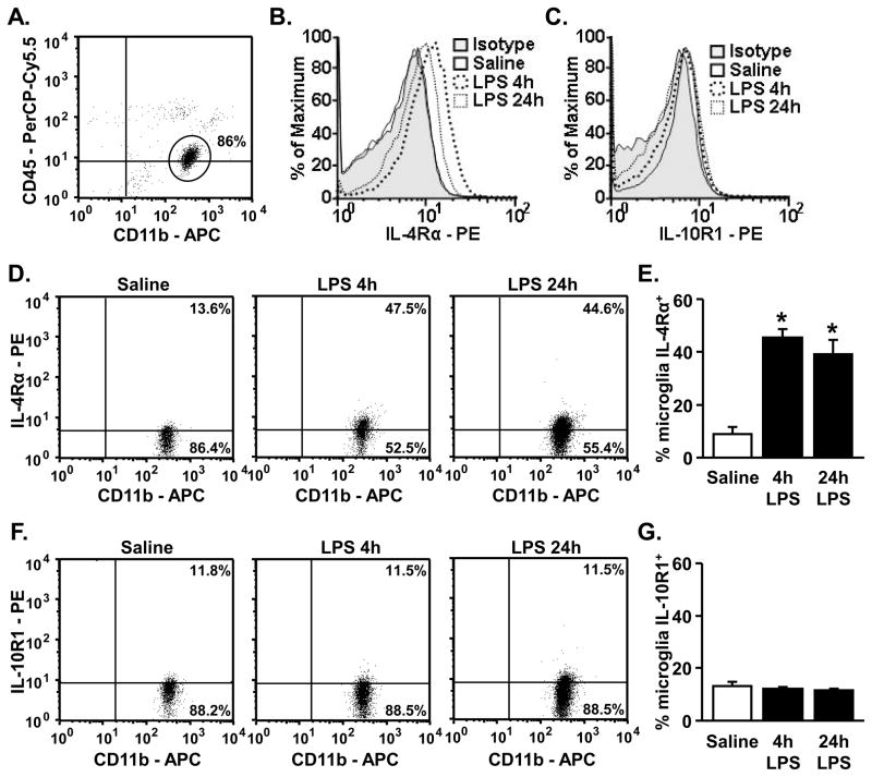
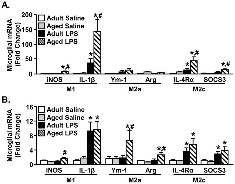
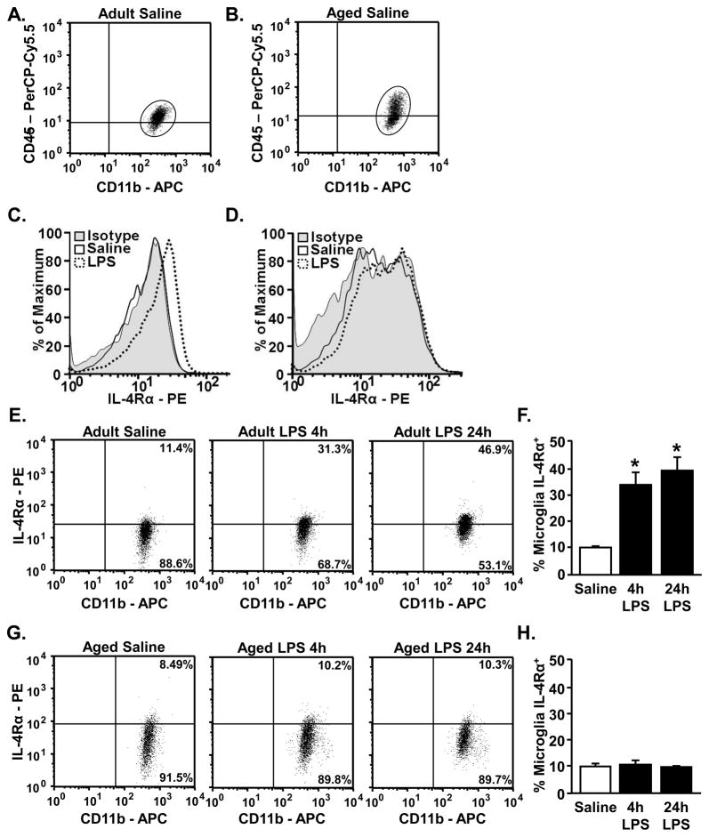
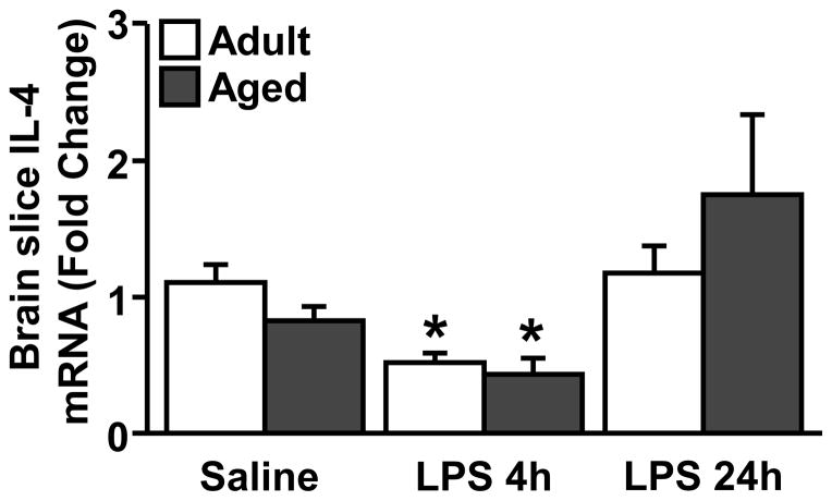
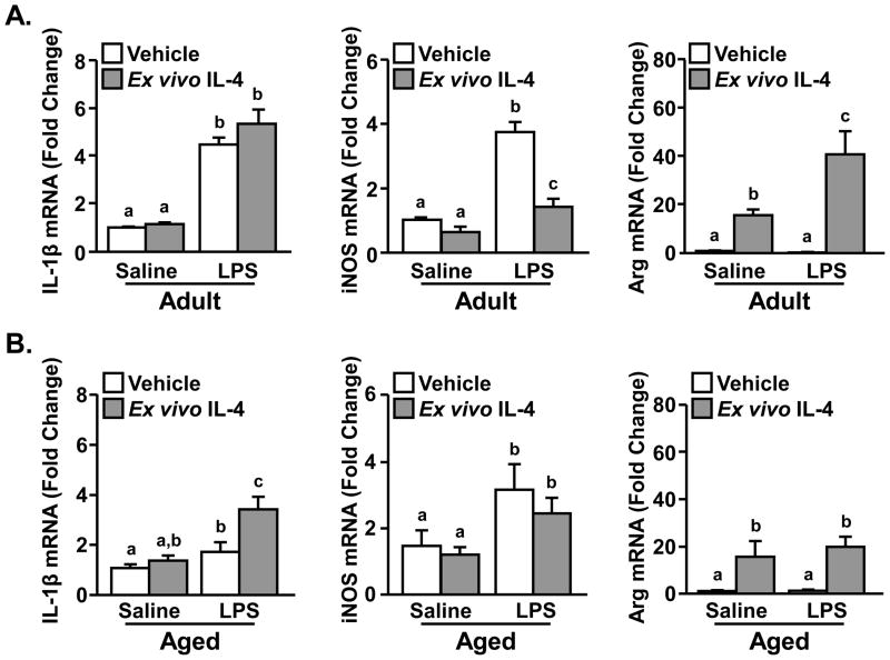
In several models of aging, microglia become more inflammatory and reactive to immune challenges. For example, peripheral LPS injection causes exaggerated microglial activation associated with prolonged sickness and depressive-like behavior in aged BALB/c mice. Therefore, the purpose of this study was to determine the extent to which age-related amplified microglial activation was associated with reduced sensitivity to the anti-inflammatory and M2 promoting cytokines interleukin (IL)-10 and IL-4. In initial studies with adult mice, LPS induced a time-dependent increase in M1 and M2 mRNA profiles in microglia. Furthermore, peripheral LPS injection markedly increased surface expression of IL-4 receptor-alpha (IL-4Rα), but not IL-10 receptor-1 (IL-10R1) on microglia. In BV-2 cells, IL-4, but not IL-10, re-directed LPS-activated microglia towards an M2 phenotype. Based on these findings, comparisons of M1 and M2 activation profiles, induction of IL-4Rα, and sensitivity to IL-4 were determined in microglia from adult (3-4 mo) and aged (18-22 mo) mice. In aged microglia, LPS promoted an exaggerated and prolonged M1 and M2 profile compared to adults. Moreover, IL-4Rα protein was not increased on aged microglia following LPS injection. To determine the consequence of impaired IL-4Rα upregulation, adult and aged mice were injected with LPS and activated microglia were then isolated and treated ex vivo with IL-4. While ex vivo IL-4 induced an M2 profile in activated microglia from adult mice, activated microglia from aged mice retained a prominent M1 profile. These data indicate that activated microglia from aged mice are less sensitive to the anti-inflammatory and M2-promoting effects of IL-4.


Figures

Figure 1
Figure 1. Peripheral LPS injection promoted an M1 and M2c mRNA profile in microglia
Adult (3–4 mo) BALB/c mice were injected ip with saline or LPS and M1, M2a and M2c related genes were determined from enriched microglia isolated A) 4 h or B) 24 h later (n=6). Bars represent the mean ± SEM. Means with * are significantly different (p<0.05) from Saline controls. C) Adult (3–4 mo) BALB/c mice were injected with vehicle or minocycline for 3 consecutive days. On the third day mice were injected ip with saline or LPS and M1-, M2a-, and M2c-related genes were determined from enriched microglia isolated 4 h later (n=9). Bars represent the mean ± SEM. Means with * are significantly different (p<0.05) from Vehicle Saline and means with # are significantly different (p<0.05) from Vehicle LPS.
Figure 2
Figure 2. Peripheral LPS injection increased microglial surface expression of IL-4Rα, but not IL-10R1
Adult (3–4 mo) BALB/c mice were injected ip with saline or LPS and CD11b, CD45, IL-4Rα and IL-10R1 were determined from enriched microglia isolated 4 h or 24 h later. A) A representative bivariate dot plot of Percoll isolated cells gated on CD11b+/CD45low expression for microglia. All analyses were completed using the CD11b+/CD45low microglia. Representative histograms of mean fluorescent intensity (MFI) for B) IL-4Rα and C) IL-10R1. Representative bivariate dot plots of microglia stained for D) IL-4Rα (n=4–7) and F) IL-10R1 (n=4). Average percentage of microglia that were E) IL-4Rα+ and G) IL-10R1+. Bars represent the mean ± SEM. Means with * are significantly different (p<0.05) from Saline controls.
Figure 3
Figure 3. M1, M2a, and M2c profile in adult and aged microglia after peripheral LPS injection
Adult (3–4 mo) and aged (18–22 mo) BALB/c mice were injected ip with saline or LPS and M1-, M2a-, and M2c-related genes were determined from enriched microglia isolated A) 4 h or B) 24 h later (n=6). Bars represent the mean ± SEM. Means with * are significantly different (p<0.05) from Adult Saline and means with # are significantly different (p<0.05) from Adult LPS.
Figure 4
Figure 4. Microglia from aged mice do not have increased surface expression of IL-4Rα following LPS
Adult (3–4 mo) and aged (18–22 mo) BALB/c mice were injected ip with saline or LPS and CD11b, CD45, and IL-4Rα were determined from enriched microglia isolated 4 h and 24 h later (n=4). Representative bivariate dot plots fro microglia from A) adult and B) aged mice showing the gated microglia (CD11b+/CD45low) that were used for IL-4Rα analysis. Representative histograms of the MFI for IL-4Rα in saline and LPS treated C) adult and D) aged mice as well as the age-matched isotype control. Representative bivariate dot plots for microglial IL-4Rα expression from E) adult and G) aged mice. Average percentage of microglia from F) adult and H) aged mice that were IL-4Rα+. Bars represent the mean ± SEM. Means with * are significantly different (p<0.0001) from Saline controls.
Figure 5
Figure 5. IL-4 mRNA is decreased in the brain of adult and aged mice after LPS
Adult (3–4 mo) and aged (18–22 mo) BALB/c mice were injected ip with saline or LPS and IL-4 mRNA was determined from a coronal brain slice 4 h and 24 h later (n=5). Means with * are significantly different (p<0.05) from Adult Saline.
Figure 6
Figure 6. Ex vivo treatment with IL-4 re-directed active microglia from adult, but not aged, mice towards an M2 profile
Adult (3–4 mo) and aged (18–22 mo) BALB/c mice were injected ip with saline or LPS. Four hours later microglia were isolated, plated for 1 h, and treated ex vivo with IL-4 (20 ng/mL). RNA was isolated 3 h and expression of IL-1β, iNOS and Arg were determined from A) adult and B) aged mice (n=9). Bars represent the mean ± SEM. Means with different letters (a,b,c) are different (p<0.07) from each other.

References

    1. Abraham J, Jang S, Godbout JP, Chen J, Kelley KW, Dantzer R, Johnson RW. Aging sensitizes mice to behavioral deficits induced by central HIV-1 gp120. Neurobiol Aging. 2008;29:614. - PMC - PubMed
    1. Abraham J, Johnson RW. Consuming a diet supplemented with resveratrol reduced infection-related neuroinflammation and deficits in working memory in aged mice. Rejuv Res. 2009;12:445. - PMC - PubMed
    1. Alexopoulos GS. Depression in the elderly. Lancet. 2005;365:1961. - PubMed
    1. Allen JB, Wong HL, Costa GL, Bienkowski MJ, Wahl SM. Suppression of monocyte function and differential regulation of IL-1 and IL-1ra by IL-4 contribute to resolution of experimental arthritis. J Immunol. 1993;151:4344. - PubMed
    1. Bachstetter AD, Morganti JM, Jernberg J, Schlunk A, Mitchell SH, Brewster KW, Hudson CE, Cole MJ, Harrison JK, Bickford PC, Gemma C. Fractalkine and CX3CR1 regulate hippocampal neurogenesis in adult and aged rats. Neurobiol Aging. 2011;32:2030. - PMC - PubMed

Publication types

MeSH terms