. 2011 Jul 26;2(4):e00098-11.
doi: 10.1128/mBio.00098-11. Print 2011.

ChePep controls Helicobacter pylori Infection of the gastric glands and chemotaxis in the Epsilonproteobacteria

Affiliations

ChePep controls Helicobacter pylori Infection of the gastric glands and chemotaxis in the Epsilonproteobacteria

Michael R Howitt et al. mBio. .

Abstract

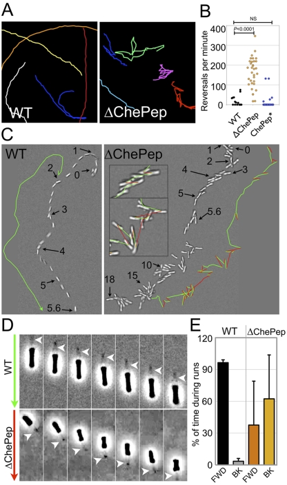
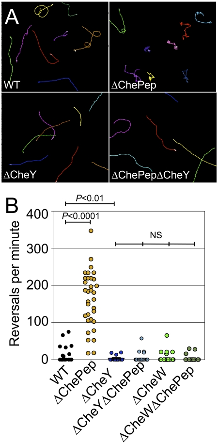
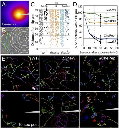
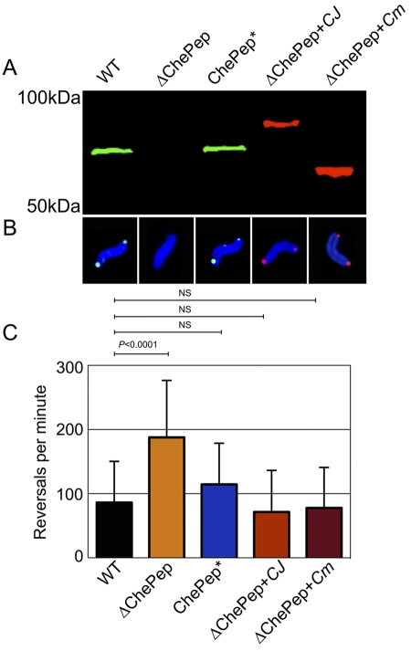
Microbes use directed motility to colonize harsh and dynamic environments. We discovered that Helicobacter pylori strains establish bacterial colonies deep in the gastric glands and identified a novel protein, ChePep, necessary to colonize this niche. ChePep is preferentially localized to the flagellar pole. Although mutants lacking ChePep have normal flagellar ultrastructure and are motile, they have a slight defect in swarming ability. By tracking the movement of single bacteria, we found that ΔChePep mutants cannot control the rotation of their flagella and swim with abnormally frequent reversals. These mutants even sustain bursts of movement backwards with the flagella pulling the bacteria. Genetic analysis of the chemotaxis signaling pathway shows that ChePep regulates flagellar rotation through the chemotaxis system. By examining H. pylori within a microscopic pH gradient, we determined that ChePep is critical for regulating chemotactic behavior. The chePep gene is unique to the Epsilonproteobacteria but is found throughout this diverse group. We expressed ChePep from other members of the Epsilonproteobacteria, including the zoonotic pathogen Campylobacter jejuni and the deep sea hydrothermal vent inhabitant Caminibacter mediatlanticus, in H. pylori and found that ChePep is functionally conserved across this class. ChePep represents a new family of chemotaxis regulators unique to the Epsilonproteobacteria and illustrates the different strategies that microbes have evolved to control motility.

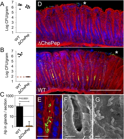
Importance: Helicobacter pylori strains infect half of all humans worldwide and contribute to the development of peptic ulcers and gastric cancer. H. pylori cannot survive within the acidic lumen of the stomach and uses flagella to actively swim to and colonize the protective mucus and epithelium. The chemotaxis system allows H. pylori to navigate by regulating the rotation of its flagella. We identified a new protein, ChePep, which controls chemotaxis in H. pylori. ChePep mutants fail to colonize the gastric glands of mice and are completely outcompeted by normal H. pylori. Genes encoding ChePep are found only in the class Epsilonproteobacteria, which includes the human pathogen Campylobacter jejuni and environmental microbes like the deep-sea hydrothermal vent colonizer Caminibacter mediatlanticus, and we show that ChePep function is conserved in this class. Our study identifies a new colonization factor in H. pylori and also provides insight into the control and evolution of bacterial chemotaxis.


Figures

FIG 1
FIG 1
ChePep localizes to the flagellar poles. (A) H. pylori cells at different stages of the cell cycle were stained with antibodies to H. pylori (red) and ChePep (green) and aligned according to their length to analyze ChePep expression and localization in individual bacteria. Scanning electron microscopy (SEM) illustrates the location of flagella in a young (left) versus septating (right; arrowheads indicate septation cleft) H. pylori cell. (For the quantitative relationship between bacterial length and ChePep intensity, see Fig. S2A in the supplemental material.) (B) Confocal 3D reconstructions of H. pylori cells at different stages of growth stained with anti-ChePep (green), anti-H. pylori (red), and DAPI for DNA (blue). Fl, flagella. Stars indicate chromosomal segregation. Scale bar, 1 µm in B.
FIG 2
FIG 2
ChePep is essential for colonizing the antral gastric glands and confers a significant advantage in colonization of the stomach. (A) Colony-forming units (CFU) of H. pylori in the stomachs of mice colonized with either the WT or ∆ChePep mutant for 2 weeks. Each marker represents an individual mouse. (B) CFU counts from mice coinfected with both the WT and ∆ChePep mutant in a 1:1 ratio for 2 weeks. The dashed red line indicates the limit of detection. P < 0.0001. (C) Volumetric analysis of bacteria colonizing the antral glands calculated from 100-µm-thick sections imaged by 3D confocal microscopy from single infections of either the WT or ∆ChePep mutant. The average number of H. pylori cells within the gastric glands per section is plotted. Data from 20 sections from three mice infected with the WT and 26 sections from three mice infected with the ∆ChePep mutant were compared. P < 0.0001. (D) 3D confocal microscopy of murine stomachs infected with either the WT or the ∆ChePep mutant. F-actin is stained with phallodin (red), and nuclei (blue) and H. pylori cells (green) are immunolabeled. Asterisks indicate H. pylori cells in the surface mucus of the stomach, while a box highlights bacterial colonies in mid-glands. (E) Magnified view of the area boxed in panel D. (F) SEM of WT-infected gland. (G) Magnified view of area boxed in panel F. Scale bars represent 100 µm in panel D, 10 µm in panels E and F, and 1 µm in G. P values are from the two-tailed Student t test.
FIG 3
FIG 3
ChePep reduces switching of the flagellar rotational direction. (A) Magnified tracings of both WT and ∆ChePep cell swimming behavior (see Movie S1 in the supplemental material). (B) Quantification of reversals per minute of the WT, ∆ChePep, and ChePep* (∆ChePep complemented in trans) strains. (C) DIC video microscopy of WT and ∆ChePep cell swimming (see Movie S2 in the supplemental material). The position of a single swimming bacterium over time is shown for the WT versus the ∆ChePep mutant. The position at a particular time (in seconds) is marked with arrows, and the swimming path is marked in green in one direction and red when the bacteria reverse swimming direction. Inset images of ∆ChePep cell movement at higher magnification are also shown. (D) High-magnification phase-contrast video microscopy shows a ∆ChePep mutant swimming backwards with the flagella “pulling” (see Movie S3 in the supplemental material). The arrows on the left indicate the direction that the bacteria are swimming. (Green indicates swimming forward, and red indicates swimming backward.) The position of the flagella is marked with an arrowhead in each panel. (E) Quantification of the percentage of time that WT and ∆ChePep cells swim forward (FWD) with the flagella “pushing” or backwards (BK) with the flagella “pulling.” n = 8 for each strain. P values are from the two-tailed Student t test.
FIG 4
FIG 4
ChePep controls flagellar switching through the chemotaxis system. (A) Motility tracings of the WT, the ∆ChePep and ∆CheY single mutants, and the ∆ChePep ∆CheY double mutant strain. (B) Quantification of reversals per minute of the WT and ∆ChePep, ∆CheY, ∆CheY ∆ChePep, ∆CheW, and ∆CheW ∆ChePep mutants. P values are from the two-tailed Student t test.
FIG 5
FIG 5
ChePep regulates H. pylori chemotaxis response in a pH gradient. (A) A microscopic pH gradient generated by a microinjection needle is visualized by Lysosensor dye fluorescence (pseudocolor image) (see Fig. S4 in the supplemental material for details). (B) A sample frame from a movie of WT bacteria and their motility tracings after 20 s in the pH gradient is shown. Rings are 10 µm apart. (C) The distance of each H. pylori cell to the acid point source is plotted before (−) or 10 s after (+) exposure to the pH gradient; ChePep* is ∆ChePep complemented in trans. NS, no statistical difference. (D) Plot of the percentage of H. pylori cells remaining within 60 µm of the micropipette tip over time. One hundred percent is defined as the number of bacteria within this radius directly before addition of acid. Error bars indicate standard deviations from the mean of five independent movies. Results for the ∆ChePep mutant and WT strains are significantly different (P < 0.0001, F test). (E) Bacterial motility paths of WT versus different mutants 2 s before (upper panels) and after 10 s in the pH gradient (bottom panels) (see Movie S5 in the supplemental material).
FIG 6
FIG 6
ChePep is functionally conserved throughout the Epsilonproteobacteria. (A) Immunoblot of the WT and ∆ChePep, ChePep* (∆ChePep complemented with H. pylori ChePep), ∆ChePep + CJ (∆ChePep complemented with FLAG-tagged C. jejuni ChePep), and ∆ChePep + Cm (∆ChePep complemented with FLAG-tagged C. mediatlanticus ChePep) strains. The blot is probed with anti-H. pylori ChePep (green) and anti-FLAG (red). (B) Immunofluorescence of H. pylori stained with anti-H. pylori (blue), anti-ChePep (green), and anti-FLAG (red). (C) Reversals per minute of the WT and ∆ChePep, ChePep*, ∆ChePep + Cj, and ∆ChePep + Cm strains. P values are from the two-tailed Student t test. NS, no statistical difference.

Comment in

References

    1. Campbell BJ, Engel AS, Porter ML, Takai K. 2006. The versatile epsilon-proteobacteria: key players in sulphidic habitats. Nat. Rev. Microbiol. 4:458–468 - PubMed
    1. Blaser MJ. 1997. Epidemiologic and clinical features of Campylobacter jejuni infections. J. Infect. Dis. 176(Suppl. 2):S103–S105 - PubMed
    1. Beswick EJ, Suarez G, Reyes VE. 2006. H pylori and host interactions that influence pathogenesis. World J. Gastroenterol. 12:5599–5605 - PMC - PubMed
    1. Schreiber S, et al. 2004. The spatial orientation of Helicobacter pylori in the gastric mucus. Proc. Natl. Acad. Sci. U. S. A. 101:5024–5029 - PMC - PubMed
    1. Larsen SH, Reader RW, Kort EN, Tso WW, Adler J. 1974. Change in direction of flagellar rotation is the basis of the chemotactic response in Escherichia coli. Nature 249:74–77 - PubMed

Publication types

MeSH terms