. 2011 May 3;108(18):7619-24.
doi: 10.1073/pnas.1104410108. Epub 2011 Apr 13.

Plant intracellular innate immune receptor Resistance to Pseudomonas syringae pv. maculicola 1 (RPM1) is activated at, and functions on, the plasma membrane

Affiliations

Plant intracellular innate immune receptor Resistance to Pseudomonas syringae pv. maculicola 1 (RPM1) is activated at, and functions on, the plasma membrane

Zhiyong Gao et al. Proc Natl Acad Sci U S A. .

Erratum in

  • Proc Natl Acad Sci U S A. 2011 May 24;108(21):8915. Gao, Zhiyoug [corrected to Gao, Zhiyong]

Abstract

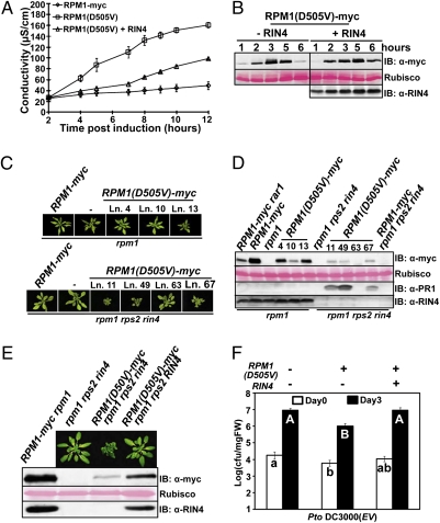
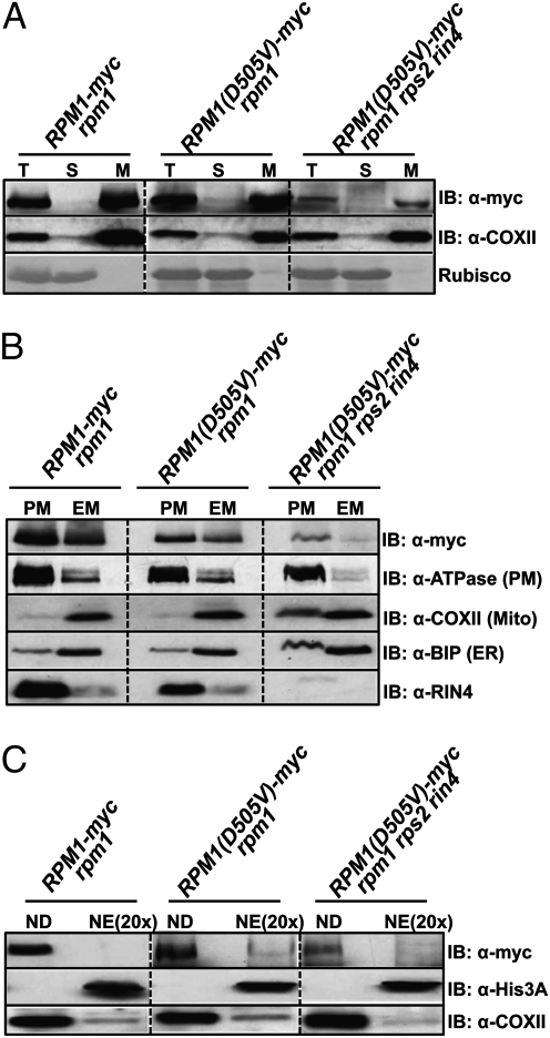
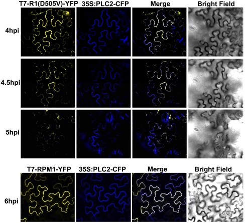
Plants deploy intracellular innate immune receptors to recognize pathogens and initiate disease resistance. These nucleotide-binding, leucine-rich repeat (NB-LRR) proteins are activated by pathogen effector proteins that are delivered into the host cell to suppress host defense responses. Little is known about the sites and mechanisms of NB-LRR activation, but some NB-LRR proteins can function inside the plant nucleus. We demonstrate that RPM1 is activated on the plasma membrane and does not relocalize to the nucleus. An autoactive RPM1(D505V) allele that recapitulates key features of normal RPM1 activation also resides on the plasma membrane. There is no detectable relocalization of activated RPM1 to the nucleus. Hindering potential nuclear entry of RPM1-Myc did not affect either its effector-triggered hypersensitive-response (HR) cell death or its disease resistance functions, further suggesting that nuclear translocation is not required for RPM1 function. RPM1 tethered onto the plasma membrane with a dual acylated N-terminal epitope tag retained the ability to mediate HR, consistent with this RPM1 function being activated on the plasma membrane. Plant NB-LRR proteins can thus function at various locations in the cell.


Conflict of interest statement

The authors declare no conflict of interest.

Figures

Fig. 1.
Fig. 1.
The MHD domain mutant RPM1(D505V) induces cell death in N. benthamiana. (A) RPM1 mutants used in this work. Wild-type sequences (Upper) and mutant sequences (Lower) are shown. (B and C) Estradiol-inducible RPM1 alleles were transiently expressed in N. benthamina (OD600 = 0.3). The HR-inducing activity of the RPM1 alleles was measured by ion leakage. (D and E) RPM1 proteins were detected by immunoblot. Samples were collected at 5 h after the induction with 40 μM estradiol and 40 μg of total extract loaded. Rubisco was used for protein loading control. Protein samples from uninfiltrated leaves were used as a control for nonspecific cross-reactivity.
Fig. 2.
Fig. 2.
The autoactivity of RPM1(D505V) is sufficient to trigger enhanced disease resistance and can be suppressed by RIN4. (A) Ion leakage induced by transient expression of RPM1(D505V) with or without RIN4 in N. benthamiana. RIN4 is expressed from its native promoter [OD600 = 0.3 for RIN4 and OD600 = 0.3 for RPM1(D505V) or RPM1]. RPM1(D505V) was induced with 40 μM estradiol. (B) RPM1(D505V)-myc expression over a time course after estradiol induction, with or without RIN4 co-expression. (C) Pictures of rosettes from RPM1(D505V)-myc transgenic plants (genetic backgrounds noted at top). The plants were grown under 16-h-long days for 6 wk. (D) Protein expression levels in the transgenic plants. Samples were collected from 4-wk-old plants grown under 9-h short days. (E) Photos and protein expression levels of isogenic RPM1(D505V)-myc line 49 plants with or without RIN4. Line 49 of RPM1(D505V)-myc rpm1 rps2 rin4 (C and D) was crossed with rpm1 rps2 to obtain isogenic RPM1(D505V)-myc rpm1 rps2 plants. (F) Enhanced disease resistance following inoculation with Pto DC3000 of the isogenic plants from E. Seedlings were dip-inoculated with 2.5 × 10⁷ cfu/mL of Pto DC3000. Pair-wise comparison for all means from day 0 or day 3 values were performed with a one-way ANOVA test coupled with Tukey–Kramer HSD with 95% confidence limits.
Fig. 3.
Fig. 3.
RPM1(D505V) localizes on the plasma membrane in Arabidopsis. (A) RPM1(D505V) resides on microsomal membranes. Total protein (T) was separated into soluble (S) and microsomal membrane (M) fractions by ultracentrifugation. COXII, a mitochondria membrane protein, was used as a membrane protein marker. Rubisco was used as a soluble protein marker. (B) RPM1(D505V) resides on the plasma membrane. The microsomal membrane fraction (M) was further separated into the plasma membrane fraction (PM) and the endomembrane fraction (EM). H+-ATPase, COXII, and Bip were used as plasma membrane, mitochondrial membrane, and endoplasmic reticulum markers, respectively. (C) RPM1(D505V) is not in the nuclear fraction. RPM1(D505V) was present in nuclear depleted (ND) but not in nuclear enriched (NE) fractions. Histone 3A was used as a nuclear protein marker. COXII was used as a non-nuclear marker. Note that NE fractions are loaded at a 20× yield equivalent compared with ND fractions. Three grams of leaves from plants grown under 16-h-long days for 6 wk were used for the experiments. The leaves of RPM1(D505V) rpm1 and RPM1(D505V) rpm1 rps2 rin4 are from line 4 and line 11, respectively (Fig. 2D).
Fig. 4.
Fig. 4.
T7-RPM1(D505V)-YFP localizes to the plasma membrane of N. benthamiana epidermal cells. Transient expression of T7-RPM1(D505V)-YFP or T7-RPM1-YFP (OD600 = 0.3) was induced with 20 μM estradiol at 48 h after agrobacteria infiltration. Images were collected at the time after induction noted at left. 35S:PLC2-CFP (OD600 = 0.3) was co-expressed as a plasma membrane marker.
Fig. 5.
Fig. 5.
Plasma membrane-tethered RPM1 retains effector-mediated HR function in N. benthamiana. (A). CBL tag contains dual-lipid modification sites. (B) The CBL tag tethers soluble RPM1(G205E/D505V)-Myc to the microsomal membrane following conditional transient expression in N. benthamiama. (C) CBL and mCBL tags do not affect the membrane localization of RPM1-Myc. (D) CBL-tagged proteins localize on the plasma membrane. For BD, proteins were transiently expressed in N. benthamiana under the control of 35S promoter (OD600 = 0.5). Samples were collected at 40 h after agrobacteria infiltration. (E) The HR function of CBL-tagged RPM1. CBL- or mCBL-tagged RPM1-myc (R1) under the control of 35S promoter, T7-RIN4 (R4) under the control of its native promoter and AvrRpm1-HA (AvrRpm1) under the control of a dexamethasone (Dex)-inducible promoter were co-expressed in N. benthamiana (OD600 = 0.5, 0.3, and 0.05, respectively). AvrRpm1 was induced with 20 μM Dex at 36 h after infiltration. (F) The expression level of the constructs. The protein levels of the tagged RPM1 and RIN4 were detected at 2 h after induction. The protein levels of AvrRpm1 were detected at 6 h after induction.

References

    1. Jones JD, Dangl JL. The plant immune system. Nature. 2006;444:323–329. - PubMed
    1. Dodds PN, Rathjen JP. Plant immunity: Towards an integrated view of plant-pathogen interactions. Nat Rev Genet. 2010;11:539–548. - PubMed
    1. Ting JP, Willingham SB, Bergstralh DT. NLRs at the intersection of cell death and immunity. Nat Rev Immunol. 2008;8:372–379. - PubMed
    1. Vance RE, Isberg RR, Portnoy DA. Patterns of pathogenesis: Discrimination of pathogenic and nonpathogenic microbes by the innate immune system. Cell Host Microbe. 2009;6:10–21. - PMC - PubMed
    1. Eitas TK, Dangl JL. NB-LRR proteins: Pairs, pieces, perception, partners, and pathways. Curr Opin Plant Biol. 2010;13:472–477. - PMC - PubMed

Publication types

MeSH terms

LinkOut - more resources