. 2011 Feb 11:11:43.
doi: 10.1186/1471-2148-11-43.

Molecular adaptation of a plant-bacterium outer membrane protease towards plague virulence factor Pla

Affiliations

Molecular adaptation of a plant-bacterium outer membrane protease towards plague virulence factor Pla

Johanna Haiko et al. BMC Evol Biol. .

Abstract

Background: Omptins are a family of outer membrane proteases that have spread by horizontal gene transfer in Gram-negative bacteria that infect vertebrates or plants. Despite structural similarity, the molecular functions of omptins differ in a manner that reflects the life style of their host bacteria. To simulate the molecular adaptation of omptins, we applied site-specific mutagenesis to make Epo of the plant pathogenic Erwinia pyrifoliae exhibit virulence-associated functions of its close homolog, the plasminogen activator Pla of Yersinia pestis. We addressed three virulence-associated functions exhibited by Pla, i.e., proteolytic activation of plasminogen, proteolytic degradation of serine protease inhibitors, and invasion into human cells.

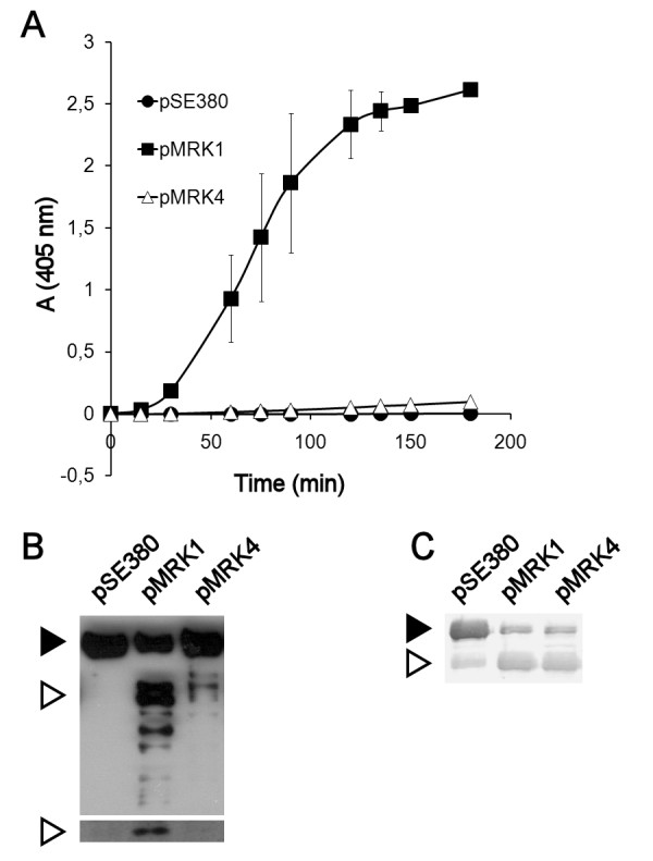
Results: Pla and Epo expressed in Escherichia coli are both functional endopeptidases and cleave human serine protease inhibitors, but Epo failed to activate plasminogen and to mediate invasion into a human endothelial-like cell line. Swapping of ten amino acid residues at two surface loops of Pla and Epo introduced plasminogen activation capacity in Epo and inactivated the function in Pla. We also compared the structure of Pla and the modeled structure of Epo to analyze the structural variations that could rationalize the different proteolytic activities. Epo-expressing bacteria managed to invade human cells only after all extramembranous residues that differ between Pla and Epo and the first transmembrane β-strand had been changed.

Conclusions: We describe molecular adaptation of a protease from an environmental setting towards a virulence factor detrimental for humans. Our results stress the evolvability of bacterial β-barrel surface structures and the environment as a source of progenitor virulence molecules of human pathogens.


Figures

Figure 1
Figure 1
Sequence alignment of Epo and Pla. The β-strands and the extracellular loops are numbered, and the 31 differing amino acids in the extracellular side of the protein are shown in bold face in gray background. The catalytic residues are indicated with asterisks. The sequences were aligned with ClustalW [50], and the image was rendered with Alscript [51]. The secondary structures are defined by Stride [52], and they are based on the known Pla structure [18].
Figure 2
Figure 2
Comparison of Pla and Epo. Pla and Epo were compared in (A) plasminogen activation, (B) degradation of plasminogen, and (C) degradation of α2-antiplasmin. pla and epo were expressed in recombinant E. coli XL1. The strains are indicated with their plasmid names (see Table 1). In (A), plasmin formation was measured using a chromogenic plasmin substrate. The data are average of two independent assays with duplicate samples, and standard deviations are shown. In (B), Western blotting with anti-plasminogen (top panel) and anti-plasminogen catalytic domain (bottom panel) antibodies is shown. The black arrowhead indicates plasminogen and the white arrowheads indicate cleaved plasminogen, the heavy chain (top panel) and the light chain (bottom panel) of plasmin. In (C), degradation of α2AP was visualized with Western blotting using α2AP antibody, and the black arrowhead indicates uncleaved α2AP and the white arrowhead indicates the degradation product.
Figure 3
Figure 3
Analysis of expression of Pla-Epo hybrid proteins in E. coli. Expression of the Pla-Epo-hybrid proteins was analyzed by Western blotting of whole cells with a mixture of anti-Pla, anti-Epo, and anti-Pla-loop antisera. The constructs are indicated with their plasmid names (see Table 1). Migration distance of the Pla isoforms are indicated; pre-Pla is the immature form of Pla, α-Pla and γ-Pla are mature Pla proteins with differing conformations, and β-Pla is the mature autocleaved form of Pla.
Figure 4
Figure 4
Cumulative plasminogen activation by Pla-Epo hybrid proteins expressed in E. coli. (A) Pla-Epo hybrids with cumulative substitutions towards Pla. (B) Pla-Epo hybrids with cumulative substitutions towards Epo. Inserts show the plasmid constructs (see Table 1). The data are average from two independent assays with duplicate samples, and standard deviations are shown.
Figure 5
Figure 5
Analysis of the ten substituted residues in Pla structure (left) and Epo model (right). L3 and its residues are colored blue, L4 red, and L5 green. The substituted residues are drawn thick and in a darker shade, and the other discussed residues thin and in lighter shade. The hydrogen bond between D212 and N263 in Pla is marked with a dashed line, and three interaction areas circled in both structures. Counting from left: a tight cluster of hydrophobic residues in L5; a polar quintet between L5, L4 and L3 in Pla and a L4 triplet in Epo; hydrophobic contacts within the barrel opening in Pla and polar residues at the outside of the barrel in Epo.
Figure 6
Figure 6
Degradation of serpins by E. coli expressing the Pla-Epo hybrid proteins. Degradation of PAI-1 (A) and α2AP (B) in a 2-h incubation was analyzed by Western blotting. The black arrowhead indicates uncleaved serpin and the white arrowhead indicates the degradation product. The strains are indicated with their plasmid names (see Table 1).
Figure 7
Figure 7
Summary of the substitution analysis. Molecular models of selected Pla-Epo hybrid proteins are shown, with the substituted sites coloured black. The outer membrane (OM; the girdle area) and loops 3 and 5 are indicated. The active site amino acids are shown in a space filling representation. The table summarizes the plasminogen (Plg) activation and invasion capacities of Epo, Pla, and the Pla-Epo hybrid proteins with 10, 31, and 42 substitutions towards Pla; in total, there are 65/292 amino acid differences between mature Pla and Epo. For clarity, also the plasmid names are shown.

References

    1. Pallen MJ, Nelson KE, Preston GM. Bacterial pathogenomics. Washington, DC, USA: ASM Press; 2007.
    1. Seifert HS, Dirita VJ. Evolution of microbial pathogens. Washington, DC, USA: ASM Press; 2006.
    1. Hacker J, Kaper JB. Pathogenicity islands and the evolution of microbes. Annual Review of Microbiology. 2000;54:641–679. doi: 10.1146/annurev.micro.54.1.641. - DOI - PubMed
    1. Yen YT, Kostakioti M, Henderson IR, Stathopoulos C. Common themes and variations in serine protease autotransporters. Trends Microbiol. 2008;16(8):370–379. doi: 10.1016/j.tim.2008.05.003. - DOI - PubMed
    1. Perry RD, Fetherston JD. Yersinia pestis - etiologic agent of plague. Clin Microbiol Rev. 1997;10(1):35–66. - PMC - PubMed

Publication types

MeSH terms