. 2011 Jan 11;6(1):e15946.
doi: 10.1371/journal.pone.0015946.

Defects in very long chain fatty acid synthesis enhance alpha-synuclein toxicity in a yeast model of Parkinson's disease

Affiliations

Defects in very long chain fatty acid synthesis enhance alpha-synuclein toxicity in a yeast model of Parkinson's disease

Yong Joo Lee et al. PLoS One. .

Abstract

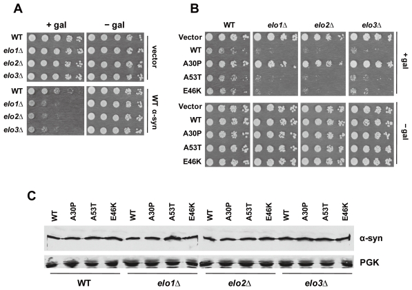
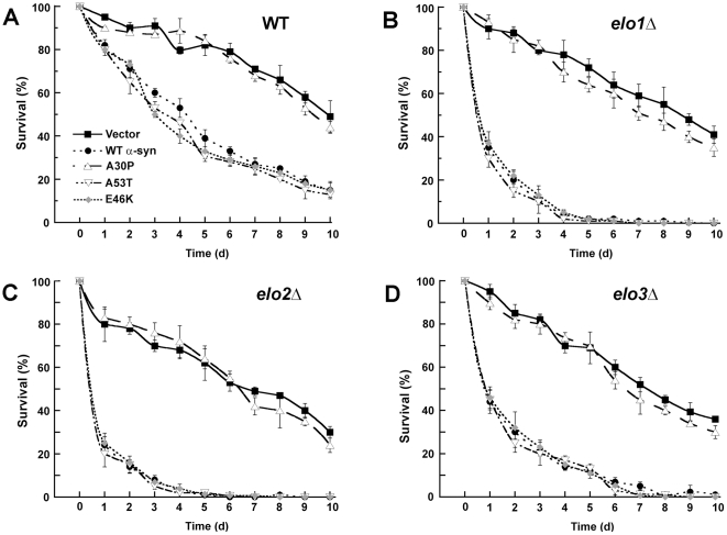
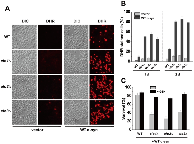
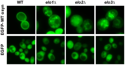
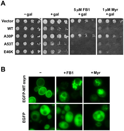
We identified three S. cerevisiae lipid elongase null mutants (elo1Δ, elo2Δ, and elo3Δ) that enhance the toxicity of alpha-synuclein (α-syn). These elongases function in the endoplasmic reticulum (ER) to catalyze the elongation of medium chain fatty acids to very long chain fatty acids, which is a component of sphingolipids. Without α-syn expression, the various elo mutants showed no growth defects, no reactive oxygen species (ROS) accumulation, and a modest decrease in survival of aged cells compared to wild-type cells. With (WT, A53T or E46K) α-syn expression, the various elo mutants exhibited severe growth defects (although A30P had a negligible effect on growth), ROS accumulation, aberrant protein trafficking, and a dramatic decrease in survival of aged cells compared to wild-type cells. Inhibitors of ceramide synthesis, myriocin and FB1, were extremely toxic to wild-type yeast cells expressing (WT, A53T, or E46K) α-syn but much less toxic to cells expressing A30P. The elongase mutants and ceramide synthesis inhibitors enhance the toxicity of WT α-syn, A53T and E46K, which transit through the ER, but have a negligible effect on A30P, which does not transit through the ER. Disruption of ceramide-sphingolipid homeostasis in the ER dramatically enhances the toxicity of α-syn (WT, A53T, and E46K).


Conflict of interest statement

Competing Interests: The authors have declared that no competing interests exist.

Figures

Figure 1
Figure 1. Enhanced α-syn toxicity in elongase null mutants.
(A) The effect of WT α-syn expression on the growth of three deletion strains (elo1Δ, elo2Δ, and elo3Δ) and the wild-type BY4741 strain was evaluated. In panels (A, B), strains were transformed with pAG426GAL-WT (WT α-syn), pAG426GAL-A30P (A30P), pAG426GAL-A53T (A53T), pAG426GAL-E46K (E46K) or pAG426GAL (empty vector) and spotted in successive 10-fold serial dilutions on solid sucrose and solid galactose plates and incubated for 3 days at 30°C. Experiments were repeated three times with similar results. (B) The effect of the various α-syns (WT, A30P, A53T, or E46K) on the growth of the three elo deletion strains was also evaluated. (C) Western blot analysis of yeast cells expressing α-syn. Lysates were prepared from cultures grown for 6 h in inducing medium and then subjected to SDS–PAGE followed by western blot analysis. The cell-signaling polyclonal antibody against α-syn was used to visualize the three α-syns. The loading control was the yeast protein Pgk1p. Identical amounts of protein were loaded per well. Plasmids: see panel (A).
Figure 2
Figure 2. elo1Δ/+α-syn slow growth phenotype is rescued by ELO1.
(A) Growth properties of wild-type cells and elo1Δ cells expressing WT α-syn (pAG426GAL-WT) and expressing Elo1p from a CEN plasmid (pAG415GPD-ELO1) or over expressing from a 2 µ plasmid (pAG425GAL-ELO1 are shown. The spot assay was conducted as described in Fig. 1. (B) The western blot analysis shows the level of Elo1p-Tap protein in ELO1-TAP cells, in elo1Δ cells transformed with pAG415GPD-ELO1 (CEN, GPD1 promoter), and in elo1Δ cells transformed with pAG425GAL-ELO1 (2 µ, GAL1 promoter). A monoclonal antibody against the tap tag was used, and Pgk1 was used as the loading control.
Figure 3
Figure 3. α-Syn is extremely toxic to aged cells.
(A) This panel shows chronological aging curves for wild-type BY4741 cells with empty vector (closed square), WT α-syn (closed circle), A30P (open triangle), A53T (inverted open triangle), or E46K (gray diamond). The various cells were grown in selective media containing sucrose to stationary phase, and then the cells were washed and resuspended in selective media containing galactose. After two days of further incubation, the aging analysis started. At the indicated times aliquots were removed form the culture, diluted onto plates containing rich media, incubated for 2 days, and then colonies were counted. Curves represent the number of colonies at the given time divided by the number of colonies at time zero. Experiments were conducted three times, and error bars represent ± s.e.m. P-values are given in the text. Plasmids used: pAG426GAL (empty vector), pAG426GAL-WT (WT α-syn), pAG426GAL-A30P (A30P), pAG426GAL-A53T (A53T), and pAG426GAL-E46K (E46K). (B), (C) and (D) show chronological aging curves for elo1Δ, elo2Δ, and elo3Δ mutants, respectively, with the various α-syns.
Figure 4
Figure 4. α-Syn triggers ROS in aged elo cells.
(A) Wild-type cells containing empty plasmid (pAG426GAL) or expressing WT α-syn (pAG426GAL-WT) were cultured for 24 h in inducing media (+gal) at 30°C. After 24 h, cells were incubated with DHR 123 (5 µg/ml) for 1 h, and then visualized by differential interference contrast (DIC) and fluorescence microscopy (DHR). Cells were counted and the percentage of cells exhibiting red fluorescence was plotted. (B) Plot of the percentage of cells (WT, elo1Δ, elo2Δ, and elo3Δ) that exhibited red fluorescence due to ROS accumulation. Cultures were grown for 24 h in inducing media at 30°C and then stained with DHR. Each value was obtained from two independent experiments, where the total number of cells counted was 600. Error bars reflect s.e.m. (C) Reduced glutathione (GSH) protects aged cells from WT α-syn. The various cells expressing WT α-syn were grown in selective media containing sucrose to stationary phase, and then cells were washed and resuspended in galactose media with or without 10 mM GSH. After 3 days (1 day on aging curve) incubation, aliquots were removed from culture, diluted onto plates containing rich media, incubated for 2 days, and then colonies were counted. Each bar represents the number of colonies at the 3 days divided by the number of colonies at time zero. Experiments were conducted three times, and error bars represent ± s.e.m.
Figure 5
Figure 5. Elongase null mutations alter the localization of WT α-syn.
Yeast cells (BY4741, elo1Δ, elo2Δ, and elo3Δ) expressing eGFP-WT-α-syn or EGFP were cultured in inducing media for 6 h at 30°C and then visualized by fluorescence microscopy. Plasmids: pAG426GAL-EGFP-WT-α-syn and pAG426GAL-EGFP.
Figure 6
Figure 6. Hypersensitivity of cells expressing α-syn (WT, A53T or E46K) to fumonisin B1 (FB1) and myriocin.
(A) The toxicity of FB1 and myriocin was evaluated by evaluating the growth properties of various cells in a serial dilution assay. Strains were transformed with empty vector or plasmids harboring the various α-syns (WT α-syn, A30P, A53T, or E46K) and spotted in successive 10-fold serial dilutions on solid sucrose and solid galactose plates and incubated for 3 days at 30°C. FB1 and myriocin were dissolved in the media at 5 µM or 1 µM, respectively. Experiments were repeated three times with similar results. The pAG426GAL set of plasmids were used. (B) FB1 and myriocin drives WT α-syn off the plasma membrane and into inclusions. Wild-type cells expressing eGFP-WT-α-syn or eGFP were incubated for 6 h in inducing media containing 5 µM FB1 or 1 µM myriocin or drug vehicle, ethanol (−) at 30°C. Images were obtained by fluorescence microscopy 6 h after adding myriocin. Approximately 85%, and 80% of the cells examined exhibited the EGFP-WT-α-syn inclusions as shown in the upper center (+Fum) and right panel (+Myr), respectively. Plasmids: pAG426GAL-EGFP-WT-α-syn and pAG426GAL-EGFP.
Figure 7
Figure 7. A plasma membrane-binding form of EGFP is toxic, like α-syn, to elo3Δ cells.
(A) Inhibiting ceramide synthetase with FB1, serine palmitoyltransferase with myriocin or blocking C26-VLCFA synthesis in elo3Δ cells drives EGFP-mts1 and EGFP-mts2 into cytoplasmic inclusions. Wild-type cells or elo3Δ cells expressing EGFP-mts1, EGFP-mts2 or EGFP as a control were incubated for 6 h in inducing media containing 5 µM FB1, 1 µM myriocin (or drug vehicle, ethanol) and then imaged by fluorescence microscopy. Approximately 80%, 68%, and 88% of the cells examined exhibited the EGFP-mts1 inclusions as shown in the wild-type strain (+Fum), (+Myr), and in the elo3Δ strain, respectively. 85%, 75%, 70% of the cells examined exhibited the EGFP-mts2 inclusion as shown in the wild-type strain (+Fum), (+Myr), and in the elo3Δ strain, respectively. Plasmids: pAG426GAL-EGFP-mts1, pAG426GAL-EGFP-mts2, and pAG426GAL-EGFP. (B) Growth properties of cells expressing EGFP-mts1, EGFP-mts2, or EGFP were evaluated in a dilution spot assay. EGFP-mts1 or EGFP-mts2 is not toxic to wild-type cells, whereas it was very toxic to FB1-treated (5 µM) wild-type cells and to elo3Δ cells. The control construct EGFP was not toxic to any of the cells. The assay was conducted according to the legend in Fig. 1.

References

    1. Moore DJ, West AB, Dawson VL, Dawson TM. Molecular pathophysiology of Parkinson's disease. Annu Rev Neurosci. 2005;28:57–87. - PubMed
    1. Cookson MR. alpha-Synuclein and neuronal cell death. Mol Neurodegener. 2009;4:9. - PMC - PubMed
    1. Waxman EA, Giasson BI. Molecular mechanisms of alpha-synuclein neurodegeneration. Biochim Biophys Acta. 2009;1792:616–624. - PMC - PubMed
    1. Abou-Sleiman PM, Healy DG, Wood NW. Causes of Parkinson's disease: genetics of DJ-1. Cell Tissue Res. 2004;318:185–188. - PubMed
    1. Recchia A, Debetto P, Negro A, Guidolin D, Skaper SD, et al. Alpha-synuclein and Parkinson's disease. Faseb J. 2004;18:617–626. - PubMed

Publication types

MeSH terms