. 2010 Dec 29;5(12):e15703.
doi: 10.1371/journal.pone.0015703.

Aureusimines in Staphylococcus aureus are not involved in virulence

Affiliations

Aureusimines in Staphylococcus aureus are not involved in virulence

Fei Sun et al. PLoS One. .

Abstract

Background: Recently, dipeptide aureusimines were reported to activate expression of staphylococcal virulence genes, such as alpha-hemolysin, and increase S. aureus virulence. Surprisingly, most of the virulence genes affected by aureusimines form part of the regulon of the SaeRS two component system (TCS), raising the possibility that SaeRS might be directly or indirectly involved in the aureusimine-dependent signaling process.

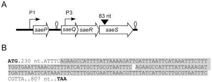
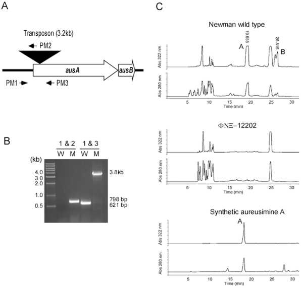
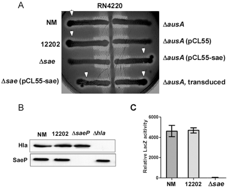
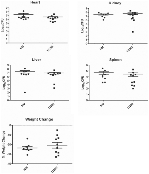
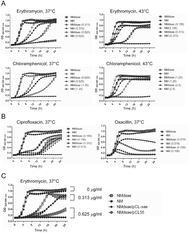
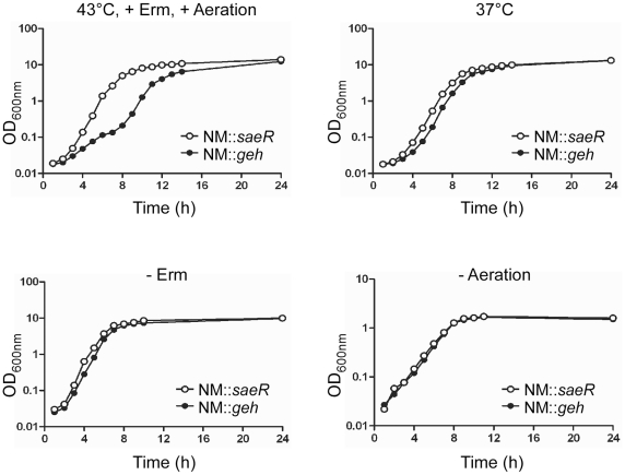
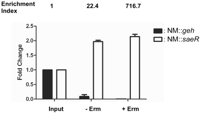
Methodology/principal findings: Using HPLC analyses, we confirmed that a transposon mutant of ausA, the gene encoding the aureusimine dipeptide synthesis enzyme, does not produce dipeptides. However, the transposon mutant showed normal hemolysis activity and alpha-hemolysin/SaeP production. Furthermore, the P1 promoter of the sae operon, one of the targets of the SaeRS TCS, showed normal transcription activity. Moreover, in contrast to the original report, the ausA transposon mutant did not exhibit attenuated virulence in an animal infection model. DNA sequencing revealed that the ausA deletion mutant used in the original study has an 83 nt-duplication in saeS. Hemolysis activity of the original mutant was restored by a plasmid carrying the sae operon. A mutant of the sae operon showed elevated resistance to chloramphenicol and erythromycin, two antibiotics widely used during staphylococcal mutagenesis. At 43°C in the presence of erythromycin and aeration, the conditions typically employed for staphylococcal mutagenesis, an saeR transposon mutant grew much faster than a control mutant and the saeR mutant was highly enriched in a mixed culture experiment.

Conclusions/significance: Our results show that the previously reported roles of aureusimines in staphylococcal gene regulation and virulence were due to an unintended mutation in saeS, which was likely selected due to elevated resistance of the mutant to environmental stresses. Thus, there is no evidence indicating that the dipeptide aureusimines play a role in sae-mediated virulence factor production or contribute to staphylococcal virulence.


Conflict of interest statement

Competing Interests: The authors have declared that no competing interests exist.

Figures

Figure 1
Figure 1. The sae locus of S. aureus (A) and saeS gene in the original ausA deletion mutant (B). A.
The four open reading frames in the sae operon are shown as open arrows with the corresponding gene names. The two sae promoters, P1 and P3, are indicated with the arrows pointing out the transcription direction. The 83 nt duplication found in ausA deletion mutant is shown as a reversed triangle. Putative transcription terminators are indicated by stem-loop structures. B. Chromosomal DNA was purified from the original ausA deletion mutant; then the saeRS region was PCR-amplified and sequenced. The 83 nt duplicated in saeS is indicated by a gray-colored shadow. The start and stop codons are bold faced. For clarity, only numbers are shown for intervening nucleotides.
Figure 2
Figure 2. The ausA transposon mutant ΦΝΞ-12202 does not produce aureusimines.
A. The ausAB operon in the transposon mutant ΦΝΞ-12202. The transposon insertion is indicated by a reversed triangle. The primers (PM1, PM2, and PM3), used for PCR-amplification, are shown as arrows. B. PCR amplification of the ausA region of wild-type Newman (W) and the transposon insertion mutant ΦΝΞ-12202 (M). The numbers at the top represent the primer pairs. The calculated size of each product is indicated on the right of the picture. Molecular size markers are shown to the left. C. The absence of aureusimines in the culture supernatant of the transposon mutant ΦΝΞ-12202. The culture supernatants from wild-type Newman and the transposon mutant were extracted with ethyl acetate and suspended in methanol. The supernatant extracts were analyzed with HPLC. As a control, synthetic aureusimine A was used. A, aureusimine A; B, aureusimine B.
Figure 3
Figure 3. The SaeRS TCS is functional in the ausA transposon mutant ΦΝΞ-12202.
A. Hemolysis patterns on sheep blood agar. The strains tested were streaked against the strain RN4220, which produces only beta-hemolysin. The white arrow indicates the hemolysis caused by alpha-hemolysin. The enhanced hemolysis at the junctions of strain RN4220 and the tested strains is due to delta-hemolysin production originating from the test strains. NM, wild type; 12202, transposon insertion mutant ΦΝΞ-12202; Δsae, a deletion mutant of the sae operon; ΔausA, original ausA deletion mutant; pCL55-sae, pCL55 plasmid containing the entire sae operon. B. Western blot analysis for alpha-hemolysis (Hla) and SaeP protein. NM, wild type; 12202, ΦΝΞ-12202; ΔsaeP, a transposon insertion mutant of saeP; Δhla, a transposon insertion mutant of hla, the gene encoding alpha-hemolysin. C. LacZ assay for P1 promoter activity. Mid-log phase cells were used to measure LacZ activity from P1-lacZ fusion in strains Newman (NM), ΦΝΞ-12202, and the sae deletion mutant (Δsae). Bar graphs depict the mean ± standard deviation for the relative LacZ activity of each indicated strain.
Figure 4
Figure 4. Aureusimines do not contribute to staphylococcal virulence.
The test strains (1×10⁷ cfu) were administered into 10 mice per strain via retro-orbital injection; then, four days later, after measuring weight losses, organs were harvested and bacterial cfu in the organs was determined. In the graph, each dot represents data from one mouse. Mean and standard deviation are indicated.
Figure 5
Figure 5. The sae deletion mutant is more resistant to certain antibiotics.
A. Effect of the sae deletion mutation on resistance to chloramphenicol and erythromycin at 37°C or 43°C. Cells were inoculated in 200 µl TSB; then the growth of the cells was measured with Bioscreen C (Growth Curves USA) for 24 h. The antibiotics and growth temperatures are shown above each graph. The concentration (µg/ml) of antibiotics is shown in parentheses. B. Effect of the sae deletion mutation on resistance to ciprofloxacin and oxacillin at 37°C. C. Complementation test for the sae deletion mutant. The used erythromycin concentration is indicated to the right of the graph. pCL55, an integration vector; pCL55-sae, pCL55 containing the entire sae operon.
Figure 6
Figure 6. An sae mutant has a growth advantage at high temperature, aeration, and presence of erythromycin.
The transposon mutants of saeR and geh (glycerol ester hydrolase) were grown under various conditions and the effects of three environmental factors (temperature, the presence of erythromycin, and aeration) on the growth pattern of the mutants were examined. For clarity, only the altered environmental factor is indicated above the graph being compared. In the test, erythromycin was added at 10 µg/ml and aeration was achieved by shaking at 250 rpm. Erm; erythromycin.
Figure 7
Figure 7. Sae mutants can be enriched under typical staphylococcal mutagenesis conditions.
Transposon insertion mutants of geh (glycerol ester hydrolase) and saeR were used in this test. Equal numbers of the two mutants were mixed and grown either in the absence or in the presence of erythromycin (10 µg/ml) with shaking for 16 h at 43°C. Chromosomal DNA was purified from each culture and the fold change of each strain was calculated by quantitative real time PCR (qPCR) as described in Materials and Methods. Bar graphs indicate the fold change of each strain. Data are presented as the mean ± standard deviation from three independent experiments. Input, the mixed culture used for inoculation; - Erm, culture grown in the absence of erythromycin; + Erm, culture grown in the presence of erythromycin.

References

    1. Archer GL. Staphylococcus aureus: a well-armed pathogen. Clin Infect Dis. 1998;26:1179–1181. - PubMed
    1. Lowy FD. Staphylococcus aureus infections. N Engl J Med. 1998;339:520–532. - PubMed
    1. Novick RP. Autoinduction and signal transduction in the regulation of staphylococcal virulence. Mol Microbiol. 2003;48:1429–1449. - PubMed
    1. Giraudo AT, Calzolari A, Cataldi AA, Bogni C, Nagel R. The sae locus of Staphylococcus aureus encodes a two-component regulatory system. FEMS Microbiol Lett. 1999;177:15–22. - PubMed
    1. Giraudo AT, Cheung AL, Nagel R. The sae locus of Staphylococcus aureus controls exoprotein synthesis at the transcriptional level. Arch Microbiol. 1997;168:53–58. - PubMed

Publication types