. 2011 Feb 10:1373:91-100.
doi: 10.1016/j.brainres.2010.12.017. Epub 2010 Dec 13.

Methamphetamine causes sustained depression in cerebral blood flow

Affiliations

Methamphetamine causes sustained depression in cerebral blood flow

Oksana Polesskaya et al. Brain Res. .

Abstract

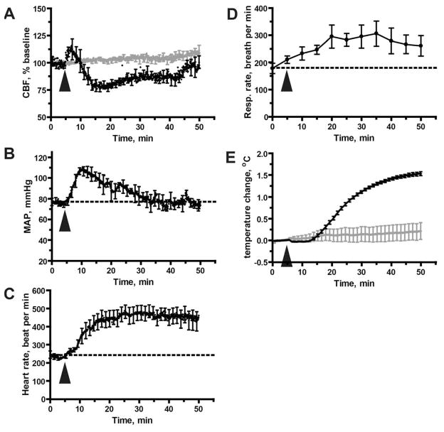
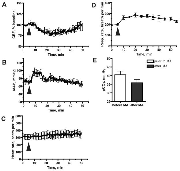
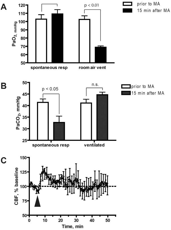
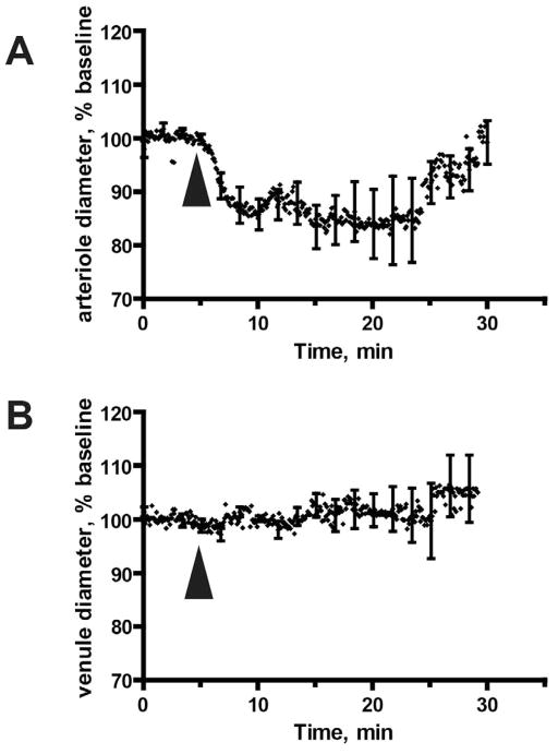
The use prevalence of the highly addictive psychostimulant methamphetamine (MA) has been steadily increasing over the past decade. MA abuse has been associated with both transient and permanent alterations in cerebral blood flow (CBF), hemorrhage, cerebrovascular accidents and death. To understand MA-induced changes in CBF, we exposed C56BL/6 mice to an acute bolus of MA (5mg/kg MA, delivered IP). This elicited a biphasic CBF response, characterized by an initial transient increase (~ 5 minutes) followed by a prolonged decrease (~ 30 minutes) of approximately 25% relative to baseline CBF--as measured by laser Doppler flowmetry over the somatosensory cortex. To assess if this was due to catecholamine derived vasoconstriction, phentolamine, an α-adrenergic antagonist was administered prior to MA treatment. This reduced the initial increase in CBF but failed to prevent the subsequent, sustained decrease in CBF. Consistent with prior reports, MA caused a transient increase in mean arterial blood pressure, body temperature and respiratory rate. Elevated respiratory rate resulted in hypocapnia. When respiratory rate was controlled by artificially ventilating mice, blood PaCO(2) levels after MA exposure remained unchanged from physiologic levels, and the MA-induced decrease in CBF was abolished. In vivo two-photon imaging of cerebral blood vessels revealed sustained MA-induced vasoconstriction of pial arterioles, consistent with laser Doppler flowmetry data. These findings show that even a single, acute exposure to MA can result in profound changes in CBF, with potentially deleterious consequences for brain function.


Conflict of interest statement

Disclosure/conflict of interests: No conflict of interest is reported.

Figures

Figure 1
Figure 1. Acute MA exposure (5 mg/kg) disregulates CBF and modulates other physiological parameters
A. Disregulation of CBF as measured by laser Doppler flowmetry. Values are expressed as a percentage of baseline CBF, defined here as the mean CBF measured during the 5 minute period immediately preceding delivery of MA (or saline, shown in grey). Results represent mean from 5 mice treated with MA and 3 mice treated with saline. B. MA-induced hypertension. Mean arterial blood pressure was measured via a catheter inserted into the femoral artery; results shown represent mean data values from 6 mice per group. C. Increase in heart rate. Results represent data from the same mice shown in (B). D. Increase in respiratory rate; results shown are mean data values from 8 mice. E. MA-induced hyperthermia. Body temperature was measured with a rectal probe, and results are presented relative to baseline temperature (calculated as in (A)). Injection of saline did not induce hyperthermia (shown in grey). Results represent data from the same mice shown in (A). (A–E) All data represent mean ± SEM. The arrowhead indicates the time at which MA (or saline) was injected.
Figure 2
Figure 2. Blockade of α-adrenoreceptors does not prevent the MA-mediated reduction in CBF
Mice were injected with the α-blocker phentolamine (10 mg/kg, i.p.), 30 min prior to MA exposure. A. Phentolamine pre-treatment does not prevent MA-mediated inhibition of CBF. CBF was measured with laser Doppler flowmetry in 6 mice. B. Effect of phentolamine pre-treatment on MA-mediated changes in MAP. MAP results represent mean values from 4 mice per group. C. Effect of phentolamine pre-treatment on MA-mediated changes in heart rate. Heart rate was measured in the same mice shown in (A). D. Effect of phentolamine pre-treatment on MA-mediated changes in respiratory rate. Respiratory rate measured in the same mice shown in (B). E. Effect of phentolamine pre-treatment on MA-mediated changes in PaCO2. PaCO2 was measured at baseline and 15 min after exposure to MA. Data were obtained from 5 mice. (A–E) Results represent mean ±SEM. The arrowhead indicates the time at which MA was injected.
Figure 3
Figure 3. Mechanical ventilation prevents CBF decrease in response to MA
A. Additional O2 is needed to provide adequate oxygenation in artificially ventilated mice. Oxygenation of arterial blood was within normal range before and after exposure to MA when mice were allowed to breathe spontaneously (note that RR reached 300 bpm after exposure to MA). In contrast, exposure to MA when mice were artificially ventilated with room air at 150 bpm resulted in a significant decrease in PaO2, relative to baseline levels (p = 0.0057, two-tailed t-test). Therefore, to avoid this non-physiological hypoxia, mice were ventilated in subsequent experiments with air containing 50% oxygen. B. Mechanical ventilation prevents the MA-mediated decrease in PaCO2. PaCO2 was measured prior to MA injection or 15 min after MA injection. Results represent mean ± SEM from 7 mice (not ventilated) and 5 mice (ventilated). The asterisk denotes that pCO2 in spontaneously breathing animals was significantly reduced at 15 min following exposure to MA, as compared to baseline (p = 0.017, two-tailed t-test). C. Mechanical ventilation prevents the MA-mediated decrease in CBF. CBF was measured in mechanically ventilated mice. Value are expressed as a percentage of baseline CBF, and represent mean ± SEM from 5 mice. Arrowheads indicate MA injection.
Figure 4
Figure 4. Acute MA exposure causes constriction of pial arterioles but does not affect pial veins
The diameter of pial blood vessels was determined by analysis of two- photon imaging data over a 30 minute time course (5 minutes preceding MA injection, and 25 minutes thereafter). The data represent results from (A) 9 arterioles and (B) 4 veins. Images were collected from 9 animals. (A, B) Data represent mean ± SD for vessel diameter, calculated every 5 seconds. Arrowheads indicate MA injection.

References

    1. Alhassoon OM, Dupont RM, Schweinsburg BC, Taylor MJ, Patterson TL, Grant I. Regional cerebral blood flow in cocaine- versus methamphetamine-dependent patients with a history of alcoholism. Int J Neuropsychopharmacol. 2001;4:105–12. - PubMed
    1. Brassard P, Seifert T, Secher NH. Is cerebral oxygenation negatively affected by infusion of norepinephrine in healthy subjects? Br J Anaesth. 2009;102:800–5. - PubMed
    1. Busija DW, Heistad DD, Marcus ML. Effects of sympathetic nerves on cerebral vessels during acute, moderate increases in arterial pressure in dogs and cats. Circ Res. 1980;46:696–702. - PubMed
    1. Chang L, Ernst T, Speck O, Patel H, DeSilva M, Leonido-Yee M, Miller EN. Perfusion MRI and computerized cognitive test abnormalities in abstinent methamphetamine users. Psychiatry Res. 2002;114:65–79. - PubMed
    1. Cook CE, Jeffcoat AR, Sadler BM, Hill JM, Voyksner RD, Pugh DE, White WR, Perez-Reyes M. Pharmacokinetics of oral methamphetamine and effects of repeated daily dosing in humans. Drug Metab Dispos. 1992;20:856–62. - PubMed

Publication types

MeSH terms