. 2009 Dec;47(12):3887-94.
doi: 10.1128/JCM.01533-09. Epub 2009 Oct 14.

Diagnostic bioluminescent phage for detection of Yersinia pestis

Affiliations

Diagnostic bioluminescent phage for detection of Yersinia pestis

David A Schofield et al. J Clin Microbiol. 2009 Dec.

Abstract

Yersinia pestis is the etiological agent of the plague. Because of the disease's inherent communicability, rapid clinical course, and high mortality, it is critical that an outbreak, whether it is natural or deliberate, be detected and diagnosed quickly. The objective of this research was to generate a recombinant luxAB ("light")-tagged reporter phage that can detect Y. pestis by rapidly and specifically conferring a bioluminescent signal response to these cells. The bacterial luxAB reporter genes were integrated into a noncoding region of the CDC plague-diagnostic phage phiA1122 by homologous recombination. The identity and fitness of the recombinant phage were assessed through PCR analysis and lysis assays and functionally verified by the ability to transduce a bioluminescent signal to recipient cells. The reporter phage conferred a bioluminescent phenotype to Y. pestis within 12 min of infection at 28 degrees C. The signal response time and signal strength were dependent on the number of cells present. A positive signal was obtained from 10(2) cells within 60 min. A signal response was not detectable with Escherichia coli, although a weak signal (100-fold lower than that with Y. pestis) was obtained with 1 (of 10) Yersinia enterocolitica strains and 2 (of 10) Yersinia pseudotuberculosis strains at the restrictive temperature. Importantly, serum did not prevent the ability of the reporter phage to infect Y. pestis, nor did it significantly quench the resulting bioluminescent signal. Collectively, the results indicate that the reporter phage displays promise for the rapid and specific diagnostic detection of cultivated Y. pestis isolates or infected clinical specimens.


Figures
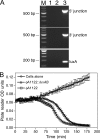
FIG. 1.
FIG. 1.
φA1122::luxAB identity and fitness. (A) PCR identification of φA1122::luxAB. PCR primers were designed against luxA or to span the 5′ or 3′ integration sites. For the 5′ and 3′ integration sites, primers were designed to bind either within the recombination cassette (luxAB) or in the phage φA1122 genome either 5′ or 3′ of the cassette. PCR analysis was performed in the absence of template (lane 1), with wild-type φA1122 DNA (lane 2), or with recombinant φA1122::luxAB DNA (lane 3). The predicted sizes of PCR products for the 5′ junction, 3′ junction, and luxA were 591, 521, and 163 bp, respectively. PCR analysis indicated the presence of luxA and integration of the luxAB into the φA1122 genome at the expected location. M, 100-bp marker DNA ladder. (B) Phage lysis assay. An exponentially growing Y. pestis culture was divided equally and left untreated or infected at a multiplicity of infection of ∼5 with φA1122 or φA1122::luxAB. Cultures were monitored for growth (optical density [OD] units) every 3 min across the lysis window at 28°C using a SpectraMax Plus plate reader. The graph is representative of multiple independent experiments (values are the means [n = 3] ± standard errors of the means).
FIG. 2.
FIG. 2.
Phage-mediated bioluminescent detection of Y. pestis. (A) Signal response time. Y. pestis was grown at 28°C or 37°C until an A600 of approximately 0.2 was reached. At time zero, cells were mixed with the reporter phage and incubated at 28°C or 37°C. Bioluminescence (RLU) was measured over time following the addition of n-decanal. Values are the means (n = 3) ± SD. (B) Dose-dependent detection. Tenfold serial dilutions of a Y. pestis culture were mixed with the reporter phage and incubated at 28°C. Bioluminescence (RLU) was measured over time following the addition of n-decanal. Values are the means (n = 3) ± SD. *, significant increase (P < 0.05) compared to controls (phage or cells alone) at the designated time points.
FIG. 3.
FIG. 3.
Phage-mediated detection of Y. pestis at different stages of cell growth. (A) An overnight culture of Y. pestis was diluted (1:30) into fresh LB, grown at 28°C, and monitored for growth (optical density at 600 nm [OD600] readings). At the designated stages during the growth curve (arrows 1, 2, 3, and 4 approximately denote the lag, early exponential, late exponential, and early stationary phases, respectively), cells were harvested (3,500 × g, 10 min) and “normalized” in LB to an approximate OD600 of 0.3 (the CFU/ml for arrows 1, 2, 3, and 4 were 5.3 × 10⁷, 6.9 × 10⁷, 1.3 × 10⁸, and 1.4 × 10⁸, respectively). (B) Normalized Y. pestis cells, which were harvested at various points during growth (arrows in panel A), were mixed at similar concentrations (CFU/ml) with the reporter phage and incubated at 28°C. Bioluminescence (RLU) was measured over time following the addition of n-decanal. Values are the means (n = 3) ± SD.
FIG. 4.
FIG. 4.
Reporter phage-mediated detection of Y. pestis in human serum. (A) Serum does not quench Y. pestis-mediated bioluminescence. Y. pestis, harboring a plasmid-borne copy of luxAB (p5′0.3-luxAB-3′0.3-SK), was grown at 28°C to an A600 of 0.2. The cells were divided equally, collected by centrifugation (3,500 × g, 10 min), and resuspended in LB or human serum (Sigma catalog no. H4522). Cells were serially diluted 10-fold in LB or serum, and aliquots were measured for bioluminescence after the addition of n-decanal. Values are the means (n = 3) ± SD. (B) The ability of the reporter phage to transduce a bioluminescent signal response to Y. pestis in LB or serum was assessed. Y. pestis was grown at 28°C to an A600 of 0.15, collected by centrifugation (3,500 × g, 10 min), and resuspended in either LB or serum. Cells (in LB or serum) were mixed to give various serum concentrations (24, 49, 74, or 99% final), ensuring that the cell number was consistent between the sample sets (final concentration of 6.9 × 10⁶ CFU/ml). Reporter phage was added, and the cultures were incubated at 28°C. Bioluminescence (RLU) was measured over time following the addition of n-decanal. Values are the means (n = 3) ± SD.

References

    1. Abshire, T. G., J. E. Brown, and J. W. Ezzell. 2005. Production and validation of the use of gamma phage for identification of Bacillus anthracis. J. Clin. Microbiol. 43:4780-4788. - PMC - PubMed
    1. Advier, M. 1933. Etude d'un bacteriophage antipesteux. Bull. Soc. Path. Exotique. 26:94-99.
    1. Banaiee, N., M. Bobadilla-Del-Valle, S. Bardarov, Jr., P. F. Riska, P. M. Small, A. Ponce-De-Leon, W. R. Jacobs, Jr., G. F. Hatfull, and J. Sifuentes-Osornio. 2001. Luciferase reporter mycobacteriophages for detection, identification, and antibiotic susceptibility testing of Mycobacterium tuberculosis in Mexico. J. Clin. Microbiol. 39:3883-3888. - PMC - PubMed
    1. Brigati, J. R., S. A. Ripp, C. M. Johnson, P. A. Iakova, P. Jegier, and G. S. Sayler. 2007. Bacteriophage-based bioluminescent bioreporter for the detection of Escherichia coli O157:H7. J. Food Prot. 70:1386-1392. - PubMed
    1. Calendar, R., and S. T. Abedon. 2005. The bacteriophages, 2nd ed. Oxford University Press, Oxford, United Kingdom.

Publication types

LinkOut - more resources