. 2009 May;58(5):1058-66.
doi: 10.2337/db08-1237. Epub 2009 Feb 10.

GPR119 is essential for oleoylethanolamide-induced glucagon-like peptide-1 secretion from the intestinal enteroendocrine L-cell

Affiliations

GPR119 is essential for oleoylethanolamide-induced glucagon-like peptide-1 secretion from the intestinal enteroendocrine L-cell

Lina M Lauffer et al. Diabetes. 2009 May.

Abstract

Objective: Intestinal L-cells secrete the incretin glucagon-like peptide-1 (GLP-1) in response to ingestion of nutrients, especially long-chain fatty acids. The Galphas-coupled receptor GPR119 binds the long-chain fatty acid derivate oleoylethanolamide (OEA), and GPR119 agonists enhance GLP-1 secretion. We therefore hypothesized that OEA stimulates GLP-1 release through a GPR119-dependent mechanism.

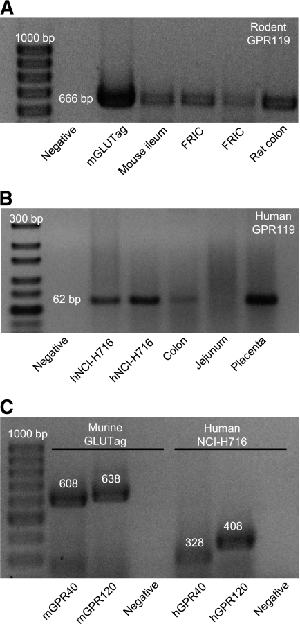
Research design and methods: Murine (m) GLUTag, human (h) NCI-H716, and primary fetal rat intestinal L-cell models were used for RT-PCR and for cAMP and GLP-1 radioimmunoassay. Anesthetized rats received intravenous or intraileal OEA, and plasma bioactive GLP-1, insulin, and glucose levels were determined by enzyme-linked immunosorbent assay or glucose analyzer.

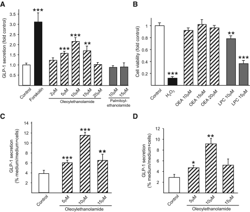
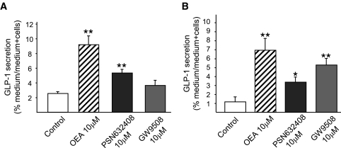
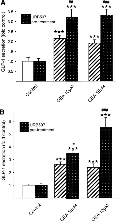
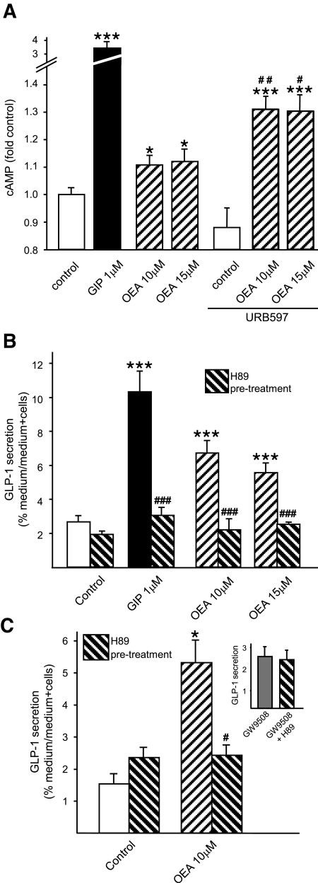
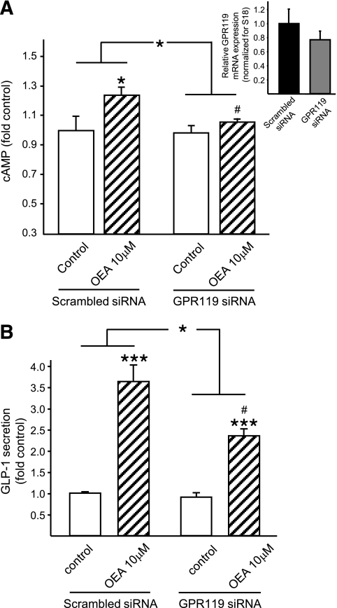
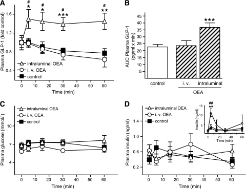
Results: GPR119 messenger RNA was detected in all L-cell models. OEA treatment (10 micromol/l) of mGLUTag cells increased cAMP levels (P < 0.05) and GLP-1 secretion (P < 0.001) in all models, with desensitization of the secretory response at higher concentrations. GLP-1 secretion was further enhanced by prevention of OEA degradation using the fatty acid amide hydrolase inhibitor, URB597 (P < 0.05-0.001 vs. OEA alone), and was abolished by H89-induced inhibition of protein kinase A. OEA-induced cAMP levels and GLP-1 secretion were significantly reduced in mGLUTag cells transfected with GPR119-specific small interfering RNA (P < 0.05). Application of OEA (10 micromol/l) directly into the rat ileum, but not intravenously, increased plasma bioactive GLP-1 levels in euglycemic animals by 1.5-fold (P < 0.05) and insulin levels by 3.9-fold (P < 0.01) but only in the presence of hyperglycemia.

Conclusions: The results of these studies demonstrate, for the first time, that OEA increases GLP-1 secretion from intestinal L-cells through activation of the novel GPR119 fatty acid derivate receptor in vitro and in vivo.


Figures

FIG. 1.
FIG. 1.
Expression of GPR119 mRNA in L-cell models. A: Total RNA from mGLUTag cells, FRICs, and murine intestinal tissues was analyzed for expression of GPR119 mRNA by RT-PCR. B: Total RNA from hNCI-H716 cells, human intestinal tissues, and human placenta was analyzed for expression of GPR119 mRNA by RT-PCR. C: Total RNA from mGLUTag cells and hNCI-H716 cells was analyzed for expression of GPR40 and GPR120 mRNA by RT-PCR. All products were separated on agarose gels and visualized with ethidium bromide, with the molecular-size ladder on the left. Negative controls did not include RNA template. The anticipated band sizes of products are indicated in base pairs (bp).
FIG. 2.
FIG. 2.
Effects of OEA on GLP-1 secretion. mGLUTag cells (n = 9–12) (A), hNCI-H716 cells (n = 8) (C), and FRIC cells (n = 4) (D) were incubated with medium alone (1% DMSO, negative control), forskolin (10 μmol/l, positive control), OEA (2–20 μmol/l), or PEA (10–15 μmol/l, negative control) for 2 h. GLP-1 content of media and cells was determined by RIA. B: To determine potential effects on cell viability, mGLUTag cells were incubated with medium alone (1% DMSO, negative control), H2O2 (5 mmol/l, positive control), OEA (10–20 μmol/l), or LPC (10–15 μmol/l) for 2 h, followed by MTT assay (n = 8–16). *P < 0.05; **P < 0.01; ***P < 0.001 vs. control.
FIG. 3.
FIG. 3.
Effects of GPR119 and GPR40/120 agonists on GLP-1 secretion. mGLUTag (n = 4) (A) and hNCI-H716 (n = 4) (B) cells were incubated with medium alone (1% DMSO, negative control), OEA (10 μmol/l), the GPR119 agonist PSN632408 (10 μmol/l), or the combined GPR40/GPR120 agonist GW9508 (10 μmol/l) for 2 h. GLP-1 content of media and cells was determined by RIA. *P < 0.05; **P < 0.01 vs. control.
FIG. 4.
FIG. 4.
Effect of inhibition of OEA degradation on OEA-induced GLP-1 secretion. mGLUTag (n = 6–18) (A) and hNCI-H716 (n = 12) (B) cells were pretreated for 30 min with URB597 (1 μmol/l) to inhibit FAAH and prevent OEA degradation before incubation with medium alone (1%DMSO, negative control) or OEA (10–15 μmol/l) for 2 h. GLP-1 content of media and cells was determined by RIA. ***P < 0.001 vs. control; #P < 0.05; ##P < 0.01; ###P < 0.001 vs. OEA treatment alone.
FIG. 5.
FIG. 5.
Effect of PKA inhibition on OEA-induced GLP-1 secretion. mGLUTag (n = 6–9) (A and B) and hNCI-H716 (n = 4–6) (C) cells were pretreated for 30 min with medium alone (1% DMSO, negative control), H89 (10 μmol/l for mGLUTag and 30 μmol/l for hNCI-H716), or URB597 (1 μmol/l) to inhibit PKA or FAAH, respectively, followed by incubation with medium alone (1% DMSO, negative control), GIP (1 μmol/l, positive control), or OEA (10–15 μmol/l) for 2 h. C, inset: hNCI-H716 cells were pretreated for 30 min with media alone (1% DMSO) or with H89 (30 μmol/l), followed by incubation with the GPR40/120 agonist GW9508 (10 μmol/l). cAMP content of cells and GLP-1 content of media and cells were determined by RIA. *P < 0.05; ***P < 0.001 vs. appropriate control; #P < 0.05; ##P < 0.01; ###P < 0.001 vs. paired treatment alone.
FIG. 6.
FIG. 6.
Effect of GPR119 knockdown on OEA-induced GLP-1 secretion. mGLUTag cells were transfected with scrambled siRNA (20 pmol/l, control) or GPR119 siRNA (20 pmol/l) 2 days before the experiment. Cells were then incubated with medium alone (1% DMSO, negative control) or OEA (10–15 μmol/l) for 2 h. cAMP content of cells (n = 6) (A) and GLP-1 content of media and cells (n = 9) (B) were determined by RIA. A, inset: GPR119 mRNA transcript levels were determined by quantitative RT-PCR relative to 18S transcript levels. *P < 0.05; ***P < 0.001 vs. control or vs. the Δ change in control cells, as indicated by the lines. #P < 0.05 vs. OEA treatment with scrambled siRNA.
FIG. 7.
FIG. 7.
In vivo effect of OEA on GLP-1 secretion. Anesthetized rats received intraluminal or intravenous injections of vehicle (saline/10% Tween 80; combined controls), intraluminal OEA (2 ml of 10 μmol/l), or intravenous OEA (5 mg/kg), and blood samples were collected over a 1-h period. Plasma concentrations of bioactive GLP-1 (A), glucose (C), and insulin (D) were determined by ELISA and glucose analyzer, as appropriate (n = 5–11). B: AUC for the absolute plasma bioactive GLP-1 concentrations was determined using the trapezoidal rule and is expressed per min. D, inset: Rats (n = 4–5) were maintained at 13 mmol/l plasma glucose (hyperglycemic clamp) for a minimum of 30 min, and this was maintained throughout the OEA treatment procedure. Plasma insulin levels were determined by RIA. A: To reduce interassay variations due to use of separate kits, bioactive GLP-1 concentrations were calculated as fold increase over basal GLP-1 levels (control: 29.4 ± 8.6 pg/ml; intraluminal OEA: 18.9 ± 4.9 pg/ml; and intravenous OEA: 32.7 ± 3.4 pg/ml; P = NS between the basal values). *P < 0.05 vs. control; **P < 0.01 vs. control; ***P < 0.001 vs. control; #P < 0.05 and ##P < 0.01 vs. basal values.

References

    1. Drucker DJ: The biology of incretin hormones. Cell Metab 2006; 3: 153– 165 - PubMed
    1. Wang Q, Brubaker PL: Glucagon-like peptide-1 treatment delays the onset of diabetes in 8 week-old db/db mice. Diabetologia 2002; 45: 1263– 1273 - PubMed
    1. Farilla L, Bulotta A, Hirshberg B, Li Calzi S, Khoury N, Noushmehr H, Bertolotto C, Di Mario U, Harlan DM, Perfetti R: Glucagon-like peptide 1 inhibits cell apoptosis and improves glucose responsiveness of freshly isolated human islets. Endocrinology 2003; 144: 5149– 5158 - PubMed
    1. Sokos GG, Nikolaidis LA, Mankad S, Elahi D, Shannon RP: Glucagon-like peptide-1 infusion improves left ventricular ejection fraction and functional status in patients with chronic heart failure. J Card Fail 2006; 12: 694– 699 - PubMed
    1. Ban K, Noyan-Ashraf MH, Hoefer J, Bolz SS, Drucker DJ, Husain M: Cardioprotective and vasodilatory actions of glucagon-like peptide 1 receptor are mediated through both glucagon-like peptide 1 receptor-dependent and -independent pathways. Circulation 2008; 117: 2340– 2350 - PubMed

Publication types

MeSH terms