. 2009 Mar 6;284(10):6476-85.
doi: 10.1074/jbc.M806599200. Epub 2009 Jan 5.

Formation of a stabilized cysteine sulfinic acid is critical for the mitochondrial function of the parkinsonism protein DJ-1

Affiliations

Formation of a stabilized cysteine sulfinic acid is critical for the mitochondrial function of the parkinsonism protein DJ-1

Jeff Blackinton et al. J Biol Chem. .

Abstract

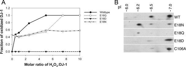
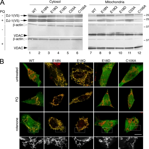
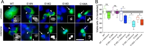
The formation of cysteine-sulfinic acid has recently become appreciated as a modification that links protein function to cellular oxidative status. Human DJ-1, a protein associated with inherited parkinsonism, readily forms cysteine-sulfinic acid at a conserved cysteine residue (Cys106 in human DJ-1). Mutation of Cys106 causes the protein to lose its normal protective function in cell culture and model organisms. However, it is unknown whether the loss of DJ-1 protective function in these mutants is due to the absence of Cys106 oxidation or the absence of the cysteine residue itself. To address this question, we designed a series of substitutions at a proximal glutamic acid residue (Glu18) in human DJ-1 that alter the oxidative propensity of Cys106 through changes in hydrogen bonding. We show that two mutations, E18N and E18Q, allow Cys106 to be oxidized to Cys106-sulfinic acid under mild conditions. In contrast, the E18D mutation stabilizes a cysteine-sulfenic acid that is readily reduced to the thiol in solution and in vivo. We show that E18N and E18Q can both partially substitute for wild-type DJ-1 using mitochondrial fission and cell viability assays. In contrast, the oxidatively impaired E18D mutant behaves as an inactive C106A mutant and fails to protect cells. We therefore conclude that formation of Cys106-sulfinic acid is a key modification that regulates the protective function of DJ-1.


Figures

FIGURE 1.
FIGURE 1.
Structural effects of mutations designed to test the hypothesis that Cys106-sulfinic acid formation is critical to DJ-1 function. A, a ribbon representation of the DJ-1 dimer, with one monomer in brown and the other in green. The dimer 2-fold axis is perpendicular to the page and indicated by an ellipse. The oxidationprone cysteine (C106) and the interacting glutamic acid (E18) are represented in each monomer. B, electron density for the 1.15 Å resolution structure of E18Q DJ-1 around Cys106 is shown at the 1σ contour level and calculated with σA weighted coefficients 2mFo - DFc. In E18Q DJ-1, Cys106 is oxidized to the cysteine-sulfinic acid, where stabilizing hydrogen bonds are shown as dotted lines with distances given in Å. C, a superposition of oxidized E18Q (darker model) and wild-type DJ-1 (lighter model) shows that the key stabilizing hydrogen bond between residue 18 and Cys106-formula image is lengthened in E18Q DJ-1, weakening this interaction. D, 2mFo - DFc electron density contoured at 1σ is shown in blue for the 1.20 Å resolution crystal structure of E18D DJ-1. Cys106 is oxidized to the easily reduced Cys106-SO- oxidation product in this variant. In addition, there is minor electron density that is consistent with either Cys106-formula image or an alternate conformation for Cys106-SO-. E, a superposition of residues in the vicinity of Cys106 in E18D DJ-1 (darker model) and the corresponding region in oxidized wild-type DJ-1 (lighter model). The E18D substitution results in structural perturbations at Cys106 that stabilize the Cys106-SO- oxidation product and hinder further oxidation. All figures were created using POVscript+ (40).
FIGURE 2.
FIGURE 2.
Substitutions at position 18 of DJ-1 impact Cys106-formula image formation. A, oxidation of Cys106 in vitro to Cys106-formula image. Mass spectrometry was used to monitor the oxidation of DJ-1 as a function of hydrogen peroxide concentration in solution. The fraction of protein oxidized was calculated as a ratio of the integrated area of the oxidized protein peak to the total area of both the oxidized and reduced peaks. A comparison of the oxidation curves of these proteins shows that every substitution at position 18 results in diminished oxidation compared with the wild-type protein, although the extent of this diminution varies among the three substitutions. E18D abolishes the ability of Cys106 to be oxidized to cysteine-sulfinic acid, and E18N oxidizes very easily at low H2O2 levels. B, oxidation of DJ-1 in vivo. Human M17 neuroblastoma cells were transfected with V5-tagged versions of the indicated DJ-1 constructs (wild type (WT), E18N, E18Q, E18D, and C106A, from top to bottom) and exposed to 300 μm paraquat for 24 h. Protein extracts were separated on two-dimensional gels and blotted for DJ-1. Estimated pI values for each isoform are indicated above the blots. Images are representative of duplicate experiments for each construct.
FIGURE 3.
FIGURE 3.
DJ-1 mutants are present in mitochondrial and cytosolic pools. A, M17 neuroblastoma cells were transfected with V5-tagged WT (lane 1), E18N (lane 2), E18Q (lane 3), E18D (lane 4), C53A (lane 5), or C106A (lane 6) DJ-1 variants. Cells were also subjected to oxidative stress by exposure to 300 μm paraquat (PQ) for 24 h as indicated. Cytosolic fractions (left, top two blots) or mitochondrial proteins retained after carbonate extraction (right, top two blots) were probed for V5-DJ-1. Mitochondrial enrichment was confirmed by simultaneously reprobing the same blots with monoclonal antibodies to VDAC1 and to cytosolic β-actin, as indicated. B, subcellular localization of the V5-tagged DJ-1 variants was verified using transiently transfected M17 neuroblastoma cells that were stained for V5 (green) and mitochondria using Mitotracker (red). Upper panels, untreated cells; middle panels, cells treated with 300 μm paraquat (PQ); lower panels, cells treated with 100 nm rotenone. The scale bar in the lower right panel of the merged images represents 10 μm and applies to all. To show mitochondrial morphology, a higher magnification view of the boxed areas of the rotenone-treated cells is shown in black and white below each set. Although cells transfected with WT, E18N, or E18Q DJ-1 maintained elongated and connected mitochondria in the presence of rotenone, cells transfected with E18D or C106A DJ-1 showed mitochondrial fragmentation.
FIGURE 4.
FIGURE 4.
DJ-1 Glu18 substitutions affect mitochondrial function in living cells. A-C, living mouse embryonic fibroblasts were imaged after transfection with mitochondrial matrix-localized yellow fluorescent protein (A) to reveal mitochondrial morphology. FRAP was used to measure mitochondrial connectivity. B, time course in wild-type (WT; brown) or DJ-1 knock-out (KO; black) mouse embryonic fibroblasts and knock-out fibroblasts stably expressing human DJ-1 (green). Three independent experiments were performed, each with 30 cells measured; hence, overall n = 90. C, mobile fractions were calculated from FRAP time course experiments. Horizontal lines, median values; boxes, upper and lower quartiles. Range bars show 10-90% intervals. Individual values outside of the range are shown as single dots. Differences were analyzed by one-way ANOVA; *, p < 0.05; **, p < 0.01 by Newman-Kuell's post hoc tests. D and E, mutations around the oxidation sensor residue Cys106 have varied impact on the ability of human DJ-1 to rescue mouse DJ-1 deficiency phenotypes. DJ-1 knock-out fibroblasts were transfected with vector alone (black) or with the indicated human DJ-1 variants (green, WT; magenta, E18N; cyan, E18Q; blue, E18D; red, C106A). Mitochondrial connectivity was assessed by FRAP. Time course is shown in D, and mobile fractions are shown in E. Each point is the average of 60 cells combined from two independent experiments. Differences were analyzed comparing the indicated constructs with WT DJ-1; *, p < 0.05; ***, p < 0.01 by one-way ANOVA with Newman-Kuell's post hoc tests. ns, not significant.
FIGURE 5.
FIGURE 5.
DJ-1 Glu18 substitutions affect cellular resistance to rotenone-induced toxicity. A, nuclear morphology as a marker of rotenone-induced loss of cell viability. M17 neuroblastoma cells were transiently transfected with V5-tagged WT, E18N, E18Q, E18D, or C106A DJ-1 constructs, as indicated, and either left untreated (upper panels) or exposed to 200 nm rotenone for 24 h (lower panels). Cells were stained for V5 (green) and counterstained with Hoechst 33342 (blue) and scored as having intact nuclei (arrowheads) or fragmented/shrunken nuclei (arrows). The insets show examples of nuclei from the blue channel at higher magnification. The scale bar in the bottom right panel represents 20 μm and applies to all images. B, cells were transfected as in A with WT (green), E18N (magenta), E18Q (blue), E18D (cyan), and C106A (red) DJ-1 variants. Cell viability is expressed as the percentage of transfected (V5-positive) cells that had intact nuclei compared with all transfected cells. Each box plot represents data from three randomly selected microscope fields (between 26 and 75 cells/field) counted in each of three independent experiments for an overall n = 9 per construct. Horizontal lines, median values; boxes, upper and lower quartiles; range bars, the range of percentage viabilities for all fields counted. The dotted line represents mean viability counted in untransfected cells from the same cultures, with the shaded box indicating one S.D. value (84.6 ± 2.7% viability, n = 9 fields, mean of 28 cells/field). Differences were analyzed comparing the indicated untreated and rotenone-treated cells for the same construct; *, p < 0.05 by one-way ANOVA (p < 0.001 overall) with Newman-Kuell's post hoc tests. ns, not significant.

References

    1. Reddie, K. G., and Carroll, K. S. (2008) Curr. Opin. Chem. Biol. 12 746-754 - PubMed
    1. Biteau, B., Labarre, J., and Toledano, M. B. (2003) Nature 425 980-984 - PubMed
    1. Jonsson, T. J., Murray, M. S., Johnson, L. C., Poole, L. B., and Lowther, W. T. (2005) Biochemistry 44 8634-8642 - PMC - PubMed
    1. Annesi, G., Savettieri, G., Pugliese, P., D'Amelio, M., Tarantino, P., Ragonese, P., La Bella, V., Piccoli, T., Civitelli, D., Annesi, F., Fierro, B., Piccoli, F., Arabia, G., Caracciolo, M., Cirò Candiano, I. C., and Quattrone, A. (2005) Ann. Neurol. 58 803-807 - PubMed
    1. Bonifati, V., Rizzu, P., van Baren, M. J., Schaap, O., Breedveld, G. J., Krieger, E., Dekker, M. C., Squitieri, F., Ibanez, P., Joosse, M., van Dongen, J. W., Vanacore, N., van Swieten, J. C., Brice, A., Meco, G., van Duijn, C. M., Oostra, B. A., and Heutink, P. (2003) Science 299 256-259 - PubMed

Publication types

MeSH terms

LinkOut - more resources