. 2008 Dec;295(6):R2034-40.
doi: 10.1152/ajpregu.00118.2008. Epub 2008 Oct 8.

Adverse effects of chronic circadian desynchronization in animals in a "challenging" environment

Affiliations

Adverse effects of chronic circadian desynchronization in animals in a "challenging" environment

Fabian Preuss et al. Am J Physiol Regul Integr Comp Physiol. 2008 Dec.

Abstract

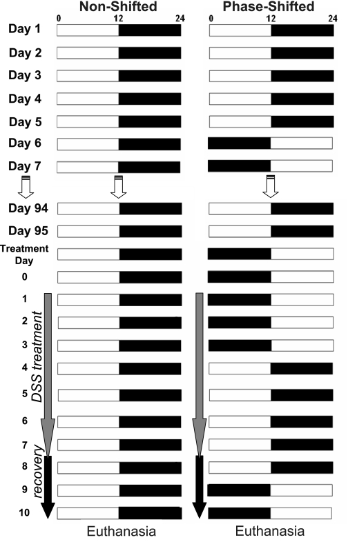
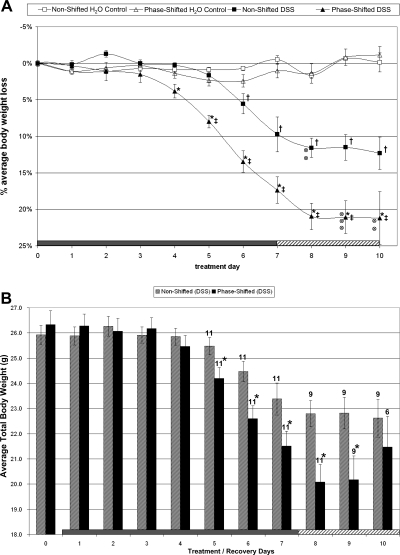
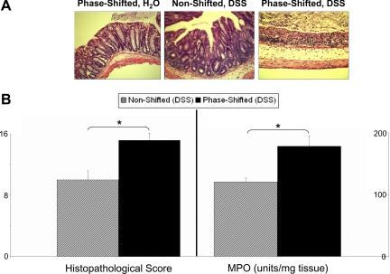
Continuous disruption of circadian rhythms, as seen in human shift workers, has been associated with the development of a number of adverse mental and physiological conditions. However, scientific evidence linking circadian disruption to overall health, particularly in animal models, is not well documented. In this study, we have demonstrated that exposing C57BL/6J mice to 12-h phase shifts every 5 days for 3 mo had no effect on body weight or intestinal physiology. However, when animals were further challenged with dextran sodium sulfate to induce colitis, chronic shifting of the light-dark cycle led to a dramatic increase in the progression of the colitis as indicated by reduced body weight, abnormal intestinal histopathology, and an exacerbated inflammatory response. These data indicate that circadian disruption is an important predisposing factor that may provoke the onset or worsening of various disease states such as inflammatory disorders. This study provides further evidence for continued investigations using animal models of circadian disruption to examine the consequences of circadian disruption on health when organisms are faced with a "challenging" environment.


Figures

Fig. 1.
Fig. 1.
Protocol for phase shifting the light-dark (LD) cycle and colitis induction. The diagram illustrates the applied phase-shift paradigm over the 107 days of the study. Fifteen animals (n = 15) were kept continuously in a 12:12-h LD environment (nonshifted), whereas 16 mice were subjected to 12-h reversals of the LD cycle every 5 days continuously for 3 mo (phase shifted). Two percent dextran sodium sulfate (DSS) was added to the drinking water to a subset of animals (n = 11) from both the nonshifted and phase-shifted groups at the beginning of day 98 (treatment day 1). After 7 days of DSS administration, the water supply was replaced with DSS-free water for a 3-day recovery period before euthanasia and tissue collection. The phase-shifting LD cycle continued during the DSS treatment and recovery period (right).
Fig. 2.
Fig. 2.
Body weight change during DSS challenge and recovery period. A: ANOVA for body weight for all mice alive at the end of study. The progression of weight loss (%change from day 0, means ± SE) during a 7-day inflammatory challenge (DSS was added to water supply from days 1 to 7) and a 3-day recovery period (animals received pure drinking water) is presented. Before this 10-day protocol, mice were maintained on either a stable LD schedule or a 12-h phase-shifting schedule every 5 days for 3 mo, as shown in Fig. 1. Body weight was maintained in the phase-shifted and nonshifted control groups on pure drinking water throughout the 10-day period. The effects of the DSS challenge were more pronounced in the phase-shifted group as demonstrated by an earlier onset and significantly increased loss of body weight compared with the nonshifted group. Two animals in the DSS nonshifted group and 5 animals in the DSS-treated shifted group (indicated by ⊗) died during the recovery period and were not included in the repeated-measures ANOVA analysis of body weight. *P < 0.05, phase shifted (DSS) vs. nonshifted (DSS). ‡P < 0.05, phase shifted (DSS) vs. phase-shifted H2O control. †P < 0.05, nonshifted (DSS) vs. nonshifted H2O control. B: t-test analysis of body weight for all surviving animals on each day of treatment and recovery: This graph depicts the body weight averages (means ± SE) for all DSS-challenged mice in the nonshifted and phase-shifted groups. The 2 nonshifted and 5 shifted animals that died during the recovery period were removed from analysis only on the respective days of their death. Significance was determined by a t-test comparing the body weights of all living mice on each individual day. *P < 0.05, non shifted (DSS) vs. phase shifted (DSS). The number of animals in each group beginning on treatment day 5 is indicated above each bar. Significant differences in body weight between surviving DSS-challenged animals on the nonshifted LD cycle and phase-shifted cycle were observed on days 5–9.
Fig. 3.
Fig. 3.
Intestinal pathophysiology. A: histological damage stains. Representative histological stains are from phase-shifted animals with no DSS challenge (left), nonshifted animals challenged with 2% DSS (middle), and phase-shifted 2% DSS-challenged animals (right). There was no significant histology change in the colon of phase-shifted mice (left) compared with that of nonshifted H2O control mice (stain not shown). The colitis induced by 2% DSS in mice is evidenced by mild mucosal infiltration of inflammatory cells and a reduction of goblet cells (middle). The DSS-induced colitis is exacerbated as evidenced by more extensive destruction of mucosal layer and mucosal ulceration (right). These stains were used for quantitative histological analysis of inflammation (see B). B: histological damage score and myeloperoxidase (MPO) activity. The quantitative measurement for tissue damage was achieved by blindly scoring 3 independent colon sections from each animal (representative stains are shown in A) collected posteuthanasia for visual characterization of destruction, inflammation, and repair using multiple subcategories. The total score (see materials and methods) for each animal was calculated and averaged for the nonshifted and phase-shifted groups. No histological damage could be observed in any of the non-DSS-treated animals, and thus no scores are presented. The DSS challenge led to increased damaged tissue in both groups, but significantly higher levels of tissue damage were observed in the phase-shifted colons (left). MPO activity was determined from 3 colon sections from each animal. Activity levels of both H2O control groups were indistinguishable and at baseline levels of neutrophil activity (data not shown). Two percent DSS-challenged animals in both groups showed a significant 20- to 40-fold increase in MPO activity in all samples compared with the baseline activity, indicating increased neutrophil infiltration into the colon. The neutrophil infiltration process was significantly increased in the phase-shifted mice (right) compared with nonshifted mice, suggesting higher levels of inflammation in these animals. The standard errors shown indicate the variation between animals in each group, not between individual colon sections (*P < 0.01).

References

    1. Bradley PP, Priebat DA, Christensen RD, Rothstein G. Measurement of cutaneous inflammation: estimation of neutrophil content with an enzyme marker. J Invest Dermatol 78: 206–209, 1982. - PubMed
    1. Costa G Shift work and occupational medicine: an overview. Occup Med (Lond) 53: 83–88, 2003. - PubMed
    1. Davidson AJ, Sellix MT, Daniel J, Yamazaki S, Menaker M, Block GD. Chronic jet-lag increases mortality in aged mice. Curr Biol 16: R914–R916, 2006. - PMC - PubMed
    1. Haus E, Smolensky M. Biological clocks and shift work: circadian dysregulation and potential long-term effects. Cancer Causes Control 17: 489–500, 2006. - PubMed
    1. Hayashi M, Shimba S, Tezuka M. Characterization of the molecular clock in mouse peritoneal macrophages. Biol Pharm Bull 30: 621–626, 2007. - PubMed

Publication types

MeSH terms