. 2008 May;3(3):309-323.
doi: 10.2217/14796708.3.3.309.

Autophagy in neuroprotection and neurodegeneration: A question of balance

Affiliations

Autophagy in neuroprotection and neurodegeneration: A question of balance

Salvatore J Cherra 3rd et al. Future Neurol. 2008 May.

Abstract

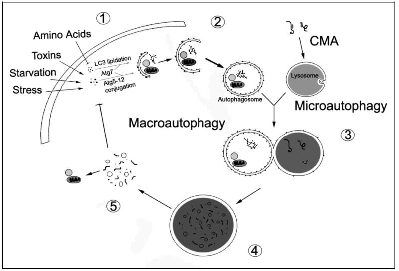
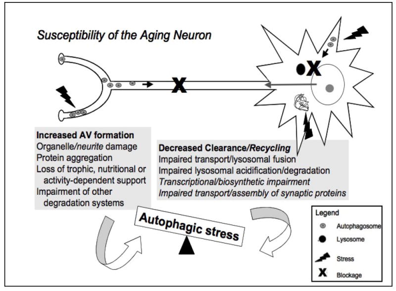
A central issue in developing therapies for neurodegenerative diseases involves understanding why adaptive responses to stress or injury fail to prevent synaptic dysfunction and neuronal cell death. Macroautophagy is a major, evolutionarily conserved response to nutrient and bioenergetic stresses, which has the capacity to remove aggregated proteins and damaged organelles such as mitochondria. This has prompted intense interest in autophagy-related therapies for Huntington's, Alzheimer's, Parkinson's, stroke and other neurological diseases. However, excessive or imbalanced induction of autophagic recycling can actively contribute to neuronal atrophy, neurite degeneration and cell death. Oxidative-, aging- and disease-related increase in demand for autophagy, coupled with declining axonal trafficking, lysosomal degradation or biosynthetic efficiencies promote increased susceptibility to a harmful state of autophagic stress. A more complete understanding of dysfunction along the entire spectrum of autophagic recycling, from autophagosome formation through clearance and regeneration of new cellular components is necessary to restore balance to the system, promote neuronal health and maximize therapeutic potentials.


Figures

Figure 1
Figure 1. Major Phases of Autophagic Recycling
The cell induces macroautophagy in response to a number of physiological and pathological signals. A series of Atg proteins transduce these signals for the execution of macroautophagy, resulting in the conjugation of Atg5 to Atg12 and the lipidation of LC3 (conversion of LC3-I to LC3-II) by Atg7 (1). While many other molecules are involved in autophagy regulation [90], these conjugation reactions are now widely used to monitor and manipulate autophagy induction. Sequestration of cytosolic components into autophagosomes follows Atg5-12 conjugation and LC3 lipidation. The autophagic cargo may include long-lived proteins, misfolded or damaged proteins, and dysfunctional organelles (2). The maturing autophagosome, which has lost the Atg5-12 complex, fuses with the lysosome (3). Successful fusion may depend upon microtubule-dependent trafficking, intermediate fusion with acidified endocytic vesicles to form amphisomes or multivesicular bodies, lysosomal pH or other unknown lysosomal membrane properties (not illustrated). Material delivered to the lysosome is degraded by lysosomal hydrolases that function at acidic pH (4). The degraded contents of the lysosome are released into the cytosol for reutilization in the biosynthesis of new proteins and organelles (5). The released biomolecules (i.e. amino acids) can also regulate macroautophagy through effects on protein synthesis [103] or through modulation of signaling pathways. The processes of chaperone-mediated autophagy (CMA) and microautophagy converge with macroautophagy at the lysosome. In CMA, molecular chaperones select proteins by their KFERQ-like motif and shuttle them to the lysosome for degradation. In microautophagy, the lysosome invaginates to remove cytosolic components for degradation.
Figure 2
Figure 2. Physiologic and Pathologic Alterations in Autophagy
Neurons require a basal degree of autophagic degradation to mediate replacement of damaged or effete cellular components or to facilitate neuritic/synaptic remodeling. Physiologic induction of increased autophagic degradation (a) may be balanced by increased biosynthetic activity to regenerate the degraded components, or result in atrophy of soma and/or neuritic arbor. By definition, physiologic atrophy is reversible upon restoration of anabolic signals. Insufficient autophagy (b) results in accumulation of ubiquitinated proteins and neurodegeneration in experimental systems, suggesting that therapeutic interventions to increase autophagic degradation may be effective for neurodegenerative diseases. However, diseased neurons often demonstrate increased autophagosomes or autolysosomes reflective of autophagic stress (see-saws). Autophagic stress is defined as an imbalance between rates of autophagosome formation and the capacity of the cell to complete the process of degradation and autophagic recycling [2](Box A). The imbalance may occur due to age- or disease-related decreases in the efficacy of autophagosome trafficking and lysosomal fusion/degradation (c), and/or excessive induction of autophagy due to disease-related injury and increased demand for autophagy (d). We hypothesize that the outcome of autophagic stress is determined by the balance between the rates of sequestration versus degradation and the transcriptional/biosynthetic capacities of the stressed neuron. New synthesis of essential components sequestered but not degraded in autophagosomes, upregulation of lysosomal and heat shock proteins, and/or regeneration of essential proteins and organelles that have been degraded all represent compensatory responses that would favor survival. However, age- and disease-related impairment of biosynthetic/regenerative capacity would exacerbate autophagic stress, leading to neurodegeneration and cell death. Thus, strategies that robustly induce autophagy may backfire in the disease context. Instead, modest levels of autophagy induction combined with therapies to promote successful completion of autophagic recycling may be necessary in aged or diseased subjects.
Figure 3
Figure 3. Factors proposed to increase the susceptibility of the aging neuron to autophagic stress and neurodegeneration
The neuron represents the most highly polarized cell type in the human body. It is exquisitely sensitive to disturbances in protein, organelle and membrane transport, and is crucially dependent upon activity-dependent signals derived from its contacts with other cells. The grey boxes summarize potential factors promoting increased autophagy induction (AV formation) and decreased autophagy completion (clearance/recycling), which can both contribute to autophagic imbalances. The high metabolic demand of maintaining functional synapses, which can be meters away from the cell body, and relatively low antioxidant defenses in the brain contribute to oxidative damage and protein aggregation, increasing demand for autophagy [10]. Additionally, aging- and disease-related deficits in the ubiquitin-proteasome system and other pathways of lysosomal degradation (chaperone-mediated autophagy) result in further induction of (macro)autophagy [58, 61, 104, 105]. At the same time, progressive accumulation of damage in non-mitotic cells due to aging/disease mechanisms results in impaired retrograde trafficking, lysosomal fusion and degradation of AVs [104]. Decreased transcriptional and biosynthetic efficiency observed with oxidative stress and aging and impaired anterograde delivery of synaptic components result in failure to compensate for autophagic stress, leading to neurite degeneration and neuronal cell death. Figure modified from Reference [2] and reproduced with permission from the Journal of Neuropathology and Experimental Neurology.

References

    1. Nixon RA. Autophagy, amyloidogenesis and Alzheimer disease. J Cell Sci. 2007;120:4081–91. - PubMed
    1. Chu CT. Autophagic stress in neuronal injury and disease. J Neuropathol Exp Neurol. 2006;65:423–32. - PMC - PubMed
    1. Rubinsztein DC, Gestwicki JE, Murphy LO, Klionsky DJ. Potential therapeutic applications of autophagy. Nat Rev Drug Discov. 2007;6:304–12. - PubMed
    1. Bandhyopadhyay U, Cuervo AM. Chaperone-mediated autophagy in aging and neurodegeneration: lessons from alpha-synuclein. Exp Gerontol. 2007;42:120–8. - PubMed
    1. Farre JC, Subramani S. Peroxisome turnover by micropexophagy: an autophagy-related process. Trends Cell Biol. 2004;14:515–23. - PubMed

LinkOut - more resources