. 2008 Jan 8;105(1):145-50.
doi: 10.1073/pnas.0710685105. Epub 2007 Dec 27.

The Parkinson's disease protein alpha-synuclein disrupts cellular Rab homeostasis

Affiliations

The Parkinson's disease protein alpha-synuclein disrupts cellular Rab homeostasis

Aaron D Gitler et al. Proc Natl Acad Sci U S A. .

Abstract

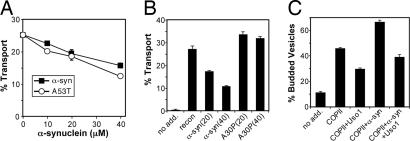
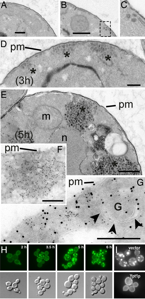
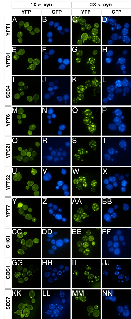
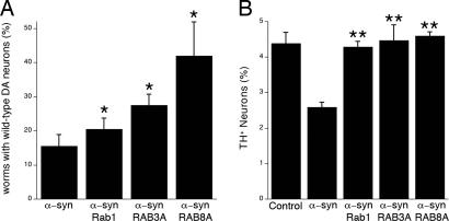
alpha-Synuclein (alpha-syn), a protein of unknown function, is the most abundant protein in Lewy bodies, the histological hallmark of Parkinson's disease (PD). In yeast alpha-syn inhibits endoplasmic reticulum (ER)-to-Golgi (ER-->Golgi) vesicle trafficking, which is rescued by overexpression of a Rab GTPase that regulates ER-->Golgi trafficking. The homologous Rab1 rescues alpha-syn toxicity in dopaminergic neuronal models of PD. Here we investigate this conserved feature of alpha-syn pathobiology. In a cell-free system with purified transport factors alpha-syn inhibited ER-->Golgi trafficking in an alpha-syn dose-dependent manner. Vesicles budded efficiently from the ER, but their docking or fusion to Golgi membranes was inhibited. Thus, the in vivo trafficking problem is due to a direct effect of alpha-syn on the transport machinery. By ultrastructural analysis the earliest in vivo defect was an accumulation of morphologically undocked vesicles, starting near the plasma membrane and growing into massive intracellular vesicular clusters in a dose-dependent manner. By immunofluorescence/immunoelectron microscopy, these clusters were associated both with alpha-syn and with diverse vesicle markers, suggesting that alpha-syn can impair multiple trafficking steps. Other Rabs did not ameliorate alpha-syn toxicity in yeast, but RAB3A, which is highly expressed in neurons and localized to presynaptic termini, and RAB8A, which is localized to post-Golgi vesicles, suppressed toxicity in neuronal models of PD. Thus, alpha-syn causes general defects in vesicle trafficking, to which dopaminergic neurons are especially sensitive.


Conflict of interest statement

Conflict of interest statement: S.L. is a cofounder of and owns stock in FoldRx Pharmaceuticals, a company developing therapies for diseases of protein misfolding, including Parkinson's disease. A.D.G. and S.L. are inventors on patents and patent applications that have been licensed to FoldRx Pharmaceuticals. J.-C.R. receives funds from FoldRx Pharmaceuticals as part of a compound-testing agreement between the company and his laboratory.

Figures

Fig. 1.
Fig. 1.
α-Syn accumulation inhibits in vitro ER→Golgi vesicular transport. (A) Reconstituted transport reactions in washed semiintact yeast cells containing [35S]glyco-pro-α-factor were incubated with increasing amounts of purified α-syn or the α-syn A53T variant. After 60 min at 25°C, the amount of Golgi-modified [35S]glyco-pro-α-factor was measured to determine transport efficiency. Transport in the absence of reconstitution factors was 2.4%, and plots represent levels minus this background. (B) In vitro reactions as in A comparing the influence of α-syn and the A30P variant on reconstituted transport when added at 20 μM and 40 μM. (C) Semiintact cells as in A were incubated with COPII proteins or COPII plus Uso1 to measure budding and tethering in the presence or absence of α-syn (40 μM). After 25 min at 25°C, freely diffusible vesicles containing [35S]glyco-pro-α-factor were separated from semiintact cells by centrifugation, and percentages of budded vesicles were determined.
Fig. 2.
Fig. 2.
EM and fluorescence microscopy of α-syn overexpression reveals a time- and dosage-dependent accumulation of transport vesicles. (A and B) Control strain (A) and one-copy (1×) α-syn strain (B) at 3 h. (C) Higher magnification of vesicles accumulated near peripheral ER at 3 h in the one-copy strain. (D and E) Time course of α-syn expression [HiTox two-copy (2×) strain] beginning at 3 h (D) and proceeding through 5 h (E). (F and G) Indirect immunolabeling of ultrathin cryosections at 5 h after induction demonstrates localization of α-syn-GFP with accumulated vesicle cluster (F; 10 nm Au, α-syn-GFP) and colocalization with α-1,6-mannose (G; 6 nm Au, α-syn-GFP; 12 nm Au, α-1,6-mannose). m, mitochondria; n, nucleus; pm, plasma membrane; G, Golgi. Asterisks indicate vesicle clusters in D and E. (H) Visualization of α-syn dynamics over time. At early time points (2 h) α-syn-GFP (IntTox 2× strain) localized to the plasma membrane. By 3.5 h many small α-syn-GFP-positive inclusions formed adjacent to the plasma membrane and over time (5 h) began to coalesce into a few large inclusions localized close to the plasma membrane as well as in the interior of the cell (6 h). (I) Ypt1 overexpression eliminates α-syn-YFP inclusions. At 6 h, α-syn-YFP-expressing cells contain many vesicular inclusions when transformed with an empty vector. These are eliminated in cells transformed with a Ypt1 expression plasmid. (Scale bars: 0.5 μm.)
Fig. 3.
Fig. 3.
α-Syn perturbs Rab GTPase homeostasis. Fluorescence microscopy of 1× or 2× α-syn-YFP-expressing cells in the presence of CFP-tagged Rab proteins. In 1× α-syn cells (A, E, I, M, Q, U, Y, CC, GG, and KK), each CFP-Rab fusion localized properly based on published reports (B, F, J, N, R, V, and Z). In 2× α-syn cells (C, G, K, O, S, W, and AA), each Rab was sequestered into the α-syn inclusions (D, H, L, P, T, X, and BB). The localization of a CFP fusion to the clathrin heavy chain protein Chc1 (DD) was not altered in 2× α-syn cells (EE and FF), indicating that the presence of a CFP tag was not responsible for the relocalization of the Rab proteins. The Golgi vesicle-localized v-SNARE Gos1 (HH) colocalized with α-syn inclusions (II and JJ), but Sec7p (KK and LL), an ARF GTP exchange factor, did not (MM and NN).
Fig. 4.
Fig. 4.
Rab1, RAB3A, and RAB8A protect against α-syn-induced dopaminergic neuron loss. (A) Multiple Rab GTPases ameliorate α-syn-induced neurodegeneration in C. elegans. DA neurons of 7-day-old transgenic nematodes overexpressing α-syn along with Rab1, RAB3A, or RAB8A were analyzed. Each Rab tested significantly suppressed α-syn toxicity in worm DA neurons (*, P < 0.05, Student's t test). For each gene tested, three transgenic lines were analyzed; a worm was scored as WT when all six anterior DA neurons (four CEP and two ADE neurons) were intact. (B) Primary rat midbrain cultures were transduced with A53T lentivirus (multiplicity of infection = 5) in the absence or presence of lentivirus encoding RAB3A, RAB8A, or Rab1 (multiplicity of infection of each Rab virus = 2). Control cells were incubated in the absence of lentivirus. Dopaminergic cell viability was determined by staining with antibodies specific for MAP2 and TH and is expressed as the percentage of MAP2-positive neurons that were also TH-positive (two to three independent experiments; at least 100 cells counted per experiment for each treatment). The data are plotted as the mean ± SEM. **, P < 0.001 vs. A53T virus alone, one-way ANOVA with Newman–Keuls posttest.

References

    1. Lee VM, Trojanowski JQ. Mechanisms of Parkinson's disease linked to pathological alpha-synuclein: New targets for drug discovery. Neuron. 2006;52:33–38. - PubMed
    1. Spillantini MG, et al. Alpha-synuclein in Lewy bodies. Nature. 1997;388:839–840. - PubMed
    1. Kontopoulos E, Parvin JD, Feany MB. Alpha-synuclein acts in the nucleus to inhibit histone acetylation and promote neurotoxicity. Hum Mol Genet. 2006;15:3012–3023. - PubMed
    1. Lee HJ, Patel S, Lee SJ. Intravesicular localization and exocytosis of alpha-synuclein and its aggregates. J Neurosci. 2005;25:6016–6024. - PMC - PubMed
    1. Fortin DL, et al. Lipid rafts mediate the synaptic localization of alpha-synuclein. J Neurosci. 2004;24:6715–6723. - PMC - PubMed

Publication types