. 2007 Feb;132(2):562-75.
doi: 10.1053/j.gastro.2006.11.022. Epub 2006 Nov 17.

Soluble proteins produced by probiotic bacteria regulate intestinal epithelial cell survival and growth

Affiliations

Soluble proteins produced by probiotic bacteria regulate intestinal epithelial cell survival and growth

Fang Yan et al. Gastroenterology. 2007 Feb.

Abstract

Background & aims: Increased inflammatory cytokine levels and intestinal epithelial cell apoptosis leading to disruption of epithelial integrity are major pathologic factors in inflammatory bowel diseases. The probiotic bacterium Lactobacillus rhamnosus GG (LGG) and factors recovered from LGG broth culture supernatant (LGG-s) prevent cytokine-induced apoptosis in human and mouse intestinal epithelial cells by regulating signaling pathways. Here, we purify and characterize 2 secreted LGG proteins that regulate intestinal epithelial cell antiapoptotic and proliferation responses.

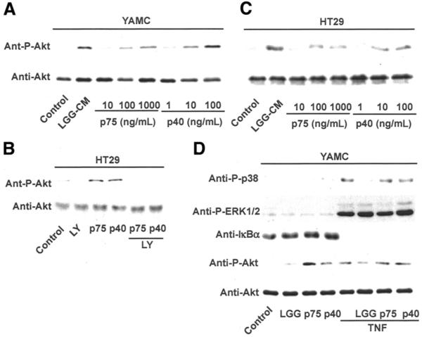
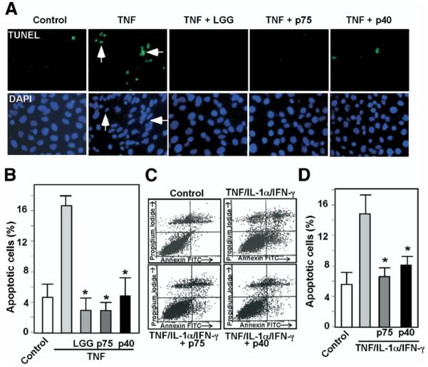
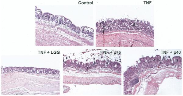
Methods: LGG proteins were purified from LGG-s, analyzed, and used to generate polyclonal antibodies for immunodepletion of respective proteins from LGG-conditioned cell culture media (CM). Mouse colon epithelial cells and cultured colon explants were treated with purified proteins in the absence or presence of tumor necrosis factor (TNF). Akt activation, proliferation, tissue injury, apoptosis, and caspase-3 activation were determined.

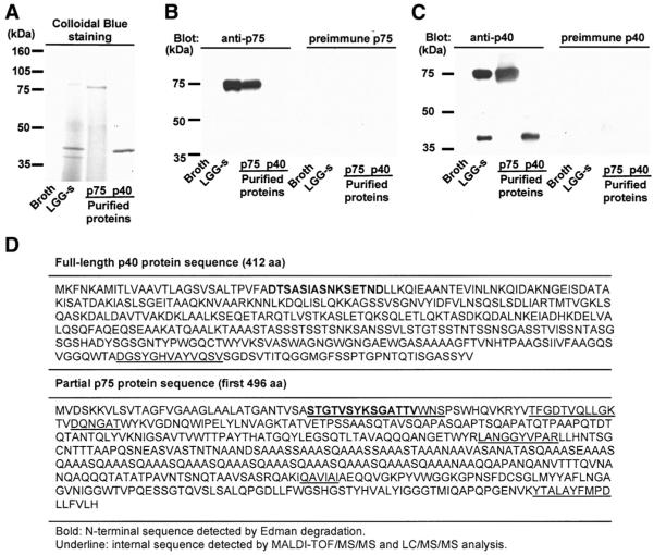
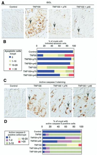
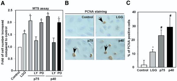
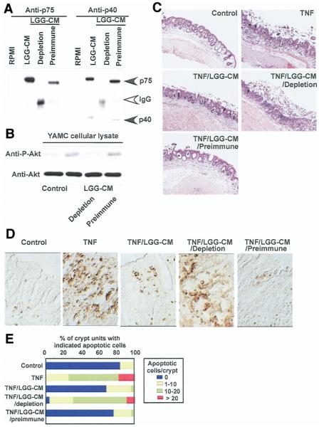
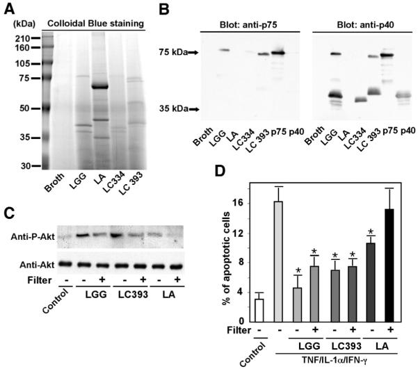
Results: We purified 2 novel proteins, p75 (75 kilodaltons) and p40 (40 kilodaltons), from LGG-s. Each of these purified protein preparations activated Akt, inhibited cytokine-induced epithelial cell apoptosis, and promoted cell growth in human and mouse colon epithelial cells and cultured mouse colon explants. TNF-induced colon epithelial damage was significantly reduced by p75 and p40. Immunodepletion of p75 and p40 from LGG-CM reversed LGG-CM activation of Akt and its inhibitory effects on cytokine-induced apoptosis and loss of intestinal epithelial cells.

Conclusions: p75 and p40 are the first probiotic bacterial proteins demonstrated to promote intestinal epithelial homeostasis through specific signaling pathways. These findings suggest that probiotic bacterial components may be useful for preventing cytokine-mediated gastrointestinal diseases.


Figures

Figure 1
Figure 1
Purification and sequencing of p75 and p40 purified from LGG-s. Filtered LGG culture supernatant was loaded onto a cation exchange column. Proteins bound to exchange media were eluted using Tris buffer containing sequential concentrations of NaCl (100–800 mmol/L). Eluted proteins were separated by SDS-PAGE and stained with Colloidal Blue Staining Kit (A, lanes 3 and 4). Proteins present in concentrated fractions of broth and LGG-s using a 5-kilodalton cut-off filter are shown (A, lanes 1 and 2, respectively). Polyclonal antibodies against p75 or p40 were generated as detailed in the Materials and Methods section and used in Western blot analysis (B and C). N-terminal sequences (bold) and internal peptide sequences (underline) of p75 and p40 were detected by Edman degradation or MALDI-TOF/MS/MS and LC/MS/MS analysis, respectively (D). LGG genetic sequences encoding p75 and p40 were determined as described in the Materials and Methods section, and predicted amino acid sequences were deduced from the nucleotide sequences (D).
Figure 2
Figure 2
p75 and p40 Stimulate Akt activation in mouse and human colon epithelial cells. YAMC (A and D) and HT29 (B and C) cells were treated with purified p75 or p40 at the indicated concentrations for 2 hours in the presence or absence of 30-minute pretreatment of PI3K inhibitor LY294002 (10 μmol/L; C). Akt, p38, ERK1/2 MAPK activation, and IκBα degradation were detected by Western blot analysis of cellular lysates with indicated antibodies. Data are representative of 5 separate experiments.
Figure 3
Figure 3
p75 and p40 Inhibit cytokine-induced apoptosis in intestinal epithelial cells. KSRI−/− MCE cells (A and B) or HT29 cells (C and D) were treated with TNF (100 ng/mL) for 6 hours or the “cytokine cocktail” combination of TNF (100 ng/mL), IL-1-α (10 ng/mL), and IFN-γ (100 ng/mL) for 16 hours, respectively, in the presence or absence of 1-hour pretreatment with viable LGG, p75 (100 ng/mL), or p40 (10 ng/mL). LGG, p75, and p40 were maintained during the entire course of cytokine treatment in all experiments shown in this paper. KSR−/− MCE cells were fixed for TUNEL with apoptotic nuclei labeled with FITC and DAPI staining (A). FITC and DAPI labeled images were taken from the same field. Arrows indicate representative apoptotic nuclei. The percentage of cells undergoing apoptosis is shown (B). HT29 cells were dissociated and stained with Annxin V-FITC and propidium iodide and analyzed by flow cytometry (C). Results are shown as density plots with Annxin V-FITC vs propidium iodide (D). Viable cells have low Annexin V-FITC and low propidium iodide staining (lower left quadrant); early apoptotic cells have high Annexin V-FITC and low propidium iodide staining (lower right quadrant); late apoptotic cells have high Annexin V-FITC and high propidium iodide staining (upper right quadrant); and necrotic cells have low Annexin V-FITC and high propidium iodide staining (upper left quadrant). The early apoptotic cell populations in the lower right quadrant are shown in D. *P < .01 compared with TNF (B) or the “cytokine cocktail” (D), respectively. Experiments were performed on at least 3 separate occasions.
Figure 4
Figure 4
p75 and p40 Rescue TNF-induced epithelial damage in cultured mouse colon explants. Colon explants derived from 6- to 8-week old C57BL/6 mice were cultured in DMEM containing 0.5% FBS and treated with TNF (100 ng/mL) for 24 hours in the presence or absence of LGG, p75 (100 ng/ mL), or p40 (10 ng/mL). Paraffin-embedded tissue sections were stained with H&E for light microscopic assessment of epithelial damage (original magnification, ×10). Images shown are representative of 7 mice in each group.
Figure 5
Figure 5
p75 and p40 Inhibit TNF-induced apoptosis in cultured mouse colon explants. Mouse colon explants prepared as in Figure 4 were treated with TNF (50 ng/mL, TNF50 or 100 ng/mL, TNF100) for 24 hours in the presence or absence of LGG, p75 (100 ng/mL), or p40 (10 ng/mL). Paraffin-embedded tissue sections were studied for apoptosis using ISOL staining. Apoptotic nuclei labeled with peroxidase were visualized using DIC microcopy (A). Caspase-3 activity was determined by immunohistochemistry using antiactive caspase-3 antibody (C). The percentage of crypts with indicated apoptotic nuclei (B) or positive active caspase-3 (D) cells is shown. Arrows indicate examples of ISOL or caspase-3-positive cells. The percentage shown here is the average representing 5 independent experiments. All images were taken with ×40 magnification.
Figure 6
Figure 6
p75 and p40 Promote intestinal epithelial cell proliferation. YAMC cells plated in 96-well dish were treated with LGG, p74 (100 ng/mL), or p40 (10 ng/mL) for 24 hours. At the end of treatment, viable cells were counted using MTS-based assays. The change in the number of control cells from the start to the end of an experiment was standardized as 100%. Changes in the treated cells were reported as a percentage relative to the untreated control (A). Cells cultured on chamber slides treated as indicated were immunostained with anti-PCNA antibody. Peroxidase-labeled positive cells, indicated by arrows, were observed by DIC microscopy (B). At least 500 cells were counted to determine the percentage of PCNA-positive cells (C). *P < .01, #P < .001 compared with control. Data represent at least 3 separate experiments.
Figure 7
Figure 7
Immunodepletion of p75 and p40 blocks LGG-CM’s antiapoptotic effects on colon epithelial cells. Immunodepletion of p75 and p40 was performed by sequential immunoprecipitation of LGG-CM with anti-p75 and p40 antibodies, characterized in Figure 1, to remove both p75 and p40 from LGG-CM. Preimmune sera were used as a control. Proteins present in LGG-CM, LGG-CM immunodepleted with antibodies (LGG-CM depletion), or preimmune sera (LGG-CM preimmune) were separated by SDS-PAGE for Western blot analysis with anti-p75 and p40 antibodies (A). LGG-CM, LGG-CM depletion, and LGG-CM preimmune were used to treat YAMC cells to detect Akt activation as shown in Figure 2 (B) or C57BL/6 mouse colon explants described as in Figure 5 in the presence or absence of TNF (100 ng/mL) for 24 hours. Paraffin-embedded tissue sections were stained with H&E for light microscopic assessment of epithelial damage (C; original magnification, ×10) and ISOL staining to detect epithelial cell apoptosis using DIC microscopy (D; original magnification, ×40). The percentage of crypts with indicated apoptotic cells is shown (E). Data represent mean scores from at least 3 experiments.
Figure 8
Figure 8
Production of p75 and p40 by Lactobacilli is strain specific. Concentrated proteins recovered from indicated bacterial-conditioned cell culture media using a 5-kilodalton cut-off filter were separated by SDS-PAGE and stained with Colloidal Blue Staining Kit (A), and Western blot analysis of these proteins with anti-p75 and anti-p40 antibodies was performed (B). To test the effects of bacteria-derived soluble factors on colon cells, the indicated bacteria were separated from YAMC cells by 0.2-μmol/L filters during coculture experiments for 2 hours. Cellular lysates were collected and Akt activation determined as in Figure 2 (C). To test these soluble factors’ effects on preventing apoptosis, bacteria were separated in transwell cocultures as in C, with HT29 cells for 1 hour followed by 16-hour cotreatment with “cytokine cocktail” containing TNF (100 ng/mL), IL-1α (10 ng/mL), and IFN-γ (100 ng/mL). Cells were then prepared for apoptosis assays using Annexin V-FITC staining as in Figure 3. The percentage of the early apoptotic cell populations is shown (D). LC334, Lactobacillus casei 334; LC393, Lactobacillus casei 393; LA, Lactobacillus acidophilus. *P < .01, compared with TNF/IL-1α/IFN-γ. Data represent 3 separate experiments.

References

    1. Sartor RB. Mucosal immunology and mechanisms of gastrointestinal inflammation. In: Feldman M, Friedman LS, Sleisenger MH, editors. Sleisenger & Fordtran’s gastrointestinal and liver disease: pathophysiology, diagnosis, management. 7th ed. I. Saunders; Philadelphia: 2002. pp. 21–51.
    1. El-Assal ON, Besner GE. HB-EGF enhances restitution after intestinal ischemia/reperfusion via PI3K/Akt and MEK/ERK1/2 activation. Gastroenterology. 2005;129:609–625. - PubMed
    1. Matsuura M, Okazaki K, Nishio A, Nakase H, Tamaki H, Uchida K, Nishi T, Asada M, Kawasaki K, Fukui T, Yoshizawa H, Ohashi S, Inoue S, Kawanami C, Hiai H, Tabata Y, Chiba T. Therapeutic effects of rectal administration of basic fibroblast growth factor on experimental murine colitis. Gastroenterology. 2005;128:975–986. - PubMed
    1. McCole DF, Rogler G, Varki N, Barrett KE. Epidermal growth factor partially restores colonic ion transport responses in mouse models of chronic colitis. Gastroenterology. 2005;129:591–608. - PubMed
    1. Sinha A, Nightingale J, West KP, Berlanga-Acosta J, Playford RJ. Epidermal growth factor enemas with oral mesalamine for mild-to-moderate left-sided ulcerative colitis or proctitis. N Engl J Med. 2003;349:350–357. - PubMed

Publication types

MeSH terms