Comparative Study
. 2007 Jan 24;27(4):796-807.
doi: 10.1523/JNEUROSCI.3501-06.2007.

Abeta oligomer-induced aberrations in synapse composition, shape, and density provide a molecular basis for loss of connectivity in Alzheimer's disease

Affiliations
Comparative Study

Abeta oligomer-induced aberrations in synapse composition, shape, and density provide a molecular basis for loss of connectivity in Alzheimer's disease

Pascale N Lacor et al. J Neurosci. .

Abstract

The basis for memory loss in early Alzheimer's disease (AD) seems likely to involve synaptic damage caused by soluble Abeta-derived oligomers (ADDLs). ADDLs have been shown to build up in the brain and CSF of AD patients and are known to interfere with mechanisms of synaptic plasticity, acting as gain-of-function ligands that attach to synapses. Because of the correlation between AD dementia and synaptic degeneration, we investigated here the ability of ADDLs to affect synapse composition, structure, and abundance. Using highly differentiated cultures of hippocampal neurons, a preferred model for studies of synapse cell biology, we found that ADDLs bound to neurons with specificity, attaching to presumed excitatory pyramidal neurons but not GABAergic neurons. Fractionation of ADDLs bound to forebrain synaptosomes showed association with postsynaptic density complexes containing NMDA receptors, consistent with observed attachment of ADDLs to dendritic spines. During binding to hippocampal neurons, ADDLs promoted a rapid decrease in membrane expression of memory-related receptors (NMDA and EphB2). Continued exposure resulted in abnormal spine morphology, with induction of long thin spines reminiscent of the morphology found in mental retardation, deafferentation, and prionoses. Ultimately, ADDLs caused a significant decrease in spine density. Synaptic deterioration, which was accompanied by decreased levels of the spine cytoskeletal protein drebrin, was blocked by the Alzheimer's therapeutic drug Namenda. The observed disruption of dendritic spines links ADDLs to a major facet of AD pathology, providing strong evidence that ADDLs in AD brain cause neuropil damage believed to underlie dementia.


Figures

Figure 1.
Figure 1.
Characterization of ADDL-bound neurons. ADDLs (red) bind to neurons expressing NMDA-R subunits, such as NR1 (A, green) and NR2B (B, green) but not to GABAergic neurons identified as GAD-positive cells (C, green). Cultured hippocampal cells exposed to 500 nm ADDLs for 15 min were fixed and immunolabeled using an oligomer-raised antibody and NNDA-R subunits 1 and 2B or glutamic acid decarboxylase (GAD65/67) antibodies. ADDLs are highly concentrated along dendritic processes rather than on the cell soma, suggesting preferential binding to excitatory synapses. Images are representative of several replicate experiments. Scale bar, 30 μm.
Figure 2.
Figure 2.
ADDL binding sites are postsynaptic. A, High-magnification image demonstrates that ADDLs bind to dendritic spines, protrusions receiving excitatory inputs. Mature hippocampal neurons exposed to 500 nm ADDLs for 15 min were double immunolabeled for bound ADDLs (cyan) with a monoclonal oligomer-selective antibody (NU-2) and drebrin (red), a dendritic spine marker highly expressed in dendritic spines. Merged image demonstrates a high level of colocalization (white represents the overlap, arrow) of ADDL puncta with dendritic spines. B, ADDL binding is region specific. Different amounts of forebrain or cerebellar crude synaptosomes (5–125 μg) were incubated with 500 nm ADDLs. ADDL-bound synaptosomes were immunoprecipitated using Dynabeads coupled to a polyclonal anti-oligomer-selective antibody (M71/2). The amount of immunoprecipitated material was assayed using PSD-95 antibody. Increasing PSD-95 immunoreactivity was observed from the forebrain synaptosomes, whereas cerebellar synaptosomes generated no signal. This suggests that ADDLs strongly bind to forebrain synapses but poorly to cerebellar synapses and demonstrates region specificity reminiscent of the brain area sensitivity to AD. Inset, ADDL-treated (AD) or vehicle-treated (VEH) synaptosomes (syn) were immunoprecipitated using M71/2-coupled Dynabeads. Bound (B) and unbound (UnB) fractions generated by the immunoprecipitation was assessed for the presence of synaptic markers (PSD-95 and SVP-38, respectively as postsynaptic and presynaptic markers). Only the “AD-syn” bound fraction showed immunoreactivity for both synaptic markers, and immunoprecipitation was specific and dependent on ADDL binding, confirming that ADDLs bind to a portion of the isolated synapses. OD, Optical Density. C, ADDL-treated synaptosomes (AD), labeled with anti-ADDLs (NU-2), incubated with Triton X-100 and DOC, and immunoprecipitated with anti-mouse IgG Dynabeads (as described in Materials and Methods) show a coisolation of ADDLs (M69–2) with postsynaptic proteins (PSD-95, NR1, and NR2A/B), all released in second Sarkosyl (SKL-2) and SDS detergent extraction. No syntaxin band was detected in these fractions (data not shown). No immunoreactive material appears in the vehicle-treated synaptosomes (VEH). Left line represents standard marker molecular weights. ADDL binding complex segregates with postsynaptic markers. D, Presynaptic and postsynaptic compartments of ADDL-bound synaptosomes were dissociated according to the protocol of Phillips et al. (2001). Dissociation demonstrates that ADDLs, detected by dot blot (0.5 μg of protein) using a selective anti-oligomer antibody (NU-1), cofractionate with synaptic junctions (SJ) and PSDs, and both fractions assayed by Western blot (3 μg of proteins) were immunopositive for PSD-95, a postsynaptic scaffolding protein. However, the active zone (AZ) fraction, which is immunoreactive for syntaxin, an integral membrane protein of the presynaptic exocytic fusion complex, did not contained ADDL-immunoreactive signal. These results confirm that ADDLs bind to excitatory synapses at postsynaptic sites.
Figure 3.
Figure 3.
NMDA-R and EphB2 surface expressions are decreased after ADDL exposure. Surface expression of NR1 and NR2B subunits and EphB2 were measured using antibodies against an extracellular epitope at the N-terminal portion of the receptor for immunolabeling under nonpermeabilizing conditions and analyzed using MetaMorph. A, Quantification represents the number of NR1-labeled puncta per length of dendrite (number of NR1 puncta/10 μm dendrite). Patterned bars represent ADDL treatment at the indicated incubation time, and black bar represents vehicle treatment at 3 h. Density of NR1 tends to decrease after AD 1 h (10.1 ± 2.2; n = 11; NS) and highly decrease after AD 3 h (3.3 ± 0.7; n = 11; p = 0.002) compared with controls represented by AD 2 min (14.3 ± 4.8; n = 9) and VEH 3 h (13.1 ± 1.4; n = 9). The follwoing dendritic lengths were measured over the different groups of treatment: AD 2 min, 383 ± 36 μm; AD 1 h, 325 ± 25 μm; AD 3 h, 348 ± 32 μm; VEH 3 h, 252 ± 29 μm. B, A significant decrease in the number of NR2B puncta per field was observed after AD 3 h (p < 0.001; n = 10). The average number of puncta for treated neurons was normalized against the mean value of vehicle-treated ones. A similar decrease was observed after 1 h biotinylated ADDL (bADDLs) treatment. Black bar represents vehicle, and white bar represents ADDL or bADDL. C, A significant decrease in the number of EphB2-labeled puncta per length of dendrite was observed after AD 6 h (p = 0.0005; n = 15), although the difference at 3 h was not significant (p = 0.55). The average number of puncta per length of dendrite for ADDL treated was normalized against the mean value of vehicle treated. Black bar represents vehicle, and white bar represents ADDL. Right panel shows representative images of dendritic branches labeled with NR1 (A), NR2B (B), and EphB2 (C) antibodies at indicated time points of treatment. Scale bar, 8 μm. **p = 0.0005.
Figure 4.
Figure 4.
Time-dependent changes in drebrin distribution attributable to ADDLs. Cells treated with ADDL for 5 min (A), 6 h (B), and 24 h (C) and vehicle for 24 h (D) and immunolabeled for drebrin, an actin binding protein enriched in dendritic spines, were imaged by confocal laser-scanning microscope. A reduction in drebrin labeling is particularly pronounced at 24 h after ADDL treatment. Scale bar, 30 μm.
Figure 5.
Figure 5.
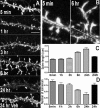
ADDL-induced aberrations in dendritic spine morphology and density. A, Confocal microscopy images representative of individual dendritic branches decorated with spiny protrusions immunolabeled for drebrin after ADDL or vehicle (Veh) treatment. Longer and more irregularly shaped spines appear after as early as 3 h treatment and are more pronounced after 6 h. Also note the fewer amount of dendritic spines present after 24 h ADDL. Scale bar, 5 μm. B, Illustration of zoomed dendritic branches harboring “spines” demonstrates the dramatic lengthening of dendritic protrusions after 6 h of ADDL treatment. The line marks the dendritic shaft. C, D, Histograms represent average length and density of drebrin-labeled dendritic spines after ADDL (patterned bars) or vehicle (black bars) incubation at various times. Spine measurements were obtained from a minimum of five separate images per group of treatment and from two independent experiments. Spine lengths were as follows (in μm): AD 5 min, 1.1 ± 0.02, n = 365 spines; Veh 24 h, 1.2 ± 0.03, n = 315, NS vs AD 5 min; AD 1 h, 1.3 ± 0.05, n = 219, p > 0.05 vs controls; AD 3 h, 1.3 ± 0.04, n = 233, p < 0.01; AD 6 h, 1.8 ± 0.06, n = 322, p < 0.001; AD 24 h, 2.0 ± 0.10, n = 110, p < 0.001. Spine densities per 10 μm of dendrite were as follows: AD 5 min, 9.2 ± 0.6, n = 18 dendritic branches; AD 1 h, 7.9 ± 0.7, n = 10, p > 0.05; AD 3 h, 7.8 ± 0.6, n = 9, p < 0.01; AD 6 h, 5.7 ± 0.3, n = 18, p < 0.0001; AD 24 h, 4.0 ± 0.7, n = 10, p < 0.001; Veh 24 h, 7.7 ± 0.7, n = 16, p > 0.05.
Figure 6.
Figure 6.
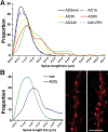
Shift in dendritic spine length monitored by drebrin (A) and spinophilin (B). A, Spine length distribution in hippocampal neurons incubated with 500 nm ADDLs (AD) or vehicle (VEH) at different times. The curves represent the observed shift in dendritic spine length after ADDL treatment. Although most of the spines have a length of 0.5–2 μm in AD 5 min, AD 1 h, and VEH 24 h, occurrence of longer spines is significantly increased in AD 6 h and AD 24 h. B, Labeling of spinophilin, another cytoplasmic scaffolding protein highly concentrated in spines, showed that dendritic spine length was increased after AD 3 h (average spine length was 1.99 ± 0.1 μm) compared with time-matched vehicle (1.23 ± 0.04 μm) (p < 0.005, 10 dendritic branches from different neurons imaged, ∼200 spine length measurements per group of treatment). High-magnification images of spinophilin IF show dendritic branches harboring spines under vehicle (left) or ADDL (right) treatment. Scale bar, 8 μm.
Figure 7.
Figure 7.
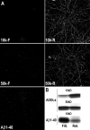
ADDLs species >50 kDa are synaptic ligands. A, Immunolabeling of Aβ-bound species with 6E10, a monoclonal antibody that recognizes monomeric form of Aβ as well as higher Aβ species, revealed that only ADDL species above 50 kDa, but not monomer, bind in a hot spot pattern on dendritic process of hippocampal cells. Cells were incubated for 15 min with either filtrate or retentate from 10 kDa cutoff filter (10k-F and 10k-R, respectively), filtrate or retentate from 50 kDa cutoff filter (50k-F and 50k-R, respectively), or Aβ1–40. B, Ultrafiltration shows that ADDL preparations contain almost no monomers. ADDLs were separated by 10 and 50 kDa cutoff filters into filtrate (Filt.) and retentate (Ret.) fractions and then analyzed by SDS-PAGE and silver stain. The bands illustrated are those migrating to the monomer position, which is the predominant silver-stained species after oligomer disassociation in SDS as described previously (Lambert et al., 1998). Material in the 10 kDa filtrate is negligible compared with the retentate fraction; to obtain the faint signal shown, the loading volume used for filtrate was 26 times greater than for retentate. For the 50 kDa cutoff fractions, equal loading volumes were used. Data show that essentially all material in ADDL preparations was larger than monomer, and approximately half was larger than 50 kDa. A control (Aβ1–40) shows that monomers, when present, pass readily through 10 kDa cutoff filters.
Figure 8.
Figure 8.
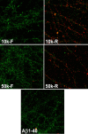
Drebrin loss is attributable to Aβ oligomers. Cells double labeled for drebrin (green) and ADDLs (M94, red) show major drebrin loss when ADDL-binding species are bound to the dendritic processes. Cells were treated for 24 h with either filtrate or retentate of 10 or 50 kDa cutoff filters (10k-F, 10k-R, 50k-F, and 50k-R, respectively). Representative images show high drebrin in the absence of ADDL binding (10k-F, 50k-F) but very low drebrin labeling in dendritic processes in the presence of ADDL labeling (10k-R, 50k-R). Note that Aβ1–40 preparations showed neither binding nor drebrin loss.
Figure 9.
Figure 9.
Memantine prevents ADDL-induced dendritic drebrin loss. A, Confocal microscopy images of drebrin immunofluorescent labeling in 21 DIV cultured rat hippocampal neurons. Grayscale intensities ranging from black to white were converted in a pseudocolor lookup table with increasing values of gray intensity. Black (0) is representing the darkest intensity, and white (255) is the brightest. Intermediary values are as follows: 20, violet; 65, cyan; 80, turquoise; 115, green; 155, yellow; 220, red. As indicated by the arrows, dendrites of neurons treated with ADDLs for 24 h (AD) exhibit decreased drebrin immunofluorescence when compared with the abundance of drebrin hot spots (yellow–red) present in the neurons treated with F-12 vehicle for 24 h (VEH). Whereas neurons treated with memantine 30 min before 24 h ADDL treatment (MEM-AD) exhibit dendritic drebrin immunofluorescence similar to that of vehicle-treated neurons and neurons treated with memantine before 24 h F-12 vehicle treatment (MEM-VH). B, Cells treated for 24 h with 500 nm ADDL (AD) or memantine before ADDLs (MEM-AD) were double labeled for ADDLs using M94 (red) and drebrin (green). Similar distribution of M94-immunoreactive hot spots is observed in both groups of treatment, demonstrating that memantine prevents ADDL-induced drebrin loss but does not alter ADDLs binding to synapses. C, Bar graph illustrating quantification of drebrin immunofluorescence integrated density from confocal image sets as shown in A. Values are normalized to vehicle-treated cells (VEH) for 24 h. Difference between VEH (n = 25) versus AD (n = 31) and AD versus MEM-AD (n = 31) are highly significant (**p < 0.001), whereas VEH versus MEM-AD are comparable (p > 0.05). Memantine by itself did not have any effect on drebrin (MEM-VEH, n = 10). D, Western blotting of hippocampal neuron extracts treated in the same manner as neurons imaged in A. Blots were probed with anti-drebrin, and antibody against cyclophilin was used as control of protein loaded in each lane.

References

    1. Allison DW, Chervin AS, Gelfand VI, Craig AM. Postsynaptic scaffolds of excitatory and inhibitory synapses in hippocampal neurons: maintenance of core components independent of actin filaments and microtubules. J Neurosci. 2000;20:4545–4554. - PMC - PubMed
    1. Alonso M, Medina JH, Pozzo-Miller L. ERK1/2 activation is necessary for BDNF to increase dendritic spine density in hippocampal CA1 pyramidal neurons. Learn Mem. 2004;11:172–178. - PMC - PubMed
    1. Aoki C, Sekino Y, Hanamura K, Fujisawa S, Mahadomrongkul V, Ren Y, Shirao T. Drebrin A is a postsynaptic protein that localizes in vivo to the submembranous surface of dendritic sites forming excitatory synapses. J Comp Neurol. 2005;483:383–402. - PubMed
    1. Boyer C, Schikorski T, Stevens CF. Comparison of hippocampal dendritic spines in culture and in brain. J Neurosci. 1998;18:5294–5300. - PMC - PubMed
    1. Buttini M, Masliah E, Barbour R, Grajeda H, Motter R, Johnson-Wood K, Khan K, Seubert P, Freedman S, Schenk D, Games D. Beta-amyloid immunotherapy prevents synaptic degeneration in a mouse model of Alzheimer's disease. J Neurosci. 2005;25:9096–9101. - PMC - PubMed

Publication types

MeSH terms

Substances