. 2007 Feb;100(4):893-904.
doi: 10.1111/j.1471-4159.2006.04162.x. Epub 2006 Dec 22.

Macrophages are comprised of resident brain microglia not infiltrating peripheral monocytes acutely after neonatal stroke

Affiliations

Macrophages are comprised of resident brain microglia not infiltrating peripheral monocytes acutely after neonatal stroke

Sheryl P Denker et al. J Neurochem. 2007 Feb.

Abstract

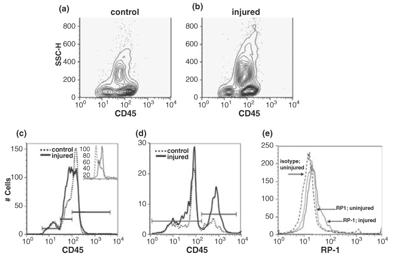
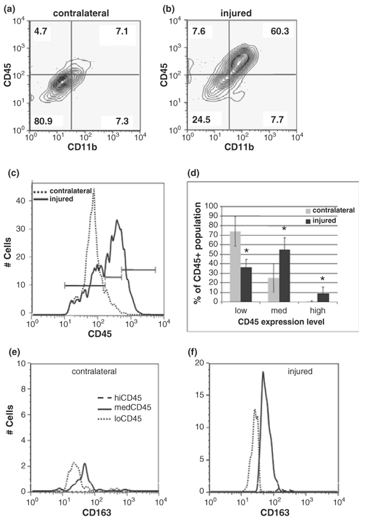
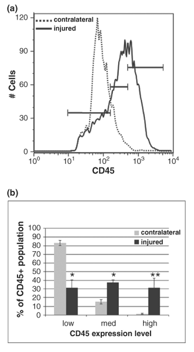
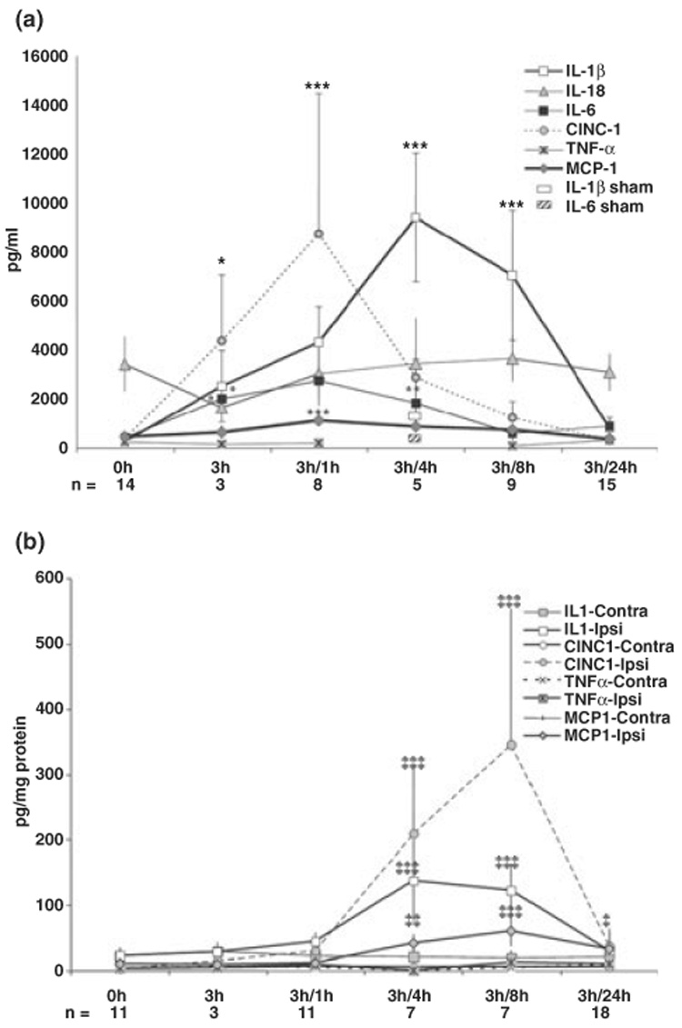
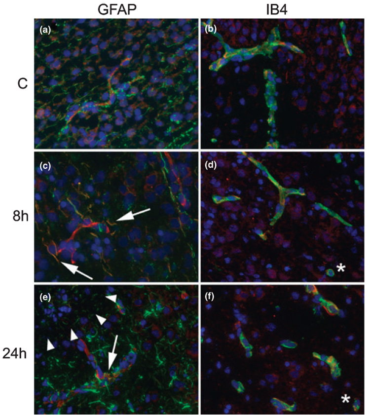
Macrophages can be both beneficial and detrimental after CNS injury. We previously showed rapid accumulation of macrophages in injured immature brain acutely after ischemia-reperfusion. To determine whether these macrophages are microglia or invading monocytes, we subjected post-natal day 7 (P7) rats to transient 3 h middle cerebral artery (MCA) occlusion and used flow cytometry at 24 and 48 h post-reperfusion to distinguish invading monocytes (CD45high/CD11b+) from microglia (CD45low/medium/CD11b+). Inflammatory cytokines and chemokines were determined in plasma, injured and contralateral tissue 1-24 h post-reperfusion using ELISA-based cytokine multiplex assays. At 24 h, the number of CD45+/CD11b+ cells increased 3-fold in injured compared to uninjured brain tissue and CD45 expression shifted from low to medium with less than 10% of the population expressing CD45high. MCA occlusion induced rapid and transient asynchronous increases in the pro-inflammatory cytokine IL-beta and chemokines cytokine-induced neutrophil chemoattractant protein 1 (CINC-1) and monocyte-chemoattractant protein 1 (MCP-1), first in systemic circulation and then in injured brain. Double immunofluorescence with cell-type specific markers showed that multiple cell types in the injured brain produce MCP-1. Our findings show that despite profound increases in MCP-1 in injured regions, monocyte infiltration is low and the majority of macrophages in acutely injured regions are microglia.


Figures

Fig. 1
Fig. 1
Peripheral leukocytes are activated 24 h after MCA occlusion in P7 rats. (a and b) Three populations of CD45+ cells are detected in CD45 vs. SSC contour plots. (c and d) Increases in CD45med (c) and CD11b (d) expression are detected with injury. In uninjured animals the percentage of CD45+ cells classified as low, med, or high (bars) is, respectively, 14 ± 7.7%, 36 ± 7.8%, and 50 ± 7.1% and in injured animals, these ratios shift to 10 ± 7%, 46 ± 4% and 44 ± 3.7. Statistically significant shifts as a result of injury occur in the CD45medium/CD11b+ positive populations (p = 0.05, n = 4). Histograms in this and all subsequent figures show CD45 gated cells. Bars in CD11b histograms represent negative and positive subpopulations. Inset in (c) shows isotype control antibody (dashed line) compared to CD45 antibody labeling of control (solid) samples. (e) MCA occlusion results in increased RP-1 expression. Quantification of RP-1 positive cells to the right of the isotype matched control antibody (dashed curve) indicates injury results in an increase in RP-1+ cells from 18 ± 9.7% to 22 ± 4.7% (solid curve, control; dotted, injured) of the CD45 population (p > 0.5, n = 3). Shown are data from a representative experiment.
Fig. 2
Fig. 2
Macrophages in the brain are predominantly activated microglia, not invading monocytes, 24 h post-ischemia reperfusion. (a and b) The number of activated leukocytes (CD45+/CD11+) increases from 7.1% in uninjured (a) to 60.3% in injured tissue (b). Values represent the percent of CD45 gated cells present in that quadrant. Note that in quadrant analysis, CD45+ cells contain both CD45med and CD45high populations. Data are representative of 4 independent experiments. (c) CD45 histograms indicate a shift towards higher intensity and a broadening of the curve with injury. Bars indicate the boundaries of low, medium and high CD45 expression levels. Dotted = contralateral, solid = injured. (d) With injury, there is a shift in the CD45 population from CD45low (36 ± 7.9%) to CD45med (55 ± 12.3%). Less than 10% (8.8 ± 6.7%) of the population in injured tissue is CD45high. Data are expressed as mean ± SD, n = 11. *p < 0.005 compared to contralateral. Grey bars = contralateral, solid = injured. (e and f) Expression of monocytic lineage marker CD163 is rapidly increased on activated microglia 24 h post-reperfusion. In contralateral hemispheres, CD163 expression is minimal (e) whereas in injured tissue CD163 expression is induced in CD45med cells (f). Dotted line, CD45low; solid line, CD45med, dashed line, CD45high. Shown are data from a representative experiment, n = 5.
Fig. 3
Fig. 3
Macrophages continue to accumulate at 48 h post-reperfusion. (a) CD45 histograms show a rightward curve shift with injury evolution. (b) CD45low cells continue to predominate in contralateral hemispheres (85 ± 3.9%, 14 ± 3.4%, 1.2 ± 0.6%, respectively, for low, med and high expression; n = 5), whereas with injury the distribution among the three subpopulations changes dramatically (31 ± 7.3%, 39 ± 3.0% and 30 ± 9%, respectively, for low, med and high; n = 4). Mean ± SD. *p < 0.005 for low, med; **p < 0.02 for high populations compared to contralateral.
Fig. 4
Fig. 4
Cytokine and chemokine levels are increased in plasma and injured brain tissue but not in contralateral hemisphere within 24 h after reperfusion. A significant transient increase in IL-1β, IL-6, CINC-1, and MCP-1 occurs first in plasma (a, 0–8 h post-reperfusion) and then in the injured brain (b, 4–8 h post-reperfusion). Measurements were performed using the multiplex cytokine kit (LINCO Research) in animals identified by DW-MRI as injured. Data are expressed as mean ± SD, n = 3–17 per time point, as indicated for individual time points. In a: *p < 0.04 vs. control; **p < 0.015 vs. control; ***p < 0.0001 vs. control. In b: §p < 0.04; §§p < 0.01; §§§p < 0.001 vs. value in the corresponding contralateral hemisphere.
Fig. 5
Fig. 5
MCP-1 expression is elevated in multiple cell types within injured regions. (a and b) MCP-1 expression is evident in the vasculature in contralateral hemisphere. (c and d) At 8 h post-reperfusion, MCP-1 is expressed in astrocyte processes (c), vessels and macrophages (d) in injured regions. (e and f) At 24 h post-reperfusion, continued expression is detected in the vasculature, astrocyte processes in the penumbra, and macrophages. Arrows in c and e indicate coexpression of MCP-1 with GFAP in astrocyte processes; asterisks in d and f indicate coexpression in cells that likely represent microglia. Arrowheads in e indicate the boundary between injury core and penumbra. All images were captured at 20 ×. In all images red = MCP-1 and blue = nuclei (DAPI), green = GFAP (a, c, e) or IB4 (b, d, f).

References

    1. Allan SM, Tyrrell PJ, Rothwell NJ. Interleukin-1 and neuronal injury. Nat Rev. Immunol. 2005;5:629–640. - PubMed
    1. Anthony DC, Bolton SJ, Fearn S, Perry VH. Age-related effects of interleukin-1 beta on polymorphonuclear neutrophil-dependent increases in blood–brain barrier permeability in rats. Brain. 1997;120:435–444. - PubMed
    1. Anthony D, Dempster R, Fearn S, Clements J, Wells G, Perry VH, Walker K. CXC chemokines generate age-related increases in neutrophil-mediated brain inflammation and blood–brain barrier breakdown. Curr. Biol. 1998;8:923–926. - PubMed
    1. Arvin KL, Du Han BHY, Lin SZ, Paul SM, Holtzman DM. Minocycline markedly protects the neonatal brain against hypoxic-ischemic injury. Ann. Neurol. 2002;52:54–61. - PubMed
    1. Back SA, Rivkees SA. Emerging concepts in periventricular white matter injury. Semin. Perinatol. 2004;28:405–414. - PubMed

Publication types

MeSH terms