. 2006 Apr;26(8):2936-46.
doi: 10.1128/MCB.26.8.2936-2946.2006.

NF-kappaB regulates spatial memory formation and synaptic plasticity through protein kinase A/CREB signaling

Affiliations

NF-kappaB regulates spatial memory formation and synaptic plasticity through protein kinase A/CREB signaling

Barbara Kaltschmidt et al. Mol Cell Biol. 2006 Apr.

Abstract

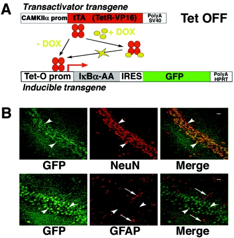
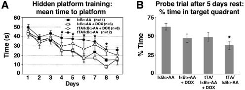
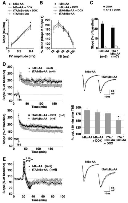
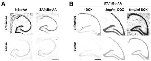
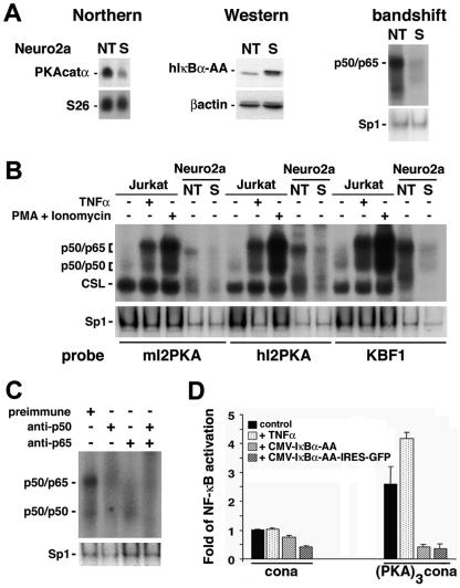
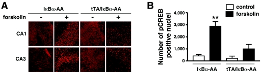
Synaptic activity-dependent de novo gene transcription is crucial for long-lasting neuronal plasticity and long-term memory. In a forebrain neuronal conditional NF-kappaB-deficient mouse model, we demonstrate here that the transcription factor NF-kappaB regulates spatial memory formation, synaptic transmission, and plasticity. Gene profiling experiments and analysis of regulatory regions identified the alpha catalytic subunit of protein kinase A (PKA), an essential memory regulator, as a new NF-kappaB target gene. Consequently, NF-kappaB inhibition led to a decrease in forskolin-induced CREB phosphorylation. Collectively, these results disclose a novel hierarchical transcriptional network involving NF-kappaB, PKA, and CREB that leads to concerted nuclear transduction of synaptic signals in neurons, accounting for the critical function of NF-kappaB in learning and memory.


Figures

FIG. 1.
FIG. 1.
Expression of the inducible transgene coding for the super-repressor and GFP in tTA/IκBα-AA double transgenic mice is restricted to neurons. (A) Schematic diagram of the binary inducible transgenic system used to inhibit NF-κB activity in the forebrain. The internal ribosome entry site has been mutated to render GFP a real tracer of super-repressor (IκBα-AA) expression. In the absence of DOX, an analog of tetracycline that passes the blood-brain barrier, super-repressor and GFP are expressed in a polycistronic transcript. SV40, simian virus 40; HPRT, hypoxanthine phosphoribosyltransferase; prom, promoter. (B) Coronal sections from the brain of an 8-week-old tTA/IκBα-AA double transgenic subjected to immunohistochemical analysis with antibodies against GFP and NeuN or GFAP and photographed in the CA1 region. Green, GFP immunoreactivity; red, NeuN immunoreactivity or GFAP immunoreactivity; yellow, double-labeled cells; white arrowheads, neuronal cells; white arrows, glial cells; scale bar, 20 μm.
FIG. 2.
FIG. 2.
Spatial memory formation in the Morris water maze task is impaired in tTA/IκBα-AA double transgenic mice. (A) Every group improved with training; however, significant, longer escape times are observed in tTA/IκBα-AA double transgenic mice in the absence of DOX compared to untreated single transgenic controls (*, P < 0.05). The number of subjects used is indicated in parentheses. DOX treatment of single or double transgenics did not significantly affect learning abilities. (B) Significantly reduced percent time in target quadrant after 5 days' rest in tTA/IκBα-AA double transgenic mice in the absence of DOX relative to untreated single transgenic controls (*, P < 0.002). P values for comparisons for all groups are the following: IκBα-AA versus IκBα-AA+DOX, P < 0.036; IκBα-AA versus tTA/IκBα-AA+DOX, P < 0.066; IκBα-AA+DOX versus tTA/IκBα-AA+DOX, P < 0.87; tTA/IκBα-AA versus IκBα-AA+DOX, P < 0.3; tTA/IκBα-AA versus tTA/IκBα-AA+DOX, P < 0.22; tTA/IκBα-AA versus IκBα-AA, P < 0.002. These values reveal that DOX treatment has by itself a significant influence on memory formation. Data are means ± standard errors of the means.
FIG. 3.
FIG. 3.
Synaptic plasticity in tTA/IκBα-AA double transgenic mice is impaired. (A) The input-output curve was obtained by plotting the fiber volley amplitude against fEPSP slope. At greater fiber volley amplitudes, the slope size in tTA/IκBα-AA double transgenic mice is significantly reduced compared to single transgenic controls (*, P < 0.05; n = 8). (B) PPF in tTA/IκBα-AA double transgenic mice is not significantly affected (P < 0.1; n = 8). (C) Pharmacological isolation of AMPA receptor and NMDA receptor currents by application of DNQX and AP-5, respectively. tTA/IκBα-AA double and IκBα-AA single transgenic mice disclose no significant difference (P > 0.1). (D) Impaired L-LTP in tTA/IκBα-AA double transgenic mice. The top left graph is a summary showing the time course of fEPSPs in tTA/IκBα-AA double transgenics (n = 8) and IκBα-AA single transgenic controls (n = 8). The difference between tTA/IκBα-AA double and IκBα-AA single transgenic mice 100 to 180 min after TBS is significant (P < 0.01). The lower left graph is a summary showing the time course of fEPSPs in tTA/IκBα-AA double transgenics treated with DOX (n = 8) or not treated (n = 8). The difference between untreated and DOX-treated tTA/IκBα-AA double transgenic mice 100 to 180 min after TBS is significant (P < 0.02). Shown at top right are single examples of superimposed traces before TBS application (thin line) and 160 min after the TBS (thick line). The lower right graph is a summary of all LTP experiments with tTA/IκBα-AA double and IκBα-AA single transgenics treated with DOX or not treated (*, P < 0.01 for tTA/IκBα-AA double transgenics versus IκBα-AA single transgenics or P < 0.02 for tTA/IκBα-AA double transgenics versus DOX-treated animals regardless of the genotype). (E) Impaired LTD in tTA/IκBα-AA double transgenic mice. Shown is a summary graph with tTA/IκBα-AA double (n = 6) and IκBα-AA single transgenic mice (n = 9). LTD was induced by ppLFS. The difference between double transgenics and controls 60 min after LFS is significant (P < 0.05). At right are single examples of superimposed traces before ppLFS (thin line) application and 60 min after the ppLFS (thick line). Data are means ± standard errors of the means.
FIG. 4.
FIG. 4.
Expression of PKA catalytic α gene is strongly reduced in tTA/IκBα-AA double transgenic mice. (A) In situ hybridization on brain sagittal sections from a tTA/IκBα-AA double transgenic mouse and an IκBα-AA single transgenic control littermate with an antisense probe specific for the mouse PKA catalytic α gene or with the sense control probe. (B) In situ hybridization with antisense or sense mouse PKA catalytic α gene probes on brain sagittal sections from tTA/IκBα-AA double transgenic mice, untreated or treated with increasing doses of DOX. Scale bar, 200 μm.
FIG. 5.
FIG. 5.
PKA catalytic α gene expression is regulated via NF-κB. (A) Northern blot analysis of total RNA (15 μg) from Neuro2a cells nontransfected (NT) or constitutively expressing the super-repressor (S). The top left blot shows hybridization with full-length mouse PKA catalytic α cDNA; in the bottom blot, the same filter was rehybridized with an S26 probe as an invariant control. Western blot (middle) analysis of protein extracts (60 μg) with a polyclonal antibody directed against IκBα (top) or a monoclonal antibody against β-actin as an internal control (bottom) confirms the overexpression of the super-repressor (human IκBα-AA [hIκBα-AA]) in the stable clone (S). Nuclear extracts (5 μg) analyzed by bandshift assay (right) for binding to a canonical κB site show high levels of nuclear p50/p65 heterodimers in nontransfected (NT) Neuro2a cells that are almost totally suppressed in the stable clone (S). The same extracts used for bandshift assays with an Sp1 site serve as an invariant control. (B) Five micrograms of nuclear extracts from Jurkat cells, untreated or treated with TNF-α (10 ng/ml) or with phorbol myristate acetate (PMA; 50 ng/ml) and ionomycin (1 μg/ml), or 0. 5 μg of nuclear extracts from nontransfected Neuro2a cells (NT) or Neuro2a cells stably expressing the super-repressor (S) were analyzed by bandshift assay for their ability to bind to κB sites identified in intron 2 of the mouse and human PKAcatα genes (mI2PKA and hI2PKA, respectively) or to a canonical κB site (KBF1). Variation in specific activities of the probes is responsible for the different intensities observed after binding with the various probes. CSL is CBF1/Su(H)L/Lag1, also known as RBP-Jk complex, which binds a half-κB site and is involved in the Notch pathway. The same extracts used for bandshift assays with an Sp1 site serve as internal controls. (C) Nuclear extracts from Neuro2a cells (5 μg) were preincubated with preimmune serum or sera directed against p50 or/and p65 and analyzed by bandshift assay for binding to the κB site from intron 2 of the mouse PKA catalytic α gene. The same extracts analyzed by bandshift assay for binding to Sp1 serve as internal controls. (D) Neuro2a cells were transfected with luciferase reporter constructs containing a minimal promoter alone (cona) or juxtaposed to a trimerized mouse intron 2 PKA κB site [(PKA)3cona] together with an EF1-lacZ normalization vector, in the absence or presence of expression vectors coding for the super-repressor (CMV-IκBα-AA or CMV-IκBα-AA-IRES-GFP, where IRES is internal ribosome entry site). Cells were left untreated or were treated with TNF-α (10 ng/ml) for 6 h and assessed for activation of the luciferase and β-galactosidase reporter genes. Data are means ± standard errors of the means.
FIG. 6.
FIG. 6.
Inhibition of NF-κB interferes with PKA-dependent CREB phosphorylation. (A) Immunocytochemistry of CREB-phosphorylation on hippocampal slice cultures from tTA/IκBα-AA double transgenic mice or IκBα-AA single transgenic littermate controls, untreated or treated with forskolin for 30 min. CA1, hippocampal region CA1; CA3, hippocampal region CA3. (B) Quantification of the total number of pCREB-positive nuclei in the hippocampal fields observed in panel A (n = 3 mice/genotype). The average values of forskolin-induced CREB phosphorylation are significantly higher in IκBα-AA single transgenic slices relative to tTA/IκBα-AA double trangenics (**, P < 0.01), whereas no significant differences could be observed between forskolin-treated tTA/IκBα-AA double transgenic slices and untreated single (IκBα-AA) or double (tTA/IκBα-AA) transgenics.

References

    1. Abel, T., P. V. Nguyen, M. Barad, T. A. Deuel, E. R. Kandel, and R. Bourtchouladze. 1997. Genetic demonstration of a role for PKA in the late phase of LTP and in hippocampus-based long-term memory. Cell 88:615-626. - PubMed
    1. Albensi, B. C., and M. P. Mattson. 2000. Evidence for the involvement of TNF and NF-kappaB in hippocampal synaptic plasticity. Synapse 35:151-159. - PubMed
    1. Balschun, D., D. P. Wolfer, P. Gass, T. Mantamadiotis, H. Welzl, G. Schutz, J. U. Frey, and H. P. Lipp. 2003. Does cAMP response element-binding protein have a pivotal role in hippocampal synaptic plasticity and hippocampus-dependent memory? J. Neurosci. 23:6304-6314. - PMC - PubMed
    1. Barco, A., J. M. Alarcon, and E. R. Kandel. 2002. Expression of constitutively active CREB protein facilitates the late phase of long-term potentiation by enhancing synaptic capture. Cell 108:689-703. - PubMed
    1. Bhakar, A. L., L. L. Tannis, C. Zeindler, M. P. Russo, C. Jobin, D. S. Park, S. MacPherson, and P. A. Barker. 2002. Constitutive nuclear factor-κB activity is required for central neuron survival. J. Neurosci. 22:8466-8475. - PMC - PubMed

Publication types

MeSH terms

Substances