Comparative Study
. 2005 Apr 6;25(14):3638-50.
doi: 10.1523/JNEUROSCI.3980-04.2005.

Astrocytes regulate inhibitory synapse formation via Trk-mediated modulation of postsynaptic GABAA receptors

Affiliations
Comparative Study

Astrocytes regulate inhibitory synapse formation via Trk-mediated modulation of postsynaptic GABAA receptors

Sarina B Elmariah et al. J Neurosci. .

Abstract

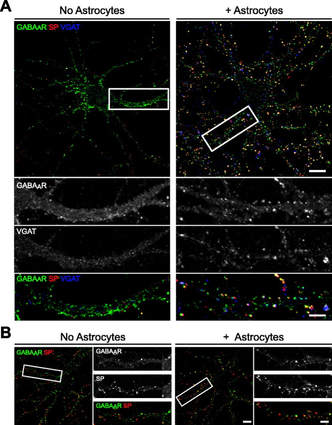
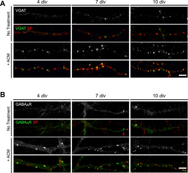
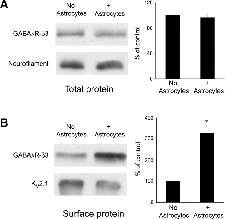
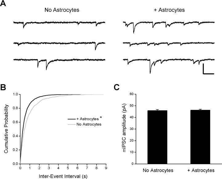
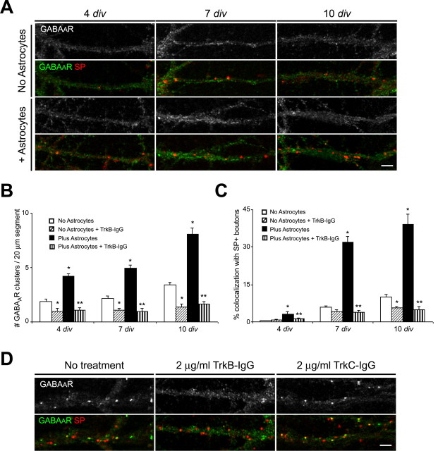
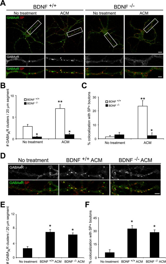
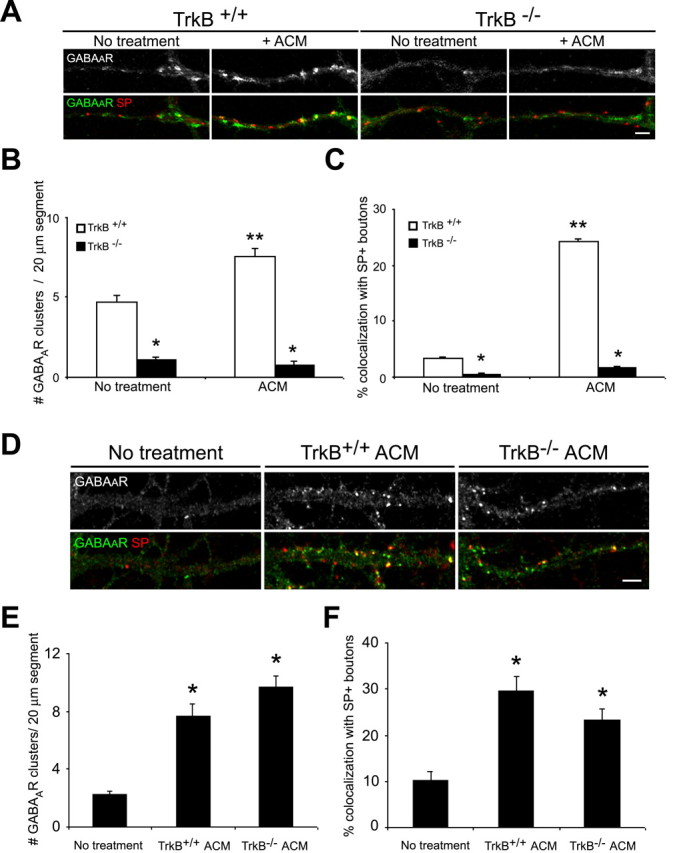
Astrocytes promote the formation and function of excitatory synapses in the CNS. However, whether and how astrocytes modulate inhibitory synaptogenesis are essentially unknown. We asked whether astrocytes regulate the formation of inhibitory synapses between hippocampal neurons during maturation in vitro. Neuronal coculture with astrocytes or treatment with astrocyte-conditioned medium (ACM) increased the number of inhibitory presynaptic terminals, the frequency of miniature IPSCs, and the number and synaptic localization of GABA(A) receptor (GABA(A)R) clusters during the first 10 d in vitro. We asked whether neurotrophins, which are potent modulators of inhibitory synaptic structure and function, mediate the effects of astrocytes on inhibitory synapses. ACM from BDNF- or tyrosine receptor kinase B (TrkB)-deficient astrocytes increased inhibitory presynaptic terminals and postsynaptic GABA(A)R clusters in wild-type neurons, suggesting that BDNF and TrkB expression in astrocytes is not required for these effects. In contrast, although the increase in the number of inhibitory presynaptic terminals persisted, no increase was observed in postsynaptic GABA(A)R clusters after ACM treatment of hippocampal neurons lacking BDNF or TrkB. These results suggest that neurons, not astrocytes, are the relevant source of BDNF and are the site of TrkB activation required for postsynaptic GABA(A)R modulation. These data also suggest that astrocytes may modulate postsynaptic development indirectly by stimulating Trk signaling between neurons. Together, these data show that astrocytes modulate inhibitory synapse formation via distinct presynaptic and postsynaptic mechanisms.


Figures

Figure 1.
Figure 1.
Astrocytes increase the number of inhibitory presynaptic terminals and postsynaptic GABAAR clusters in hippocampal neurons in vitro. Hippocampal neurons were cultured in the presence or absence of astrocytes and immunostained with antibodies against GABAAR-β2/β3 (green), SP (red), and VGAT (blue) to visualize inhibitory presynaptic terminals. A, The number of SP+ and VGAT+ presynaptic terminals and the number of GABAAR clusters increased in neuron-astrocyte cocultures (right) compared with pure neuronal cultures (left) at 10 div. The proportion of GABAAR clusters apposed to presynaptic terminals also increased in neuron-astrocyte cocultures (Table 1). Scale bar, 10 μm. Areas within white boxes are shown in the panels below at a higher magnification. Scale bar (bottom right), 2 μm. B, At 3 weeks in vitro, the proportion of GABAAR clusters localized to synapses (yellow) was approximately threefold greater in neuron-astrocyte cocultures (right) than in pure neuronal cultures (left). No differences were observed in the number of presynaptic terminals (red) or the number of GABAAR clusters (green). Scale bar, 10 μm. Areas within white boxes are shown to the right at a higher magnification. Scale bar (bottom right), 2 μm.
Figure 2.
Figure 2.
Astrocyte-conditioned medium increases the number of inhibitory synapses in hippocampal neurons in vitro. Hippocampal neurons were cultured in the presence of ACM, and inhibitory presynaptic terminals or GABAAR clusters were examined at 4, 7, and 10 div. A, Immunostaining was performed with antibodies against VGAT (green) and SP (red) to visualize inhibitory presynaptic terminals. The number of SP+ boutons and the proportion of VGAT+ inhibitory terminals increased after ACM treatment (bottom) compared with untreated controls (top). Scale bar, 2 μm. B, Immunostaining was performed with antibodies against GABAAR-β2/β3 (green) and SP (red). ACM treatment (bottom) resulted in an increase in the number and synaptic localization of GABAAR clusters (green) compared with pure neuronal cultures (top). Scale bar, 2 μm.
Figure 3.
Figure 3.
Surface GABAAR expression is increased in hippocampal neurons in the presence of astrocytes. Total cell lysates or biotinylated surface protein extracts were harvested from hippocampal neurons cultured in the presence and absence of astrocytes at 10 div. A, Western blot analysis on total cell homogenates was performed using an antibody against the GABAAR-β3 subunit (top) or neurofilament H (bottom) as a loading control. Quantification of relative band intensity compared with loading controls demonstrates no significant difference in the level of GABAAR-β3 expression in the presence of astrocytes. B, Western blot analysis on surface-biotinylated extracts was performed using an antibody against the GABAAR-β3 subunit (top) or Kv2.1 (bottom) as a loading control. Quantification of relative band intensity compared with loading controls shows that GABAAR-β3 expression at the neuronal surface is increased ∼3.5-fold when neurons are cultured in the presence of astrocytes (*p < 0.001). Error bars represent SEM.
Figure 4.
Figure 4.
Spontaneous inhibitory activity is increased in the presence of astrocytes. Whole-cell voltage-clamp recordings were performed on hippocampal pyramidal neurons at 17-18 div to examine mIPSCs in the presence and absence of astrocytes. A, Representative recordings from two neurons in the presence and absence of astrocytes. mIPSCs were recorded in the presence of the following (in μm): 1 TTX, 50 APV, and 10 CNQX. mIPSCs occurred more frequently in neuron-astrocyte cocultures (right) relative to pure neuronal cultures (left). Calibration: vertical, 0.1 nA; horizontal, 0.2 s. B, Cumulative probability distribution of the interevent interval of mIPSCs in pure neuronal and neuron-astrocyte cocultures. *p < 0.0001, significant difference compared with pure neuronal cultures (Kolmogorov-Smirnov test). C, Quantification of mIPSC amplitude in pure neuronal and neuron-astrocyte cocultures (p = 0.77; Student's t test). Error bars represent SEM.
Figure 5.
Figure 5.
Scavenging endogenous BDNF prevents the astrocyte-induced increase in the number and synaptic localization of GABAAR clusters. Hippocampal cultures with and without astrocytes were treated with 2.0 μg/ml TrkB-IgG to scavenge endogenous BDNF or with TrkC-IgG to scavenge NT3 at different ages in vitro. Immunostaining was performed with antibodies against GABAAR-β2/β3 (green) and SP (red) at 4, 7, or 10 div. A, Treatment with 2.0 μg/ml TrkB-IgG beginning at 1 div resulted in fewer postsynaptic GABAAR clusters in pure neuronal cultures (top) and neuron-astrocyte cocultures (bottom) at 4, 7, and 10 div. No change was observed in the number of SP+ boutons after BDNF scavenging. Scale bar, 2 μm. B, Quantification of TrkB-IgG effects on the number of GABAAR clusters per 20 μm dendrite segment. *p < 0.001, significant difference compared with no treatment in pure neuronal cultures; **p < 0.001, significant decrease compared with no treatment in neuron astrocyte cocultures. C, Quantification of TrkB-IgG effects on the synaptic localization of GABAAR clusters. *p < 0.001, significant difference compared with no treatment in pure neuronal cultures; **p < 0.001, significant decrease compared with no treatment in neuron astrocyte cocultures. D, Treatment with 2.0 μg/ml TrkB-IgG at 8-10 div for 48 h decreased the number of postsynaptic GABAAR clusters in neuron-astrocyte cocultures (middle) (Table 2) compared with untreated controls (left). In contrast, treatment with 2.0 μg/ml TrkC-IgG for 48 h increased the proportion of GABAAR clusters localized to synapses but had no effect on the number of GABAAR clusters (right) (Table 2). No change was observed in the number of SP+ boutons after BDNF or NT3 scavenging. Scale bar, 2 μm. Error bars represent SEM.
Figure 6.
Figure 6.
Neuronal BDNF release is required for the astrocyte-induced increase in postsynaptic GABAAR clusters. A-C, Hippocampal neurons were cultured from BDNF-/- mice and wild-type littermates in the presence and absence of ACM. Immunostaining was performed with antibodies against GABAAR-β2/β3 (green) and SP (red) at 10 div. A, GABAAR cluster number and synaptic localization were decreased in BDNF-/- neurons (right) compared with BDNF+/+ neurons (left) at 10 div. ACM treatment increased the number and synaptic localization of GABAAR clusters in BDNF+/+ neurons but had no effect on GABAAR clusters in BDNF-/- neurons. Scale bar (top right), 10 μm. Areas within white boxes are shown below at higher magnification. Scale bar (bottom right), 2 μm. B, Quantification of GABAAR cluster number per 20 μm dendrite segment. *p < 0.001, significant difference compared with BDNF+/+ neurons within a condition; **p < 0.001, significant decrease compared with no treatment in BDNF+/+ neurons. C, Quantification of GABAAR cluster synaptic localization. *p < 0.001, significant difference compared with BDNF+/+ neurons within a condition; **p < 0.001, significant decrease compared with no treatment in BDNF+/+ neurons. D-F, Hippocampal neurons were grown in the presence and absence of ACM from BDNF-/- and wild-type littermate mice, and GABAAR clusters were examined. D, ACM from BDNF+/+ (middle) and BDNF-/- (right) astrocytes increased GABAAR cluster number and synaptic localization compared with untreated controls (left) by a similar magnitude. Scale bar, 2 μm. E, Quantification of GABAAR cluster number per 20 μm dendrite segment. *p < 0.001, significant difference compared with no treatment controls. F, Quantification of GABAAR cluster synaptic localization. *p < 0.001, significant difference compared with no treatment controls. Error bars represent SEM.
Figure 7.
Figure 7.
TrkB signaling in astrocytes is not required for GABAAR cluster modulation. A-C, Hippocampal neurons were cultured from TrkB-/- mice and wild-type littermates in the presence and absence of ACM. Immunostaining was performed with antibodies against GABAAR-β2/β3 (green) and SP (red) at 10 div. A, GABAAR cluster number and synaptic localization were decreased in TrkB-/- neurons (right) compared with TrkB+/+ neurons (left) at 10 div. ACM treatment increased the number and synaptic localization of GABAAR clusters in TrkB+/+ neurons but had no effect on GABAAR clusters in TrkB-/- neurons. Scale bar, 2 μm. B, Quantification of GABAAR cluster number per 20 μm dendrite segment. *p < 0.001, significant difference compared with TrkB+/+ neurons within a condition; **p < 0.001, significant decrease compared with no treatment in TrkB+/+ neurons. C, Quantification of GABAAR cluster synaptic localization. *p < 0.001, significant difference compared with TrkB+/+ neurons within a condition; **p < 0.001, significant decrease compared with no treatment in TrkB+/+ neurons. D-F, Hippocampal neurons were grown in the presence and absence of ACM from TrkB-/- and wild-type littermate mice, and GABAAR clusters were examined. D, ACM from TrkB+/+ (middle) and TrkB-/- (right) astrocytes increased GABAAR cluster number and synaptic localization compared with untreated controls (left) by a similar magnitude. Scale bar, 2 μm. E, Quantification of GABAAR cluster number per 20 μm dendrite segment after treatment with TrkB+/+ and TrkB-/- ACM. *p < 0.001, significant difference compared with no treatment controls. F, Quantification of GABAAR cluster synaptic localization after treatment with TrkB+/+ and TrkB-/- ACM. *p < 0.001, significant difference compared with no treatment controls. Error bars represent SEM.

References

    1. Alderson RF, Curtis R, Alterman AL, Lindsay RM, DiStefano PS (2000) Truncated TrkB mediates the endocytosis and release of BDNF and neurotrophin-4/5 by rat astrocytes and Schwann cells in vitro. Brain Res 871: 210-222. - PubMed
    1. Binder DK, Routbort MJ, Ryan TE, Yancopoulos GD, McNamara JO (1999) Selective inhibition of kindling development by intraventricular administration of TrkB receptor body. J Neurosci 19: 1424-1436. - PMC - PubMed
    1. Blondel O, Collin C, McCarran WJ, Zhu S, Zamostiano R, Gozes I, Brenneman DE, McKay RD (2000) A glia-derived signal regulating neuronal differentiation. J Neurosci 20: 8012-8020. - PMC - PubMed
    1. Brunig I, Penschuck S, Berninger B, Benson J, Fritschy JM (2001) BDNF reduces miniature inhibitory postsynaptic currents by rapid downregulation of GABA(A) receptor surface expression. Eur J Neurosci 13: 1320-1328. - PubMed
    1. Christopherson KS, Ullian EM, Stokes CCA, Mullowney CE, Hell JW, Agah A, Lawler J, Mosher DF, Bornstein P, Barres BA (2005) Thrombospondins are astrocyte-secreted proteins that promote CNS synaptogenesis. Cell 120: 421-433. - PubMed

Publication types

MeSH terms

LinkOut - more resources