. 2004 Oct;114(8):1098-106.
doi: 10.1172/JCI21086.

Epithelial hypoxia-inducible factor-1 is protective in murine experimental colitis

Affiliations

Epithelial hypoxia-inducible factor-1 is protective in murine experimental colitis

Jörn Karhausen et al. J Clin Invest. 2004 Oct.

Abstract

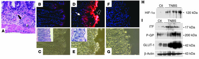
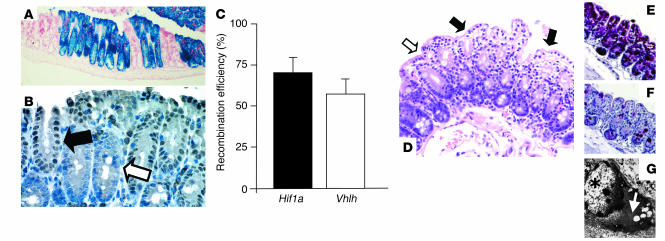
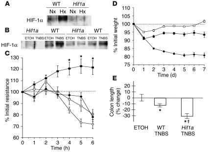
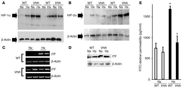
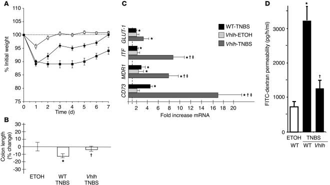
Mucosal epithelial cells are uniquely equipped to maintain barrier function even under adverse conditions. Previous studies have implicated hypoxia in mucosal tissue damage resulting from both acute and chronic inflammation. Given the importance of the transcriptional regulator hypoxia-inducible factor-1 (HIF-1) for adaptive hypoxia responses, we hypothesized that HIF-1 may serve as a barrier-protective element during mucosal inflammation. Initial studies of hapten-based murine colitis revealed extensive mucosal hypoxia and concomitant HIF-1 activation during colitis. To study this in more detail, we generated 2 mouse lines with intestinal epithelium-targeted expression of either mutant Hif1a (inability to form HIF-1) or mutant von Hippel-Lindau gene (Vhlh; constitutively active HIF-1). Studies of colitis in these mice revealed that decreased HIF-1 expression correlated with more severe clinical symptoms (mortality, weight loss, colon length), while increased HIF levels were protective in these parameters. Furthermore, colons with constitutive activation of HIF displayed increased expression levels of HIF-1-regulated barrier-protective genes (multidrug resistance gene-1, intestinal trefoil factor, CD73), resulting in attenuated loss of barrier during colitis in vivo. Taken together, these studies provide insight into tissue microenvironmental changes during model inflammatory bowel disease and identify HIF-1 as a critical factor for barrier protection during mucosal insult.


Figures

Figure 1
Figure 1
Epithelial hypoxia in TNBS colitis is associated with inflammatory lesions. (A) H&E staining in TNBS colitis (7 days after induction). In an area of relatively minor epithelial inflammation, submucosal vessels are encircled by a mixed population of inflammatory cells (filled arrow). Fibrinoid necrosis of the vessel walls (open arrow, with apoptotic bodies) and signs of luminal obliteration are observed. Magnification, ×600. (B) Localization of EF5 (nuclear counterstaining with DAPI) in a colonic section from a vehicle-control animal at day 3. Discrete immunofluorescence in surface epithelial cells and underlying crypts. (C) Corresponding phase-contrast image. Inset: H&E stain of an adjacent section. (D and E) Section taken from distal colon in TNBS-exposed animals. (D) Intense EF5 immunofluorescence overlying the area of ulceration (open arrow) and in underlying epithelial portions (filled arrow). (E) Phase contrast of D. Inset: H&E staining of an adjacent section from the same tissue, displaying erosion of the epithelial layer and beginning inflammatory infiltration. (F) Competed stain (section adjacent to that shown in D and E). Antibody specificity is documented by absence of Cy3 signal when antibody was saturated with free drug. (G) Corresponding phase-contrast image. Magnification in BG, ×400. (H) HIF-1α stabilization in TNBS colitis. Western blot analysis from TNBS and vehicle-control (Ctl) animals 7 days after induction of colitis. Blots are derived from nuclear extracts from the colon of control and TNBS-treated animals. (I) Western blot analysis of the HIF-1–responsive genes ITF, P-GP, and GLUT-1 in control and TNBS-treated animals.
Figure 2
Figure 2
Fabp-Cre–mediated recombination in the colon: analysis of Fabp-Cre activity and histological characterization of mutant mice. (A and B) Fabp-Cre activity as determined by a lacZ reporter transgene and immunohistochemical staining for Cre-recombinase. (A) Section of the cecum stained with X-gal. Recombination occurs always in all cells of an individual crypt. Magnification, ×100. (B) Paraffin-embedded section of the colon from a Vhlh mutant mouse. Consistent with the recombination process in the lacZ transgenic mice, all cells within a Cre-positive crypt display nuclear-localized Cre signal (filled arrow), while all cells in a negative crypt stain negatively (open arrow). Magnification, ×400. (C) Determination of the efficiency of recombination by quantitative PCR. Levels of expression of the recombined gene (Hif1a or Vhlh) were compared with the expression levels of a non-recombining control gene in genomic DNA from isolated colonic epithelial cells (n = 3 for each genotype). (DG) Clear-cell changes in the Vhlh mutant colon. (D) Luminal epithelial cells with pronounced clearing are indicated by filled arrows; the open arrow points to adjacent luminal cells without clearing. Clearing is a result of glycogen accumulation in Vhlh mutant cells, as shown by diastase-sensitive PAS staining. (E) PAS stain without diastase treatment. (F) PAS stain after diastase treatment of the adjacent colon. (G) Ultrastructural analysis of clear cells showed accumulation of cytoplasmic material that is morphologically consistent with glycogen (white arrow). The star indicates the nucleus of a clear cell. Magnification in DF, ×200.
Figure 3
Figure 3
Impairment of barrier function through conditional deletion of Hif1a in intestinal epithelium. (A) Lack of HIF-1 induction in conditional Hif1a mutants in whole-body hypoxia. Western blot analysis of nuclear HIF-1α levels from colonic scrapings of WT littermates or conditional Hif1a mice subjected to normoxia (Nx; 21% O2, 4 h) or hypoxia (Hx; 8% O2, 4 h). (B) Mucosal colonic scrapings 7 days after induction of TNBS colitis compared with vehicle control. HIF-1α levels in Hif1a mutants failed to respond, which indicates successful impairment of the HIF-1 pathway. (C) Ex vivo assay of barrier electrical resistance in colonic tissue explants. In samples derived from WT animals (filled squares), resistance increased in hypoxia (*P – 0.05 after 3–6 h). In contrast, both in hypoxic samples from Hif1a conditional mutant animals (filled circles) and in all normoxic samples (WT, open squares; Hif1a mutant, open circles), measurements gradually dropped. (D) Changes of body weight following induction of TNBS colitis. When compared with their WT littermates (filled squares, n = 14), Hif1a conditional mutant mice (filled circles, n = 25) displayed a more severe clinical course with significantly greater and ongoing loss of weight (P < 0.01 by ANOVA). No difference between vehicle control–treated Hif1a mutant and WT animals was observed (open squares). (E) Colon length relative to vehicle control–treated (ETOH) littermates 7 days after induction of colitis. Hif1a conditional mutant animals displayed significant shortening of the colon ( P – 0.05 vs. TNBS-treated WT, *P – 0.05 vs. vehicle-treated controls).
Figure 4
Figure 4
Conditional Vhlh deletion results in constitutive activation of the HIF-1 pathway in the whole-body hypoxia model. (A and B) Western blot analysis for HIF-1α and HIF-2α in conditional Vhlh mutant animals and WT littermates subjected to normoxia (21% O2, 4 hours) or hypoxia (8% O2, 4 hours). In contrast to WT animals, which display normal regulation, mutant animals constitutively overexpress HIF-1α (A) and HIF-2α (B). (C and D) HIF-1–dependent gene induction is exemplified by induction of ITF on both the transcriptional level (C) and the translational level (D) from colonic tissue derived from control and conditional Vhlh mutant animals subjected to normoxia or hypoxia. (E) Quantitation of serum FITC-dextran as a measure of intestinal permeability in control and conditional Vhlh mutant animals subjected to hypoxia or normoxia. Conditional deletion of Vhlh abrogated the increase in permeability observed in control animals ( P < 0.025 vs. hypoxic WT, *P – 0.05 vs. normoxic controls).
Figure 5
Figure 5
Conditional deletion of epithelial Vhlh protects from experimental colitis. (A) Vhlh mutant animals (filled circles, n = 12) recovered their initial weight loss after administration of TNBS significantly faster than WT littermates (filled squares, n = 22; P < 0.01 by ANOVA). Values for vehicle control–treated Vhlh mutant and WT animals were combined (open squares). (B) Colonic length was significantly decreased in WT TNBS-treated animals compared with conditional Vhlh mutant animals with TNBS colitis ( P – 0.05 vs. TNBS-treated WT animals, *P – 0.05 vs. vehicle-treated controls). (C) Real-time PCR analysis for GLUT-1, ITF, MDR1, and CD73. Data are presented as fold ± SEM increase over vehicle control–treated WT animals (represented by the line at 1-fold increase). While TNBS treatment significantly induced all 4 genes (*P – 0.05 vs. vehicle-treated WT), such differences were enhanced in conditional Vhlh mutant animals exposed to TNBS in regard to ITF, MDR1, and CD73 ( P – 0.05 vs. TNBS-treated WT, P – 0.05 vs. vehicle-treated Vhlh mutants). (D) Significantly lower flux of the FITC-dextran as a measure of intestinal permeability in TNBS-treated Vhlh mutant compared with TNBS-treated WT animals ( P – 0.05; P not significant for TNBS-treated Vhlh mutants vs. vehicle control–treated mice). As no difference was seen between mutant and WT vehicle control–treated animals (compare with Figure 4E), values were combined.

References

    1. Thornton M, Solomon MJ. Crohn’s disease: in defense of a microvascular aetiology. Int. J. Colorectal Dis. 2002;17:287–297. - PubMed
    1. Hatoum OA, Binion DG, Otterson MF, Gutterman DD. Acquired microvascular dysfunction in inflammatory bowel disease: loss of nitric oxide-mediated vasodilation. Gastroenterology. 2003;125:58–69. - PubMed
    1. Sands BE. From symptom to diagnosis: clinical distinctions among various forms of intestinal inflammation. Gastroenterology. 2004;126:1518–1532. - PubMed
    1. Wakefield AJ, et al. Pathogenesis of Crohn′s disease: multifocal gastrointestinal infarction. Lancet. 1989;2:1057–1062. - PubMed
    1. Yokoyama K, et al. Obliterative arteritis with nitric oxide synthase and HLA-DR expression in Crohn’s colitis. Hepatogastroenterology. 2001;48:401–407. - PubMed

Publication types

MeSH terms