. 2003 Jan;111(1):81-90.
doi: 10.1172/JCI16147.

Rapid Akt activation by nicotine and a tobacco carcinogen modulates the phenotype of normal human airway epithelial cells

Affiliations

Rapid Akt activation by nicotine and a tobacco carcinogen modulates the phenotype of normal human airway epithelial cells

Kip A West et al. J Clin Invest. 2003 Jan.

Abstract

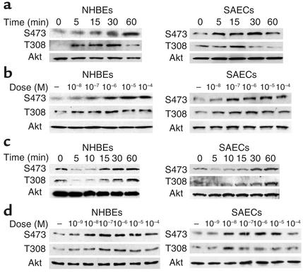
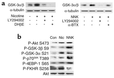
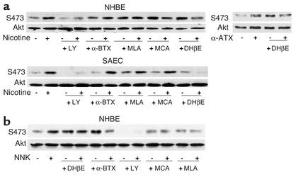
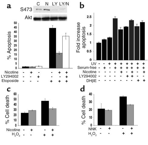
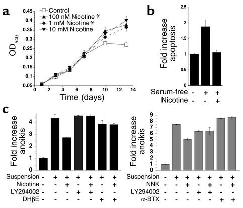
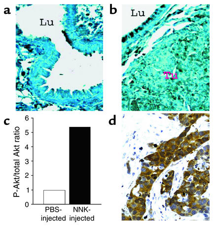
Tobacco-related diseases such as lung cancer cause over 4.2 million deaths annually, with approximately 400,000 deaths per year occurring in the US. Genotoxic effects of tobacco components have been described, but effects on signaling pathways in normal cells have not been described. Here, we show activation of the serine/threonine kinase Akt in nonimmortalized human airway epithelial cells in vitro by two components of cigarette smoke, nicotine and the tobacco-specific carcinogen 4-(methylnitrosamino)-1-(3-pyridyl)-1-butanone (NNK). Activation of Akt by nicotine or NNK occurred within minutes at concentrations achievable by smokers and depended upon alpha(3)-/alpha(4)-containing or alpha(7)-containing nicotinic acetylcholine receptors, respectively. Activated Akt increased phosphorylation of downstream substrates such as GSK-3, p70(S6K), 4EBP-1, and FKHR. Treatment with nicotine or NNK attenuated apoptosis caused by etoposide, ultraviolet irradiation, or hydrogen peroxide and partially induced a transformed phenotype manifest as loss of contact inhibition and loss of dependence on exogenous growth factors or adherence to ECM. In vivo, active Akt was detected in airway epithelial cells and lung tumors from NNK-treated A/J mice, and in human lung cancers derived from smokers. Redundant Akt activation by nicotine and NNK could contribute to tobacco-related carcinogenesis by regulating two processes critical for tumorigenesis, cell growth and apoptosis.


Figures

Figure 1
Figure 1
Phosphorylation of Akt by nicotine or NNK in NHBEs and SAECs. (a) Nicotine increased Akt phosphorylation in a time-dependent manner in NHBEs (left panels) and SAECs (right panels), as assessed by immunoblotting with anti–phospho-S473, anti–phospho-T308, and anti-Akt antibodies. (b) Nicotine-induced Akt phosphorylation was also dose-dependent. (c) NNK increased Akt phosphorylation in a time-dependent manner. (d) NNK-induced Akt phosphorylation was also dose-dependent.
Figure 2
Figure 2
Akt kinase activity and effect on downstream substrates. (a) We measured Akt kinase activity in NHBEs by immunoprecipitating active Akt and assessing phosphorylation of an exogenous peptide, GSK-3α/β, after administration of nicotine (left panels) or NNK (right panels). LY294002, DHβE (an α34 nAchR antagonist), or α-BTX (an α7 nAchR antagonist) inhibited nicotinic induction of Akt kinase activity. (b) Phosphorylation of substrates downstream of Akt in NHBEs was increased after administration of nicotine (Nic; middle lane) or NNK (right lane), compared with that in untreated cells (Con), as assessed by immunoblotting with the indicated phosphospecific antibodies.
Figure 3
Figure 3
Effect of nAchR antagonists on nicotinic activation of Akt in NHBEs and SAECs. (a) Nicotine. Only LY294002 or the α34 antagonist DHβE inhibited nicotine-induced Akt phosphorylation in NHBEs (upper left panels) and SAECs (lower panels). To confirm the role of α3 nAchRs in activating Akt in NHBEs, α-ATX (an α3 agonist) was added to NHBEs with or without DHβE (upper right panels). (b) NNK. In contrast to nicotine-mediated Akt phosphorylation, NNK-induced phosphorylation in NHBEs was inhibited by LY294002, the α7 antagonists α-BTX and MLA, and the nonspecific inhibitor MCA. DHβE was ineffective.
Figure 4
Figure 4
Nicotine-mediated Akt activation and survival of NHBEs. (a) Topoisomerase II inhibition. Nicotine (10 μM) protected against etoposide-induced apoptosis, as assessed by flow cytometry. Pretreatment with LY294002 decreased nicotine-mediated survival. Parallel samples were harvested for immunoblotting (inset; C, control; N, nicotine; LY, LY294002; LY/N, LY294002 + nicotine). (b) UV irradiation. Nicotine (10 μM) protected against UV irradiation–induced apoptosis, as measured using CellDeath ELISA kits. Pretreatment with LY294002 or DHβE attenuated nicotine-mediated survival. (c and d) H2O2 treatment. NHBEs were pretreated with nicotine (10 μM) (c) or NNK (d) as above, with or without H2O2 (200 μM). After 4 hours, cells were harvested, dead cells that exhibited cytoplasmic inclusion of 0.4% trypan blue were counted, and this number was compared with the total number of cells. At least 300 cells per sample were counted by a blinded observer.
Figure 5
Figure 5
Nicotine alters NHBE phenotype. (a) Loss of contact inhibition. NHBEs were incubated with different concentrations of nicotine (filled symbols; asterisks indicate daily dosing) or complete media alone (open squares). Cell number was measured by absorbance at 540 nm using a 96-well microplate reader. (b) Serum starvation. NHBEs were grown in DMEM with 0.1% BSA or control media for 9 days with or without nicotine (10 μM) given once on day 1. Apoptosis was assessed using CellDeath ELISA kits. (c) Anoikis. Nicotine (left panel) or NNK (right panel) decreased anoikis, as measured using CellDeath ELISA kits. Pretreatment with LY294002 decreased protection conferred by either nicotine or NNK. Pretreatment with DHβE attenuated nicotine-mediated survival (left panel), and pretreatment with α-BTX attenuated NNK-mediated survival.
Figure 6
Figure 6
Detection of Akt phosphorylation in vivo. (a) Lung tissue, including epithelial lining of airway lumen (Lu), from A/J mice given PBS orally was harvested and processed for immunohistochemistry with phosphospecific S473 antibodies as described. No staining is detectable. (b) Lung tumor (Tu) and epithelial lining of airway lumen (Lu) from A/J mice given NNK orally exhibit staining with phosphospecific S473 antibodies. (c) Quantification of phosphorylated Akt/total Akt in protein extracts derived from lung tissue from PBS- or NNK-injected mice. Quantification of immunoblots was performed using NIH Image software. (d) Phosphorylated Akt in a human lung adenocarcinoma derived from a smoker with a 48-pack-per-year smoking history.

Comment in

References

    1. Lawlor MA, Alessi DR. PKB/Akt: a key mediator of cell proliferation, survival and insulin responses? J. Cell Sci. 2001;114:2903–2910. - PubMed
    1. Scheid MP, Woodgett JR. Pkb/akt: functional insights from genetic models. Nat. Rev. Mol. Cell Biol. 2001;2:760–768. - PubMed
    1. Myers MP, et al. The lipid phosphatase activity of PTEN is critical for its tumor suppressor function. Proc. Natl. Acad. Sci. USA. 1998;95:13513–13518. - PMC - PubMed
    1. Brognard J, Clark AS, Ni Y, Dennis PA. Akt/protein kinase B is constitutively active in non-small cell lung cancer cells and promotes cellular survival and resistance to chemotherapy and radiation. Cancer Res. 2001;61:3986–3997. - PubMed
    1. Maus AD, et al. Human and rodent bronchial epithelial cells express functional nicotinic acetylcholine receptors. Mol. Pharmacol. 1998;54:779–788. - PubMed

MeSH terms

Substances