. 2001 Jul 31;98(16):9110-5.
doi: 10.1073/pnas.171300598.

alpha-Synuclein occurs in lipid-rich high molecular weight complexes, binds fatty acids, and shows homology to the fatty acid-binding proteins

Affiliations

alpha-Synuclein occurs in lipid-rich high molecular weight complexes, binds fatty acids, and shows homology to the fatty acid-binding proteins

R Sharon et al. Proc Natl Acad Sci U S A. .

Abstract

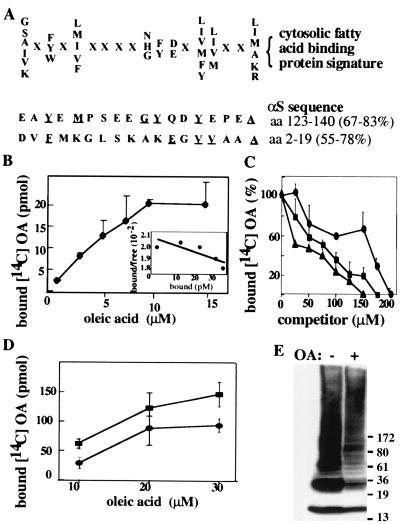
alpha-Synuclein (alphaS) is a 140-residue neuronal protein that forms insoluble cytoplasmic aggregates in Parkinson's disease (PD) and several other neurodegenerative disorders. Two missense mutations (A53T and A30P) are linked to rare forms of familial PD. The normal function of alphaS is unknown, and cultured cell systems that model its modification from soluble monomers to aggregated forms have not been reported. Through a systematic centrifugal fractionation of mesencephalic neuronal cell lines and transgenic mouse brains expressing wild-type or A53T human alphaS, we observed unusual, previously unrecognized species of alphaS that migrate well above the 17-kDa monomeric form in denaturing gels. Incubation at 65 degrees C of high-speed cytosols from cells or brains revealed a modified alphaS species migrating at approximately 36 kDa and an extensive higher molecular mass alphaS-reactive smear. Extraction of the cytosols with chloroform/methanol or with a resin (Lipidex 1000) that binds fatty acids resulted in a similar pattern of higher molecular mass alphaS forms. On the basis of this effect of delipidation, we reexamined the primary structure of alphaS and detected a motif at the N and C termini that is homologous to a fatty acid-binding protein signature. In accord, we found that purified human alphaS binds oleic acid, with an apparent K(d) of 12.5 microM. We also observed an enhanced association of A53T alphaS with microsomal membranes in both mesencephalic cells and transgenic mouse brains. We conclude that alphaS has biochemical properties and a structural motif that suggest it is a novel member of the fatty acid-binding protein family and may thus transport fatty acids between the aqueous and membrane phospholipid compartments of the neuronal cytoplasm.


Figures

Figure 1
Figure 1
Distribution of αS in subcellular fractions in normal and tg mouse brains and transfected MES cells. (A) Scheme of the centrifugal fractionation procedure. (B) Endogenous αS distribution in normal mouse brain. Whole normal mouse brain was fractionated as in A and subjected to quantitative Western blotting with H3C Ab. Results are presented as the ratio of the amounts of αS monomer (17 kDa) in each fraction to that of total αS monomer in the starting homogenate (100%) run in parallel on each gel (means ± SD of three brains). (C) Higher portion of A53T than WT αS is associated with P25 microsomes. Results are presented as the ratio of the amounts of αS monomer in P25 to that in S370 analyzed in parallel. [Means ± SD of n = 5 brains and n = 8 MES cultures each for wt (empty bars) and A53T (filled bars).]
Figure 2
Figure 2
αS forms higher Mr aggregated species in the cytosols of mouse brain and αS-transfected MES cells. (A) The appearance of higher Mr αS immunoreactive forms is temperature-dependent. Aliquots of S370 cytosol (20 μg) from normal mouse brain were incubated at the indicated temperatures for 18 h, loaded on an 8–16% Tris-Glycine SDS gel, and blotted with H3C Ab. *, Gel-excluded material. (B) Appearance of the higher Mr αS immunoreactive forms is time-dependent. Aliquots (20 μg) of S370 cytosol from human wt αS transfected MES cells were incubated at 65°C for the indicated times and blotted with human αS-specific Ab LB509. (C) The αS-reactive higher Mr material is immunospecific. The indicated αS Abs were preabsorbed (+) or not (−) at 4°C overnight with 5 μg purified αS per microliter Ab and used to blot duplicate aliquots (20 μg) of S370 (from wt human αS-transfected MES cells) that had been incubated either at 0°C or 65°C overnight. (D) αS monomer is shifted upwards by ≈1 kDa on 18 h incubation at 65°C (also visible in C). S370 aliquots (20 μg) from wt αS-transfected MES cells were blotted with LB509.
Figure 3
Figure 3
αS associates with lipids in brain and MES cells. (A) Aliquots (20 μg) of S370 cytosol from normal mouse brain were untreated (lane1), treated with chloroform/methanol and the interface between the upper and lower phases collected (lane 2) or incubated at 65°C overnight (lane 3) and then blotted with H3C. (B) Exposure of novel αS species by treatment with a FA-binding resin (Lipidex 1000), without or with heating. Aliquots (20 μg) of S370 from human wt αS-transfected MES cells were incubated overnight with or without Lipidex at the indicated temperatures, and samples were spun (10,000 × g for 5 min). The post-Lipidex supernatant (S-Lx) and the sample without Lipidex treatment (S370) were loaded directly, whereas the Lipidex pellet (P-Lx) was boiled for 10 min in 2× Laemmli buffer, and the extract was loaded. LB509 Western blots.
Figure 4
Figure 4
αS has characteristics of a FABP. (A) The cytosolic FABPs signature. The 18-residue motif comprises nine specific residues and nine undefined residues (represented by X). The homology percentage is calculated either by considering only the specific residues [67% in the amino acid 123–140 stretch (C terminus) and 55% in the amino acid 2–19 stretch (N terminus)] or by including the X residues needed to maintain proper spacing between the specific residues (83% at the C terminus and 78% at the N terminus). Underlined residues are shared with the FABP signature. (B) Purified human αS (5 μM) was incubated with increasing concentrations of 14C OA. Protein-bound OA was separated from free OA by using the Lipidex assay (see Materials and Methods). Data points are means of triplicates ± SD. (Inset) Scatchard plot. Estimated Kd and Bmax values were 12.5 μM and 1, respectively. (C) FA displacement curve: purified human αS (5 μM) was incubated with 10 μM [14C] OA and increasing concentrations of unlabeled OA (●) AA (■) or DHA (▴), as indicated. (D) Aliquots (5 μg) of S370 cytosols from untransfected or wt-αS-transfected MES cells were incubated with increasing concentrations of 14C OA, as in B. Nontransfected cells (●), αS transfected cells (■). (E) Aliquots of S370 (20 μg) were incubated at 65°C overnight without (−) or with (+) free cold OA (5 μM) and Western blotted with LB509.

References

    1. Lansbury P T. Neuron. 1997;19:1151–1154. - PubMed
    1. Spillantini M G, Schmidt M L, Lee V M-Y, Trojanowski J Q, Jakes R, Goedert M. Nature (London) 1997;388:389–390. - PubMed
    1. Baba M, Nakajo S, Tu P-H, Tomita T, Nakaya K, Lee V M-Y, Trojanowski J Q, Iwatsubo T. Am J Pathol. 1998;152:879–884. - PMC - PubMed
    1. Polymeropoulos M H, Lavedan C, Leroy E, Ide S E, Dehejia A, Dutra A, Pike B, Root H, Rubenstein J, Boyer R, et al. Science. 1997;276:2045–2047. - PubMed
    1. Kruger R, Kuhn W, Muller T, Woitalla D, Graeber M, Kosel S, Przuntek H, Epplen J T, Schols L, Riess O. Nat Genet. 1998;18:106–108. - PubMed

Publication types

MeSH terms

LinkOut - more resources